. 2012 Jul 31;51(30):6017-27.
doi: 10.1021/bi300455t. Epub 2012 Jul 16.

Specific fluorine labeling of the HyHEL10 antibody affects antigen binding and dynamics

Affiliations

Specific fluorine labeling of the HyHEL10 antibody affects antigen binding and dynamics

Mauro Acchione et al. Biochemistry. .

Abstract

To more fully understand the molecular mechanisms responsible for variations in binding affinity with antibody maturation, we explored the use of site specific fluorine labeling and (19)F nuclear magnetic resonance (NMR). Several single-chain (scFv) antibodies, derived from an affinity-matured series of anti-hen egg white lysozyme (HEL) mouse IgG1, were constructed with either complete or individual replacement of tryptophan residues with 5-fluorotryptophan ((5F)W). An array of biophysical techniques was used to gain insight into the impact of fluorine substitution on the overall protein structure and antigen binding. SPR measurements indicated that (5F)W incorporation lowered binding affinity for the HEL antigen. The degree of analogue impact was residue-dependent, and the greatest decrease in affinity was observed when (5F)W was substituted for residues near the binding interface. In contrast, corresponding crystal structures in complex with HEL were essentially indistinguishable from the unsubstituted antibody. (19)F NMR analysis showed severe overlap of signals in the free fluorinated protein that was resolved upon binding to antigen, suggesting very distinct chemical environments for each (5F)W in the complex. Preliminary relaxation analysis suggested the presence of chemical exchange in the antibody-antigen complex that could not be observed by X-ray crystallography. These data demonstrate that fluorine NMR can be an extremely useful tool for discerning structural changes in scFv antibody-antigen complexes with altered function that may not be discernible by other biophysical techniques.


Conflict of interest statement

The authors declare no competing financial interest.

Figures
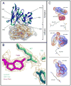
Figure 1
Figure 1
Crystal structures of the antibody–HEL complexes. (A) Ribbon depiction of the crystal structure of the scFv–HEL complex overlaid with 5FW-scFv. Antibodies are colored dark blue and cyan, and HEL is colored yellow. The two structures are virtually superimposable. The six modified tryptophan residues are annotated. (B) Depiction of the FoFc electron density map at the antibody–HEL interface (antibody colored purple, HEL colored green) showing the quality of the X-ay data. (C) Expansions of three key tryptophan residues in the overlay of the complex from panel A. The unmodified scFv is colored green and 5FW-scFv red, and HEL residues are colored gray. These magnifications show any possible steric or electronic influence on the interaction with specific HEL residues.
Figure 2
Figure 2
Comparison of the (A) UV spectra normalized to the absorption maximum and (B) CD spectra for scFv (—) and 5FW-scFv (---). (C) Comparison of CD data for the scFv–HEL (—) and 5FW-scFv–HEL (---) complexes.
Figure 3
Figure 3
Impact of complete tryptophan replacement with 5FW on antibody–antigen interaction. SPR sensograms of scFv (black) and 5FW-scFv (red). The antigen was immobilized on a carboxymethyl dextran chip surface via amine coupling, and binding (response) was monitored as the antibody was injected over this surface at a flow rate of 10 μL/min. Curves represent comparisons of one time course for injection with a group of four different injection times.
Figure 4
Figure 4
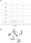
One-dimensional 19F NMR solution spectra of (A) 5FW-scFv (wild type, all six tryptophans labeled) and (B–D) three single-phenylalanine mutants (five labeled tryptophans) showing the poor sensitivity and peak overlap in the free modified antibodies. The peak marked with an asterisk is an unidentified impurity that was in all samples.
Figure 5
Figure 5
Titration of increasing amounts of HEL into 5FW-scFv (top to bottom traces). The resolution of all peaks is clear at a 1:1 5FW-scFv:HEL ratio. The peak marked with an asterisk is an unidentified impurity that was in all samples.
Figure 6
Figure 6
Assignment of fluorine signals in 5FW-scFv. (A) Stacked spectra of the complex structures of 5FW-scFv with HEL and several Phe mutants with HEL. Assignments are annotated in the spectrum of the 5FW-scFv–HEL complex (bottom). The asterisk on W35 and W36 indicates that we were not able to unequivocally assign each of these peaks, and thus, their chemical shifts may be interchanged. (B) Expansion of the six 5FW residues from the complex crystal structure in Figure 1A depicting the possible chemical and magnetic influence specific phenylalanine mutants may have on the fluorine chemical shift of nearby 5FW. The singlet peak marked with an asterisk is an unidentified impurity that was in all samples.
Figure 7
Figure 7
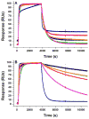
Impact of single tryptophan to phenylalanine mutations and 5FW incorporation on antibody–antigen interaction. SPR single-cycle sensorgrams are provided and were normalized to the starting point for dissociation for comparison. (A) Single-phenylalanine substitutions with 5FW biosynthetically incorporated at the five other tryptophan positions: 5FW-scFv (black), 5FW(Phe34)-scFv (red), 5FW(Phe98)-scFv (blue), 5FW(Phe36)-scFv (green), 5FW(Phe103)-scFv (orange), and 5FW(Phe94)-scFv (magenta). (B) Single-phenylalanine subsitutions with no 5FW incorporation: scFv (black), (Phe34)-scFv (red), (Phe98)-scFv (blue), (Phe36)-scFv (green), (Phe103)-scFv (orange), and (Phe94)-scFv (magenta).

References

    1. Braden BC, Poljak RJ. Structural Features of the Reactions between Antibodies and Protein Antigens. FASEB J. 1995;9:9–16. - PubMed
    1. Otlewski J, Apostoluk W. Structural and energetic aspects of protein-protein recognition. Acta Biochim Pol. 1997;44:367–387. - PubMed
    1. Lo Conte L, Chothia C, Janin J. The atomic structure of protein-protein recognition sites. J Mol Biol. 1999;285:2177–2198. - PubMed
    1. Davies DR, Cohen GH. Interactions of protein antigens with antibodies. Proc Natl Acad Sci USA. 1996;93:7–12. - PMC - PubMed
    1. Ramsland PA, Farrugia W. Crystal structures of human antibodies: A detailed and unfinished tapestry of immunoglobulin gene products. J Mol Recognit. 2002;15:248–259. - PubMed

Publication types

MeSH terms