. 2012 Jul 6;150(1):136-50.
doi: 10.1016/j.cell.2012.04.045.

Identification of stem cell populations in sweat glands and ducts reveals roles in homeostasis and wound repair

Affiliations

Identification of stem cell populations in sweat glands and ducts reveals roles in homeostasis and wound repair

Catherine P Lu et al. Cell. .

Abstract

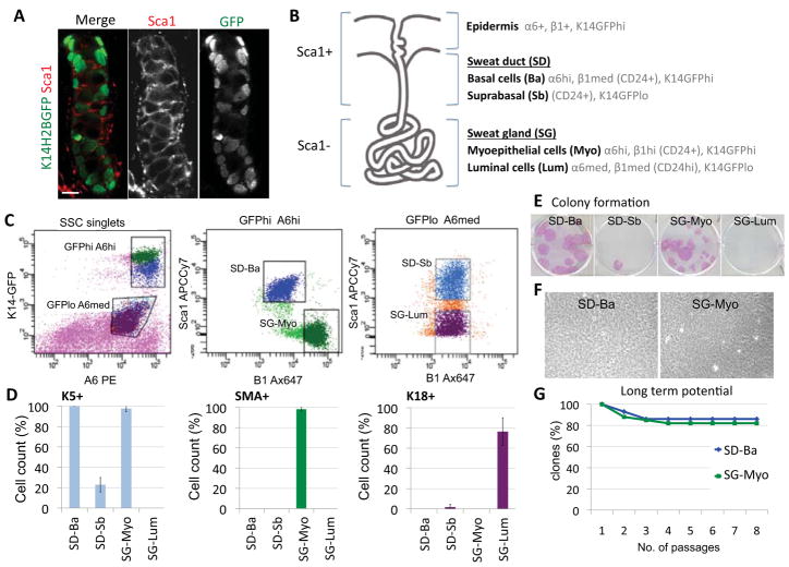
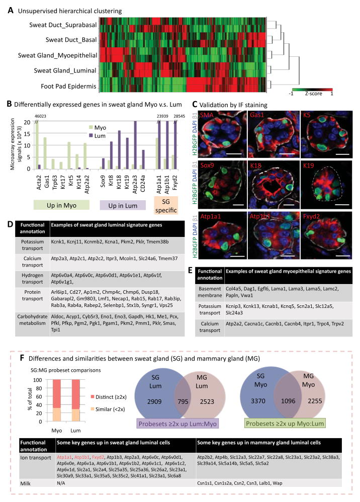
Sweat glands are abundant in the body and essential for thermoregulation. Like mammary glands, they originate from epidermal progenitors. However, they display few signs of cellular turnover, and whether they have stem cells and tissue-regenerative capacity remains largely unexplored. Using lineage tracing, we here identify in sweat ducts multipotent progenitors that transition to unipotency after developing the sweat gland. In characterizing four adult stem cell populations of glandular skin, we show that they display distinct regenerative capabilities and remain unipotent when healing epidermal, myoepithelial-specific, and lumenal-specific injuries. We devise purification schemes and isolate and transcriptionally profile progenitors. Exploiting molecular differences between sweat and mammary glands, we show that only some progenitors regain multipotency to produce de novo ductal and glandular structures, but that these can retain their identity even within certain foreign microenvironments. Our findings provide insight into glandular stem cells and a framework for the further study of sweat gland biology.


Figures

Figure 1
Figure 1. Dramatic changes in proliferation occur within developing paw skin during sweat gland morphogenesis
(A) Diagram showing orifice and intraepidermal portion of a sweat duct, which extends from epidermis (Epi) into dermis, and terminates in a coiled, secretory gland. (B) Mice constitutively expressing H2BGFP in K5 promoter-active skin epithelium were injected with EdU 4 hr prior to analyses at postnatal ages indicated. EdU+ (S-phase) cells were classified according to keratin and location (within ducts or glands) (n=100–250 EdU+ cells from 2–3 mice/stage). Overall gland but not ductal proliferation waned by maturity (P22). (C) EdU was injected daily for times indicated. Paw pads were analyzed following the last EdU injection and again at P56. Quantifications of EdU label show %myoepithelial (SMA+) and luminal (K18+) cells in EdUhi and EdUlo populations (n=3 mice, >40 pads). Representative immunofluorescence images for pulse P0–3 pawskin are shown on the left. Note: label-retaining cells were confined to glands. Scale bars, 50 μm. (D,E) H2BGFP, expressed in K5+ cells since embryogenesis, was silenced by doxy in Tet-off pTreH2BGFP/K5tTA mice as shown. (D) H2BGFP retention occurred only in glands. Scale bar, 50 μm. (E) (Left) Confocal images. Note H2BGFP label retention in SMA+ but not K18+ sweat gland cells (split channels are for boxed areas). Scale bars, 10 μm. (Right) Ultrathin sections of GFP immunogold labeled pawskin. Boxed area is magnified to see that myoepithelial cells were labeled (white arrows). Nu, nucleus. Myo, myoepithelial cells. White arrows indicate positive immunogold labeling. Scale bars, 1 μm.
Figure 2
Figure 2. Multipotent progenitors in developing sweat ducts give rise to both luminal and myoepithelial cells in sweat glands
(A) Bright field images of paw pad sections from K14Cre/RosaLacZ mice to show emerging (P3) and mature (P22) sweat glands. Sections were stained with X-gal, which turns blue in LacZ+ cells. Counter staining was done with nuclear fast red. (B) Immunofluorescence images of mature glands from K14rtta/TetOCre/RosaYFP mice induced at P1 and P28 respectively, and examined at times indicated. Some glands showed YFP-labeling of both myoepithelial (SMA+) and luminal (K8+) cells (arrow), but others only had YFP+ myoepithelial cells. (C) Cre was induced at P1–P2 in Sox9CreER X RosaLacZ or RosaYFP mice and pawskins were analyzed at times indicated. (Left), X-gal stained bright field images show that suprabasal nascent ductal cells were Sox9-promoter active and contributed to gland formation. (Right), Confocal immunofluorescence. Note: YFP+ nascent ductal cells were double positive for K14/K18. At P22, both myoepithelial and luminal glandular cells were YFP-marked (i.e. ductal-derived). Single channels of boxed areas are shown in insets. (D) Immunofluorescence images from glands of P28 K19CreER/RosaYFP mice Cre-induced at P1. Only luminal (K18+) cells were YFP-marked. (E) Cre was induced at P5–7 in K15CrePGR X RosaLacZ or RosaYFP mice and paw pads were examined at P9 and P22. Note increase over time of YFP+ cells that co-labeled with K18, indicative of clonal expansion of marked luminal cells during gland maturation. All dashed lines denote basement membrane. All scale bars are 10 μm.
Figure 3
Figure 3. Ductal but not glandular progenitors participate in epidermal wound repair
(A) Cre was induced in adult Sox9CreER/RosaLacZ mice and pawskins were either examined for LacZ expression 3d and 8wks later (unwounded, left) or subjected to epidermal scratch-wounding and examined at times indicated (wounded, right). Note: in adult mice, Sox9Cre-induction resulted in LacZ expression (blue) in all cellular compartments within the sweat duct and gland. Note expansion of βgal+ interepidermal cells surrounding the ductal orifice after wounding. Blue dashed lines, removal of epidermis. (B) Immunofluorescence images of non-wounded and 3d post-wound pawskin from Cre-induced Sox9CreER/RosaYFP mice injected with EdU for 4hr (left) or 3d (right) just prior to analysis as indicated. Magnification of boxed area is shown at its right. Quantifications are of EdU+ cells in ducts and glands (n=2 or 3 mice, >30 pads). Note that proliferation was restricted to ducts, while cells within the sweat gland remained quiescent, as depicted in the diagram in (A). Yellow dashed lines, wounded area covered by a scab. White dashed lines, basement membrane. All scale bars are 50 μm.
Figure 4
Figure 4. Myoepithelial and luminal progenitors respond as unipotent stem cells to glandular injury
(A, B) Cre was induced in K15CrePGR/RosaiDTR mice which were then treated with DT for times indicated to elicit DTR-mediated luminal cell death. EdU was given 1d before immunofluorescence analysis of pawskin, labeled with antibodies as shown (color coding is according to secondary antibodies. (A). Active caspase 3 (apoptosis) occurs only after DT. Scale bars, 50 μm. (Bottom), Sweat tests were performed on paw pads of K15CrePGR/RosaiDTR mice before, during, and after DT. Fine black dots reflect functional sweating pores, transiently reduced after glandular injury. (B) Images document luminal identity of EdU+ cells (split channels for boxed area are at right). Quantifications (n=2 mice, >80 EdU+ cells), reveal their selective proliferative response to luminal cell damage. Scale bar, 10 μm. (C) Cre was induced in K14CreER/RosaiDTR mice which were then treated with DT for 8d to induce myoepithelial cell death. EdU was given 1d before analysis. Representative fluorescence images reveal the myoepithelial identity of EdU+ cells. Quantifications (n=2 mice, >120 EdU+ cells) reveal that only myoepithelial cells proliferate in response to myoepithelial apoptosis. Scale bars, 10 μm.
Figure 5
Figure 5. Purification of distinct populations from adult sweat ducts and glands
(A) Image of sweat duct segment from K14H2BGFP pawskin, showing that cells are positive for Sca1. Scale bar, 10 μm. (B) Summary depicting localizations of markers used to isolate different populations from sweat glands and paw pad epidermis. SD, sweat duct. SG, sweat gland. (C) FACS profiles illustrating the sorting strategy. Ba, basal cells. Sb, suprabasal cells. Myo, myoepithelial cells. Lum, luminal cells. (D) Cytospin analyses for different populations isolated from the sweat ducts and glands. Percentages of K5+, SMA+, and K18+ cells in each population are shown. Quantifications were done with >150 cells from each of 3 independent sorts. (E) Colony formation assay. Representative images of colonies formed from 3000 FACS-isolated cells after 16d in culture. (F) Phase-contrast images of cells within the large holoclones formed from FACS-sorted sweat duct basal cells (SD-Ba, left) and sweat gland myoepithelial cells (SG-Myo, right). Note that both exhibit tightly packed and undifferentiated morphologies. (G) Long-term potential of purified sweat duct basal cells and sweat gland myoepithelial cells, n=28 and 34 clones, respectively. Note that for both, >80% clones survived after 8 passages (>3 months) in culture.
Figure 6
Figure 6. Expression profiling of adult sweat duct/gland populations
(A) Unsupervised Ward clustering and hierarchical analysis of the microarray profiles. (B) Examples of differentially expressed genes in myoepithelial cells and luminal cells of the sweat gland. These genes are grouped into three categories: (left) upregulated in myoepithelial cells; (middle) up-regulated in luminal cells; (right) sweat gland-specific, compared to mammary gland (see below) (C) Immunofluorescence microscopy to validate expression of sweat gland proteins whose RNA expression is up-regulated in either myoepithelial or luminal cells, or both. Sections are from K14H2BGFP adult pawskin. GFP marks myoepithelial cell nuclei. Dashed line, basement membrane. Scale bars, 10 μm. (D) Functional annotations of some of the genes up-regulated ≥2X in sweat gland luminal cells. (E) Functional annotations of some of the genes up-regulated ≥2X in sweat gland myoepithelial cells. (F) Comparisons of sweat and mammary gland mRNA profiles. Graph at left shows percentage of total probesets that are similar (<2x) or distinct (≥2x) in arrays from myoepithelial cells and luminal cells of sweat and mammary glands. Venn diagrams showing marked differences between the sweat and mammary gland molecular signatures (genes up-regulated ≥2x) of both luminal (left) and myoepithelial (right) cells. Table highlights several of the key differentially-expressed genes in luminal cells of sweat and mammary glands, respectively.
Figure 7
Figure 7. Purified progenitors from sweat ducts and glands exhibit de novo tissue morphogenesis and maintain their identity when engrafted into some (but not all) foreign microenvironments
(A) Sweat gland myoepithelial cells from K14H2BGFP mice were FACS purified and transplanted into mammary fat pads of virgin Nu/Nu mice. Engrafted tissues were analyzed at 10wks. K18, β1 and SMA immunolabelings of sections of K14H2BGFP gland-like structures reveal polarized architecture. Yellow dashed lines outline lumens. Scale bars, 10 μm. (B) Image of a 4 mo. graft of transplanted FACS-purified myoepithelial cells as in (A) but from RosaYFP mice. The entire de novo glandular structure is YFP+, demonstrating that K18+ luminal cells are derived from the donor myoepithelial population and not host mammary luminal cells. Scale bars, 10μm. (C) Ultrastructure analysis of de novo glandular structure within host mammary fat pad 10wk after transplanting donor K14H2BGFP+ myoepithelial cells. GFP+ glandular structures were identified by correlative immunofluorescence and transmission electron microscopy. I, Example of glandular structure. Cells are organized around an open luminal space. Boxed areas are enlarged in II and III. Scale bar, 10μm. II, Myoepithelial cell (Myo) beside a luminal cell (Lum). Boxed areas are magnified in insets. Boxed area (a), myoepithelial cell attachment to basement membrane (arrowheads). Boxed area (b) shows dense actin filament bundles within myoeptihelial cytoplasm. Scale bar, 500nm. III, Luminal cells within gland. Note numerous apical microvilli (Mv). Intercellular junctions are boxed, showing desmosome (De) and tight junction (Tj). Scale bar, 500nm. (D) Immunofluorescence for ATP1a1 (sweat gland-specific marker) and milk protein (mammary gland-specific marker) in de novo glandular structures from transplanted FACS-purified K14H2BGFP sweat gland myoepithelial cells and from mammary gland control tissue. Note that sweat gland myoepithelial cells retain their character even when engrafted to a mammary microenvironment. Scale bar, 10 μm. (E) Purified K14H2BGFP+ sweat duct basal cells were transplanted into mouse shoulder fat pad, and tissue was analyzed 8wks later. Left image shows a straight duct-like structure with K18+ suprabasal cells and K14H2BGFP+ basal cells. Yellow dashed lines outline lumens. Right image shows a 3D reconstruction from a Z-stack. White dashed lines demarcate the straight duct-like structure. Scale bar, 10 μm. (F–G) Purified K14H2BGFP+ sweat gland myoepithelial cells were transplanted into mammary fat pad of recipient mouse that went through pregnancy after engraftment and was lactating when the graft was taken. (F) Representative images of some sweat gland-like colonies, which still express sweat gland markers and small lumens (yellow dashed lines). Note that some glands show slight branching morphology (left). (G) Representative images of some mammary gland-like colonies that show diminished expression of sweat gland marker (ATP1a1, left) and are positive for milk proteins (right). Note they exhibit clear branching morphology and enlarged lumens, not seen in grafts from virgin hosts. Scale bars, 10 μm. (H) Graphs showing differences of sweat gland (SG) and mammary gland (MG) myoepithelial progenitors when FACS-purified from K14H2BGFP+ mice and engrafted into mammary fat pad of virgin and lactating hosts. (I) Purified K14H2BGFP+ sweat gland myoepithelial cells were combined with dermal fibroblasts and transplanted into mouse back skin. Note epidermal differentiation of engrafted cells.

References

    1. Barrandon Y, Green H. Three clonal types of keratinocyte with different capacities for multiplication. Proc Natl Acad Sci U S A. 1987;84:2302–2306. - PMC - PubMed
    1. Biedermann T, Pontiggia L, Bottcher-Haberzeth S, Tharakan S, Braziulis E, Schiestl C, Meuli M, Reichmann E. Human eccrine sweat gland cells can reconstitute a stratified epidermis. J Invest Dermatol. 2010;130:1996–2009. - PubMed
    1. Blanpain C, Lowry WE, Geoghegan A, Polak L, Fuchs E. Self-renewal, multipotency, and the existence of two cell populations within an epithelial stem cell niche. Cell. 2004;118:635–648. - PubMed
    1. Blecher SR. Anhidrosis and absence of sweat glands in mice hemizygous for the Tabby gene: supportive evidence for the hypothesis of homology between Tabby and human anhidrotic (hypohidrotic) ectodermal dysplasia (Christ-Siemens-Touraine syndrome) J Invest Dermatol. 1986;87:720–722. - PubMed
    1. Booth BW, Mack DL, Androutsellis-Theotokis A, McKay RD, Boulanger CA, Smith GH. The mammary microenvironment alters the differentiation repertoire of neural stem cells. Proc Natl Acad Sci U S A. 2008;105:14891–14896. - PMC - PubMed

Publication types

Associated data