. 2012 Jul 6;150(1):222-32.
doi: 10.1016/j.cell.2012.05.033.

Adaptive mutations that prevent crosstalk enable the expansion of paralogous signaling protein families

Affiliations

Adaptive mutations that prevent crosstalk enable the expansion of paralogous signaling protein families

Emily J Capra et al. Cell. .

Abstract

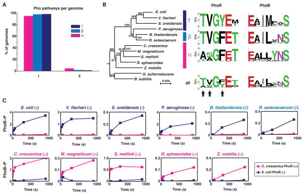
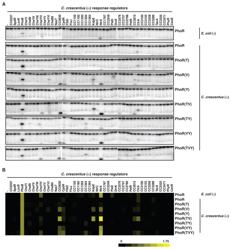
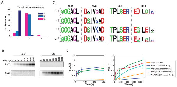
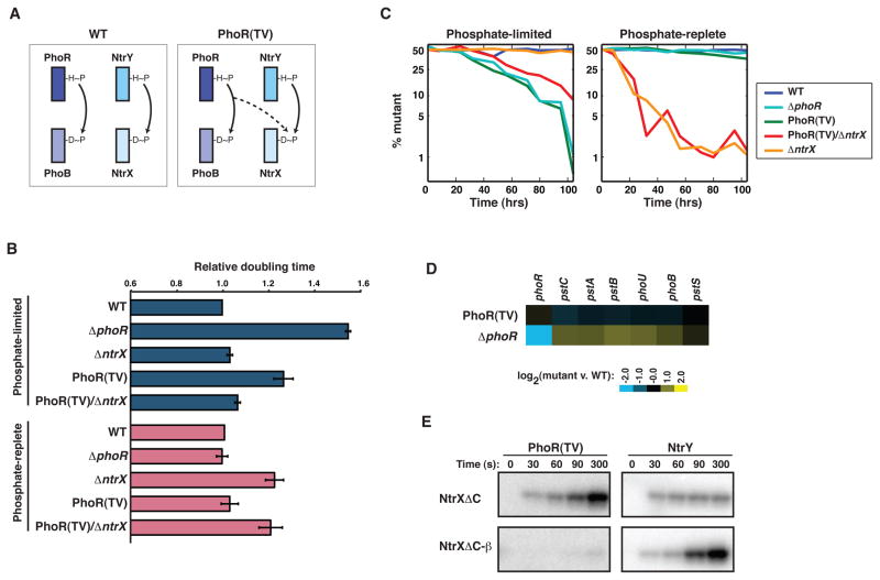
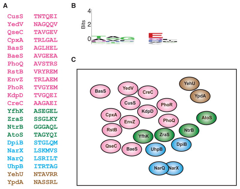
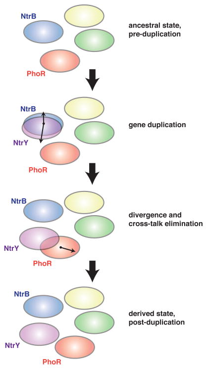
Orthologous proteins often harbor numerous substitutions, but whether these differences result from neutral or adaptive processes is usually unclear. To tackle this challenge, we examined the divergent evolution of a model bacterial signaling pathway comprising the kinase PhoR and its cognate substrate PhoB. We show that the specificity-determining residues of these proteins are typically under purifying selection but have, in α-proteobacteria, undergone a burst of diversification followed by extended stasis. By reversing mutations that accumulated in an α-proteobacterial PhoR, we demonstrate that these substitutions were adaptive, enabling PhoR to avoid crosstalk with a paralogous pathway that arose specifically in α-proteobacteria. Our findings demonstrate that duplication and the subsequent need to avoid crosstalk strongly influence signaling protein evolution. These results provide a concrete example of how system-wide insulation can be achieved postduplication through a surprisingly limited number of mutations. Our work may help explain the apparent ease with which paralogous protein families expanded in all organisms.


Figures

Figure 1
Figure 1. Phosphotransfer specificity of PhoR is different in α– and γ-proteobacteria
(A) Percentage of genomes harboring one or two Pho pathways. (B) Neighbor-joining tree for a subset of PhoR orthologs and sequence logos for specificity residues in PhoR and PhoB orthologs. Logos are shown for orthologs in each subdivision and as a combined set. Bootstrap values are out of 1000. A neighbor-joining tree for PhoB orthologs and a species tree for the species used are in Figure S1A–B. (C) Time-courses of phosphotransfer from C. crescentus PhoR (α) and E. coli PhoR (γ) to each of 11 PhoB orthologs from representative α-, γ-, and β-proteobacteria as noted above each graph. Band intensities for each PhoB were normalized in each experiment to the initial amount of autophosphorylated kinase. Values for PhoB phosphorylation can be greater than one as ATP was in excess in the reaction, allowing for re-autophosphorylation of PhoR and subsequent transfer to PhoB. For original gel images, see Figure S1D.
Figure 2
Figure 2. Substituting γ-like specificity residues into α-PhoR increases phosphorylation of NtrX
(A) Phosphotransfer profiling of E. coli PhoR, C. crescentus PhoR, and C. crescentus PhoR mutants containing γ-proteobacterial specificity residues. Each autophosphorylated kinase, indicated on the right, was incubated with each of 44 C. crescentus response regulators, indicated across the top, for 15 minutes. (B) Quantification of profiles in panel A. Band intensities for each response regulator were normalized to the level of autophosphorylated kinase and then plotted relative to PhoB. Also see Figure S2D–E.
Figure 3
Figure 3. The divergent evolution of NtrX after duplication led initially to cross-talk with PhoR in α-proteobacteria
(A) Percentage of genomes harboring one, two, or three Ntr pathways. (B) Time course of phosphotransfer from C. crescentus kinases NtrY and NtrB to C. crescentus response regulators NtrX and NtrC. The NtrY and NtrB constructs were autophosphorylated and examined for phosphotransfer at the time points indicated. Error bars represent standard deviations, n=3. (C) Sequence logos for specificity residues in NtrB-NtrC and NtrY-NtrX orthologs. Logos are shown for orthologs in each subdivision and as a combined set. (D) Time course of phosphotransfer from E. coli PhoR, C. crescentus PhoR, and C. crescentus PhoR mutants, listed in the legend, to C. crescentus PhoB and NtrX. Error bars represent standard deviations, n=3. Also see Figure S2A–C.
Figure 4
Figure 4. Cross-talk between PhoR(TV) and NtrX leads to a growth defect and fitness disadvantage in phosphate-limited media
(A) Schematic of strains examined. In the wild type, PhoR-PhoB and NtrY-NtrX are insulated, whereas the PhoR(TV) mutant leads to cross-talk between PhoR and NtrX. (B) Doubling times of C. crescentus strains ΔphoR, ΔntrX, PhoR(TV), and PhoR(TV)/ΔntrX relative to wild type in M5G (phosphate-limited) and M2G (phosphate-replete) media. Error bars indicate standard errors, n=3. For growth curves in M8G medium (5 μM), see Figure S3A–B. (C) Wild type, ΔphoR, ΔntrX, PhoR(TV), and PhoR(TV)/ΔntrX were each competed against the wild type in M2GX and M5GX. The percentage of mutant cells in the population was measured periodically for 104 hours. Curves represent the average of two independent competitions with swapped fluorophores. Also see Figure S3C–D. (D) Expression data for known members of the pho regulon in PhoR(TV) and ΔphoR in M2G (phosphate-replete) media. Data are expressed as log2 values of the ratio between a given mutant and wild-type C. crescentus, and are color-coded according to the legend. (E) Time courses of phosphotransfer from kinases PhoR(TV) and NtrY to the regulators NtrXΔC and an NtrXΔC harboring β-like specificity substitutions. Only the response regulator band is shown.
Figure 5
Figure 5. Extant two-component signaling pathways are insulated from each other at the level of phosphotransfer
(A) The six primary specificity residues are shown for each of the 22 canonical E. coli histidine kinases. Hybrid histidine kinases and the non-canonical kinases DcuS and CheA were omitted. The histidine kinases are separated into groups by color based on the family of their cognate response regulator: pink, OmpR/winged helix-turn helix; green, NtrC/AAA+ and Fis domains; blue, NarL/GerE helix-turn-helix; brown, LytR. For specificity residues from E. coli response regulators and C. crescentus histidine kinases and response regulators, see Figure S4. (B) Sequence logo for the specificity residues in panel A. (C) A qualitative two-dimensional representation of the distribution of E. coli histidine kinases in the sequence space defined by the six primary specificity-determining residues. Each oval represents the set of response regulators recognized by a histidine kinase given its specificity residues. Spheres are colored using the same scheme as in panel A. With the exception of NarQ and NarX (see text), the spheres are non-overlapping, indicating a lack of cross-talk in vivo and in vitro. Kinases were placed relative to one another based roughly on their ability to phosphorylate the cognate regulators of other histidine kinases after extended incubation times in vitro (Skerker et al., 2005; Yamamoto et al., 2005). For example, CpxA shows a strong preference for phosphotransfer to its cognate regulator CpxR, but will phosphorylate the cognate regulators of EnvZ and RstB after extended periods of time.
Figure 6
Figure 6. Adaptive divergence of duplicated signaling pathways involves the elimination of cross-talk
Ovals represent the set of response regulators recognized by a histidine kinase, as determined by its specificity residues (see also Figure 5). The NtrB-NtrC pathway is shown duplicating to produce the paralogous system NtrY-NtrX. As these pathways diverged, the specificity of NtrY overlapped that of PhoR, necessitating a change in PhoR specificity to yield the derived state with insulated pathways.

References

    1. Alm E, Huang K, Arkin A. The evolution of two-component systems in bacteria reveals different strategies for niche adaptation. PLoS Comput Biol. 2006;2:e143. - PMC - PubMed
    1. Bell CH, Porter SL, Strawson A, Stuart DI, Armitage JP. Using structural information to change the phosphotransfer specificity of a two-component chemotaxis signalling complex. PLoS Biol. 2010;8:e1000306. - PMC - PubMed
    1. Capra EJ, Perchuk BS, Lubin EA, Ashenberg O, Skerker JM, Laub MT. Systematic dissection and trajectory-scanning mutagenesis of the molecular interface that ensures specificity of two-component signaling pathways. PLoS Genet. 2010;6:e1001220. - PMC - PubMed
    1. Casino P, Rubio V, Marina A. Structural insight into partner specificity and phosphoryl transfer in two-component signal transduction. Cell. 2009;139:325–336. - PubMed
    1. Chenna R, Sugawara H, Koike T, Lopez R, Gibson TJ, Higgins DG, Thompson JD. Multiple sequence alignment with the Clustal series of programs. Nucleic Acids Res. 2003;31:3497–3500. - PMC - PubMed

Publication types

LinkOut - more resources