. 2012 Jul 6;11(1):127-35.
doi: 10.1016/j.stem.2012.04.022.

Progenitor function in self-renewing human epidermis is maintained by the exosome

Affiliations

Progenitor function in self-renewing human epidermis is maintained by the exosome

Devendra S Mistry et al. Cell Stem Cell. .

Abstract

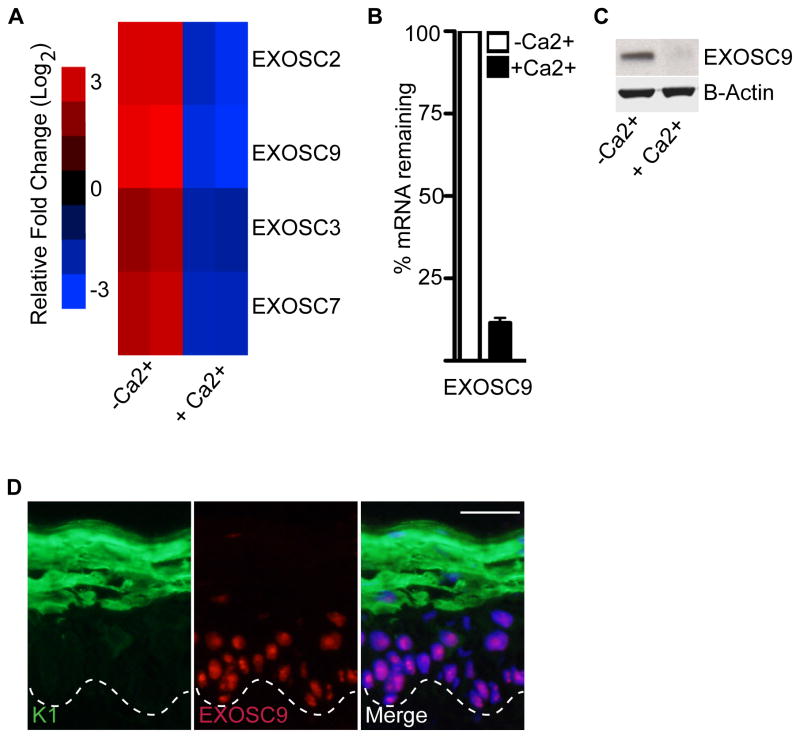
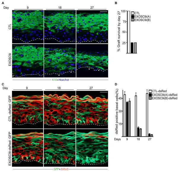
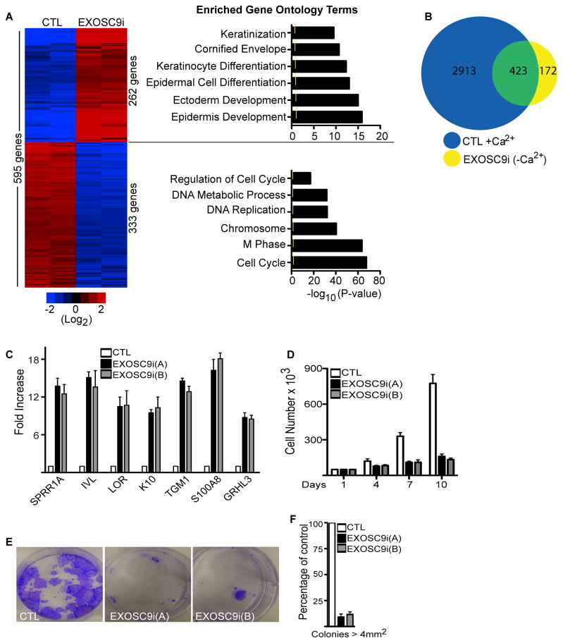
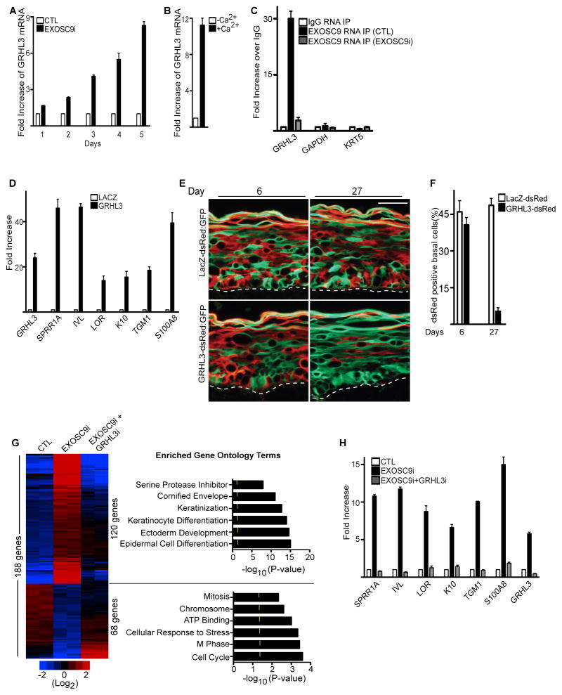
Stem and progenitor cells maintain the tissue they reside in for life by regulating the balance between proliferation and differentiation. How this is done is not well understood. Here, we report that the human exosome maintains progenitor cell function. The expression of several subunits of the exosome were found to be enriched in epidermal progenitor cells, which were required to retain proliferative capacity and to prevent premature differentiation. Loss of PM/Scl-75 also known as EXOSC9, a key subunit of the exosome complex, resulted in loss of cells from the progenitor cell compartment, premature differentiation, and loss of epidermal tissue. EXOSC9 promotes self-renewal and prevents premature differentiation by maintaining transcript levels of a transcription factor necessary for epidermal differentiation, GRHL3, at low levels through mRNA degradation. These data demonstrate that control of differentiation specific transcription factors through mRNA degradation is required for progenitor cell maintenance in mammalian tissue.


Figures

Figure 1
Figure 1. Exosome component, EXOSC9 is downregulated during epidermal differentiation
(A) Microarray analysis showing several components of the exosome being downregulated during calcium-induced differentiation (genes repressed, blue; genes induced, red; log2 scale). Heat map with (-Ca2+) denote undifferentiated and (+Ca2+) denote differentiated cells. (B–C) Downregulation of EXOSC9 during differentiation on the mRNA level and protein level. (-Ca2+) denote undifferentiated and (+Ca2+) denote differentiated cells. Error bars=mean with SEM for QRT-PCR data in (B). (D) EXOSC9 staining in adult human epidermis. EXOSC9 staining is shown in red and differentiation protein keratin 1(K1) is shown in green. Scale bar=50μm; dashed lines denote basement membrane zone.
Figure 2
Figure 2. EXOSC9 loss results in premature differentiation and loss of self-renewal in human epidermal tissue
(A) Keratinocytes expressing shRNAs for EXOSC9 (EXOSC9i) or control shRNA (CTL) were used to regenerate human epidermis on immune deficient mice. Experiments were performed using two separate shRNAs targeting different regions of EXOSC9i [EXOSC9i(A) and EXOSC9i(B)]. Tissues were harvested at 9, 18, and 27 days post grafting on mice. Keratin 10 (K10) staining shown in green marks differentiated epidermal layers. Hoechst staining in blue marks the nuclei. White arrowheads denote areas of ectopic basal layer differentiation. The dashed lines denote basement membrane zone (Scale bar=40μm; n=4 grafted mice per shRNA construct per timepoint). (B) Graft survival. Knockdown grafts that survived on mice by day 27. (C) Human epidermal progenitor competition assay. Keratinocytes were first transduced with a retrovirus to constitutively express dsRed. dsRed expressing cells were then knocked down for EXOSC9 (EXOSC9i) or control (CTL) and mixed at a 1:1 ratio with GFP expressing keratinocytes. The mixed cells were used to regenerate human epidermis on immune deficient mice. GFP expressing cells are shown in green while dsRed expressing cells are shown in red. Scale bar=40μ n=4 grafted mice per shRNA construct per timepoint. (D) Quantification of dsRed cells in the basal layer. Error bars=mean with SEM. See also Figure S1 and S2.
Figure 3
Figure 3. EXOSC9 is necessary to repress differentiation gene expression and maintain proliferation
(A) Heat map (left panel) of the 595 genes that change significantly upon EXOSC9 knockdown. Heat map is shown in red (induced genes) and blue (repressed genes) on a log2-based scale. Gene ontology analysis (right panel) of the genes with increased or decreased values upon EXOSC9 knockdown. Yellow mark in bar graphs demark p value=0.5. (B) Overlap of differentiation regulated gene set with EXOSC9i genes. 3,336 genes (blue; CTL+Ca2+) change significantly during epidermal differentiation. These 3,336 genes that change during differentiation were overlapped with the 595 genes that change when EXOSC9 is knocked down in growth conditions (EXOSC9i (–Ca2+)). Shown in the overlap (423 genes:green) are EXOSC9 regulated genes that are also differentiation regulated. (C) QRT-PCR verification of microarray data. Error bars=mean with SEM. (D) Cell proliferation assay. Cells were knocked down for EXOSC9 or control. 50,000 cells were seeded and counted over 10 days (n=3). Error bars=mean with SEM. (E) Clonogenic assay of control shRNA (CTL) and EXOSC9i keratinocytes plated at limiting dilution. (F) Quantification of colonies > 4 mm2 (n=3 per group), error bars=mean with SEM. See also Figure S3, Table S1 and Table S2.
Figure 4
Figure 4. EXOSC9 prevents premature differentiation and maintains self-renewal in progenitor cells by regulating GRHL3 transcript levels
(A) Loss of EXOSC9 results in increased levels of GRHL3. QRT-PCR on GRHL3 mRNA levels on days post EXOSC9 knockdown. (B) GRHL3 expression is increased during epidermal differentiation. QRT-PCR measurements on levels of GRHL3 increase during epidermal differentiation. Error bars=mean with SEM. (C) EXOSC9 binds to GRHL3 but not GAPDH or KRT5 transcripts. RNA immunoprecipitations (RIP) were performed using an antibody against EXOSC9 or mouse IgG on paraformaldehyde crosslinked nuclear extracts isolated from control (CTL) or EXOSC9i cells. QRTPCR was used to determine the levels of binding. Error bars=mean with SEM. (D) Forced expression of GRHL3 in epidermal progenitor cells results in premature differentiation. 4 days post-transduction of cells with a GRHL3 or LacZ encoding retrovirus, cells were harvested and assayed using QRT-PCR. Error bars=mean with SEM. (E) Epidermal progenitor competition assay. dsRed expressing epidermal cells were transduced with a retroviral construct encoding GRHL3 or LacZ. These cells were mixed at a 1:1 ratio with GFP expressing cells and used to regenerate human epidermis. GFP cells =green; dsRed cells= red. The dashed lines denote basement membrane zone (Scale bar=40μm; n=3 grafted mice per construct per timepoint). (F) Quantification of dsRed positive cells in the basal layer. (G) Knockdown of GRHL3 in EXOSC9i cells rescues the differentiation and growth arrest phenotypes of EXOSC9i cells. Heat map (left panel) of control (CTL), EXOSC9i, and EXOSC9i+GRHL3i cells. Red(induced) and blue (repressed), log2-based scale. Gene ontology analysis (right panel) of the genes rescued by double knockdown. Yellow mark in bar graphs demark p value=0.5. (H) GRHL3 is necessary for the differentiation effects of EXOSC9i cells. QRT-PCR verification of array data. Error bars=mean with SEM. See also Figure S4 and Table S3.

References

    1. Andrulis ED, Werner J, Nazarian A, Erdjument-Bromage H, Tempst P, Lis JT. The RNA processing exosome is linked to elongating RNA polymerase II in Drosophila. Nature. 2002;420:837–841. - PubMed
    1. Belostotsky D. Exosome complex and pervasive transcription in eukaryotic genomes. Curr Opin Cell Biol. 2009;21:352–358. - PubMed
    1. Belostotsky DA, Sieburth LE. Kill the messenger: mRNA decay and plant development. Curr Opin Plant Biol. 2009;12:96–102. - PubMed
    1. Blanpain C, Fuchs E. Epidermal stem cells of the skin. Annu Rev Cell Dev Biol. 2006;22:339–373. - PMC - PubMed
    1. Callahan KP, Butler JS. Evidence for core exosome independent function of the nuclear exoribonuclease Rrp6p. Nucleic Acids Res. 2008;36:6645–6655. - PMC - PubMed

Publication types