. 2012 Sep;181(3):753-60.
doi: 10.1016/j.ajpath.2012.05.014. Epub 2012 Jul 4.

Critical involvement of macrophage infiltration in the development of Sjögren's syndrome-associated dry eye

Affiliations

Critical involvement of macrophage infiltration in the development of Sjögren's syndrome-associated dry eye

Delu Zhou et al. Am J Pathol. 2012 Sep.

Abstract

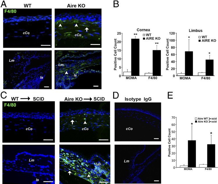
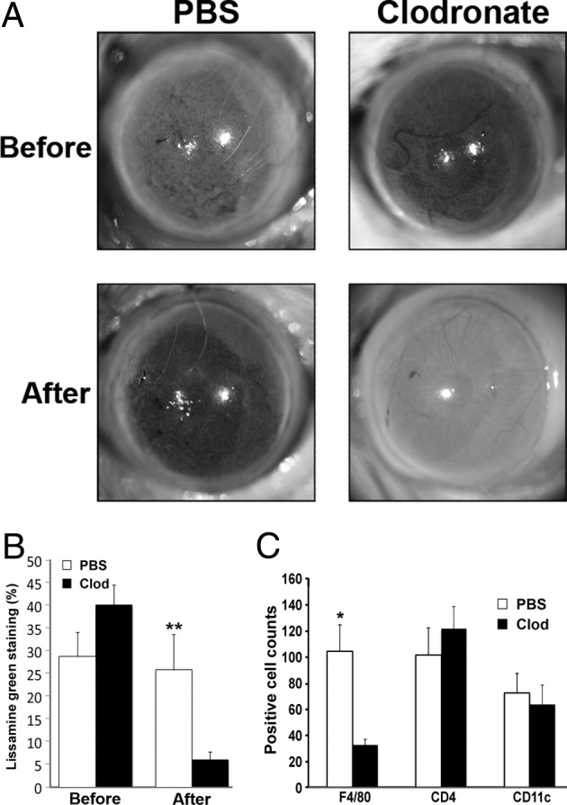
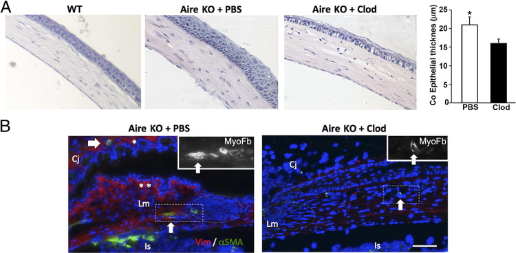
Lymphocytic infiltration of the lacrimal gland and ocular surface in autoimmune diseases such as Sjögren's syndrome (SS) causes an aqueous-deficient dry eye that is associated with significant morbidity. Previous studies from our laboratory and others have established autoimmune regulator (Aire)-deficient mice as a useful model to examine exocrinopathy and ocular surface disease associated with SS. Consistent with human SS, autoreactive CD4(+) T cells play an indispensible role in the development of exocrine and ocular surface disease in Aire knockout mice. We report that in addition to CD4(+) T cells, a large number of macrophages infiltrate the corneal stroma, limbus, and lacrimal glands of diseased mice. Adoptive transfer of autoreactive CD4(+) T cells from Aire knockout mice led to local infiltration of macrophages and ocular surface damage in immunodeficient recipients. Depletion of local macrophages, through subconjunctival injection of clodronate liposome, attenuated lissamine green staining and improved ocular phenotype. Alternatively, systemic depletion of macrophages had no effect on ocular phenotype but led to significant improvements in lacrimal gland exocrinopathy and tear secretion. Our results suggested that autoreactive CD4(+) T cells provoked macrophage infiltration to the eye and lacrimal gland, where they played a functional role in directing the development of autoimmune dry eye.


Figures

Figure 1
Figure 1
Macrophage infiltration of the central corneal (cCo) and limbus (Lm) in Aire KO mice. A: Eye tissue sections from Aire KO or WT mice were stained with antibodies directed against F4/80, MOMA-2, or IgG isotype control. F4/80+ cells infiltrating the subepithelial (arrowheads) and stromal (arrow) compartments of the cCo are indicated. The limbal region was identified using the iris insertion (Is) as an anatomical landmark. Results shown are representative of at least four independent tests. B: Quantitative analysis of MOMA-2+ and F4/80+ macrophages in the cornea and limbus of Aire KO and WT mice, shown as mean ± SE cell count. *P < 0.05 and **P < 0.01. C: CD4+ T cells from Aire KO or WT mice were adoptively transferred to immunodeficient SCID recipients. Results shown are representative of four independent tests. Mice were sacrificed at 40 days and eye sections assessed by immunofluorescence using antibodies directed against F4/80, MOMA-2, or isotype control (D). E: Quantitative analysis of corneal MOMA-2+ and F4/80+ macrophages in recipients of autoreactive CD4+ T cells (Aire KO → SCID) or WT CD4+ T cells (Aire WT → SCID), shown as mean ± SE cell number. *P < 0.05.
Figure 2
Figure 2
Effect of local macrophage depletion on ocular surface integrity and immune cell infiltration. A: Aire KO mice receiving clodronate liposome via subconjunctival injection twice a week for 2 weeks demonstrated significant improvement of ocular surface integrity as assessed by lissamine green staining. Staining was performed before and after the 2-week treatment with clodronate liposome (Clod) or PBS liposome control (PBS). B: Percentage of ocular surface staining with lissamine green is shown as mean ± SE (n = 5 to 7 per group). C: Quantification of F4/80+, CD4+, and CD11c+ cells across the ocular surface of Aire KO mice treated with Clod or PBS liposome. Data shown as mean ± SE cell number (n = 5 to 7 mice per group), *P < 0.05 and **P < 0.01.
Figure 3
Figure 3
Histologic analysis of ocular phenotype after local macrophage depletion with clodronate liposome. A: H&E staining reveals ocular surface hyperplasia as evidenced by significant thickening of the corneal (Co) epithelium, plotted as mean ± SE micrometers (n = 5 per group), P < 0.05. B: Pronounced fibrotic changes in Aire KO mice treated with PBS liposome were suggested by the increased number of subepithelial fibroblasts staining positive with vimentin (Vim+ red cells indicated by asterisks) and deeper stromal myofibroblast stained with α-smooth muscle actin (α-SMA+ green cells indicated by an arrow) adjacent to dense cellular infiltration (nuclei stained blue with DAPI, dotted circle). Aire KO mice treated with clodronate (Clod) revealed less infiltrate and lower density of fibroblasts and myofibroblast (right). Insets are high-power views of the myofibroblasts in the boxed areas. Cj, conjunctiva; Is, iris insertion; Lm, limbus. Scale bar = 50 μm. Data are representative of n = 5 per group.
Figure 4
Figure 4
Systemic depletion of macrophages increased tear secretion in Aire KO mice by preserving acinar tissues. Immunofluorescent staining of F4/80+ macrophages in the spleen (A) and lacrimal gland (B) (asterisks) are depleted after i.p. injection of clodronate liposome (Clod). F4/80+ cells appear to surround dense cellular aggregates (nuclei stained blue with DAPI, dotted circle), and bordering F4/80+ cells are depleted after i.p. clodronate (yellow box). C: Tissue lysate from WT lacrimal gland were resolved on SDS-PAGE and transferred to nitrocellulose membrane. Western blot was performed to assess the presence of lacrimal gland specific autoantibodies against an 18-kDa self-antigen, oderant binding protein, in serum samples from WT and Aire KO mice treated with Clod or PBS liposome. D: Histologic findings of lacrimal gland tissues from WT or Aire KO mice treated with Clod or PBS liposome (H&E staining, original magnification ×4). E: Percentage of immune cell infiltration in the lacrimal parenchyma. Data are presented as mean ± SE (n = 5 per group). F: Tear secretion was measured by Zone-Quick thread before and after systemic treatment with Clod or PBS. Reported as mean ± SE millimeters of wetting (n = 5 per group). *P < 0.05 and **P < 0.01.

References

    1. Kinoshita S., Nakamura T., Nishida K. Pathological keratinization of ocular surface epithelium. Adv Exp Med Biol. 2002;506:641–646. - PubMed
    1. McNamara N.A. Molecular mechanisms of keratinizing ocular surface disease. Optom Vis Sci. 2010;87:233–238. - PubMed
    1. Chen Y.T., Nikulina K., Lazarev S., Bahrami A.F., Noble L.B., Gallup M., McNamara N.A. Interleukin-1 as a phenotypic immunomodulator in keratinizing squamous metaplasia of the ocular surface in Sjögren's syndrome. Am J Pathol. 2010;177:1333–1343. - PMC - PubMed
    1. Li S., Nikulina K., DeVoss J., Wu A.J., Strauss E.C., Anderson M.S., McNamara N.A. Small proline-rich protein 1B (SPRR1B) is a biomarker for squamous metaplasia in dry eye disease. Invest Ophthalmol Vis Sci. 2008;49:34–41. - PMC - PubMed
    1. Anderson M.S., Venanzi E.S., Klein L., Chen Z., Berzins S.P., Turley S.J., von Boehmer H., Bronson R., Dierich A., Benoist C., Mathis D. Projection of an immunological self shadow within the thymus by the aire protein. Science. 2002;298:1395–1401. - PubMed

Publication types

MeSH terms