. 2012 Aug;223(2):306-13.
doi: 10.1016/j.atherosclerosis.2012.06.013. Epub 2012 Jun 16.

Copper chelation by tetrathiomolybdate inhibits vascular inflammation and atherosclerotic lesion development in apolipoprotein E-deficient mice

Affiliations

Copper chelation by tetrathiomolybdate inhibits vascular inflammation and atherosclerotic lesion development in apolipoprotein E-deficient mice

Hao Wei et al. Atherosclerosis. 2012 Aug.

Abstract

Endothelial activation, which is characterized by upregulation of cellular adhesion molecules and pro-inflammatory chemokines and cytokines, and consequent monocyte recruitment to the arterial intima are etiologic factors in atherosclerosis. Redox-active transition metal ions, such as copper and iron, may play an important role in endothelial activation by stimulating redox-sensitive cell signaling pathways. We have shown previously that copper chelation by tetrathiomolybdate (TTM) inhibits LPS-induced acute inflammatory responses in vivo. Here, we investigated whether TTM can inhibit atherosclerotic lesion development in apolipoprotein E-deficient (apoE-/-) mice. We found that 10-week treatment of apoE-/- mice with TTM (33-66 ppm in the diet) reduced serum levels of the copper-containing protein, ceruloplasmin, by 47%, and serum iron by 26%. Tissue levels of "bioavailable" copper, assessed by the copper-to-molybdenum ratio, decreased by 80% in aorta and heart, whereas iron levels of these tissues were not affected by TTM treatment. Furthermore, TTM significantly attenuated atherosclerotic lesion development in whole aorta by 25% and descending aorta by 45% compared to non-TTM treated apoE-/- mice. This anti-atherogenic effect of TTM was accompanied by several anti-inflammatory effects, i.e., significantly decreased serum levels of soluble vascular cell and intercellular adhesion molecules (VCAM-1 and ICAM-1); reduced aortic gene expression of VCAM-1, ICAM-1, monocyte chemotactic protein-1, and pro-inflammatory cytokines; and significantly less aortic accumulation of M1 type macrophages. In contrast, serum levels of oxidized LDL were not reduced by TTM. These data indicate that TTM inhibits atherosclerosis in apoE-/- mice by reducing bioavailable copper and vascular inflammation, not by altering iron homeostasis or reducing oxidative stress.


Figures

Figure 1
Figure 1. Tetrathiomolybdate reduces serum total cholesterol but increases serum triglycerides
ApoE−/− mice fed a high-fat, high-cholesterol diet were treated without or with TTM for 10 weeks as described in Methods. Serum total cholesterol (panel A) and triglycerides (panel B) were measured. Serum lipoproteins were separated by high-resolution size exclusion, fast protein liquid chromatography and cholesterol (panel C) and triglycerides (panel D) were measured in the collected fractions. VLDL elutes in fractions 12 to 20, LDL 21 to 27, and HDL 28 to 35. Data are presented as means±SEM (n=20 in each group). Asterisks denote statistical significance compared to non-TTM treated control apoE−/− mice (p<0.05).
Figure 2
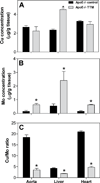
Figure 2. Tetrathiomolybdate reduces bioavailable copper in aorta, liver and heart
Animals were treated as described in the legend of Figure 1. Tissue copper and molybdenum were measured by ICP-MS as described in Methods. Concentrations of copper (panel A) and molybdenum (panel B) in aorta, liver, and heart were expressed as µg/g tissue. Copper-to-molybdenum ratios in the same tissues are shown in panel C. Data are presented as means±SEM (n=20 for heart and liver in each group, and n=4 for aorta in each group). Asterisks denote statistical significance compared to non-TTM treated control apoE−/− mice (p<0.05).
Figure 3
Figure 3. Tetrathiomolybdate increases iron levels in liver, but does not change iron and TfR mRNA and protein levels in aorta and heart
Animals were treated as described in the legend of Figure 1. Tissue iron was measured by ICP-MS as described in Methods. Concentrations of iron in heart, aorta, and liver (panel A) were expressed as µg/g tissue. Total RNA was extracted from aorta and heart. mRNA levels of TfR (panel B) were quantified using real-time RT-PCR as described in Methods and expressed as fold change of wild-type mice. Total protein was isolated from heart and immunoblotted with anti-TfR antibodies as described in Methods. GAPDH was used as internal loading control. Immunoblots show samples from three representative animals in each group (panel C). Densitometry data of TfR protein levels were generated by analyzing immunoblots with NIH ImageJ software (panel D). For panels A and B, data are presented as means±SEM (n=20 for heart and liver in each group, and n=4 for aorta in each group). For panel D, data are presented as means±SEM (n=3 for each group). Asterisks denote statistical significance compared to non- TTM-treated control apoE−/− mice (p<0.05).
Figure 4
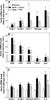
Figure 4. Tetrathiomolybdate inhibits gene expression of inflammatory mediators in aorta and heart, and macrophage markers in aorta
Animals were treated as described in the legend of Figure 1. Total RNA was extracted from aorta and heart, and mRNA levels of target genes were quantified using real-time RT-PCR as described in Methods. Aortic mRNA levels of ICAM-1, VCAM-1, MCP-1, TNFα, IL-6, CD68, F4/80, iNOS, Ym1 (Chi3l3), and Arg1 (panels A and B) and heart mRNA levels of ICAM-1, VCAM-1, TNFα, MCP-1, and IL-6 (panel C) are shown as fold change of wild-type mice. Data are presented as means±SEM (n=4 in each group for aorta mRNA and n=20 in each group for heart mRNA). Asterisks denote statistical significance compared to wild-type mice, and “#” denotes statistical significance compared to non-TTM treated control apoE−/− mice (p<0.05).
Figure 5
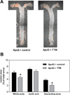
Figure 5. Tetrathiomolybdate inhibits aortic atherosclerotic lesion development
Animals were treated as described in the legend of Figure 1. Aortas were prepared and morphometric analysis of lesions was performed as described in Methods. Panel A shows surface lesions (red areas) of a pinned-out whole aorta from a representative non-TTM treated control apoE−/− mouse (left) and a TTM-treated apoE−/− mouse (right). Panel B shows percentage of aortic surface lesion areas in whole aorta, aortic arch, and descending aorta of the control and TTM-treated groups. Data are presented as means±SEM (n=12 in each group). Asterisks denote statistical significance compared to control apoE−/− mice (p<0.05).

References

    1. Lusis AJ. Atherosclerosis. Nature. 2000;407:233–241. - PMC - PubMed
    1. Davies MJ, et al. The expression of the adhesion molecules ICAM-1, VCAM-1, PECAM, and E-selectin in human atherosclerosis. J Pathol. 1993;171:223–229. - PubMed
    1. Gosling J, et al. MCP-1 deficiency reduces susceptibility to atherosclerosis in mice that overexpress human apolipoprotein B. J Clin Invest. 1999;103:773–778. - PMC - PubMed
    1. Bourdillon MC, et al. ICAM-1 deficiency reduces atherosclerotic lesions in double-knockout mice (ApoE(−/−)/ICAM-1(−/−)) fed a fat or a chow diet. Arterioscler Thromb Vasc Biol. 2000;20:2630–2635. - PubMed
    1. Collins T, Cybulsky MI. NF-kappaB: pivotal mediator or innocent bystander in atherogenesis. J Clin Invest. 2001;107:255–264. - PMC - PubMed

Publication types

MeSH terms