. 2012 Sep 15;369(2):211-22.
doi: 10.1016/j.ydbio.2012.06.021. Epub 2012 Jul 4.

The mouse Wnt/PCP protein Vangl2 is necessary for migration of facial branchiomotor neurons, and functions independently of Dishevelled

Affiliations

The mouse Wnt/PCP protein Vangl2 is necessary for migration of facial branchiomotor neurons, and functions independently of Dishevelled

Derrick M Glasco et al. Dev Biol. .

Abstract

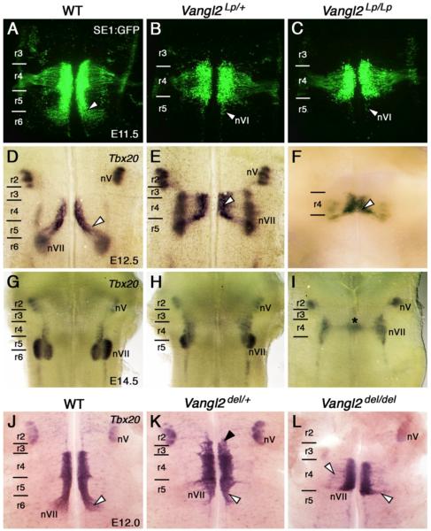
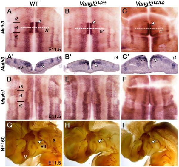
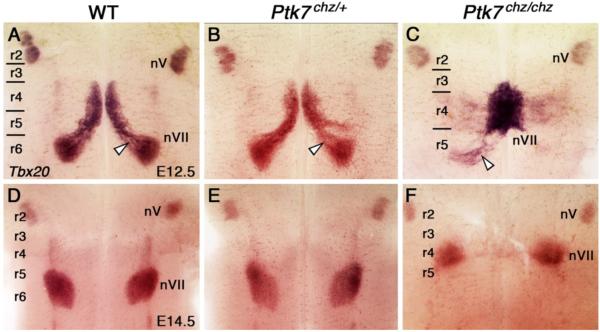
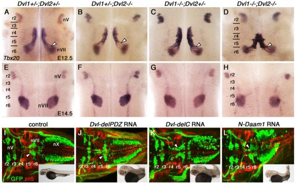
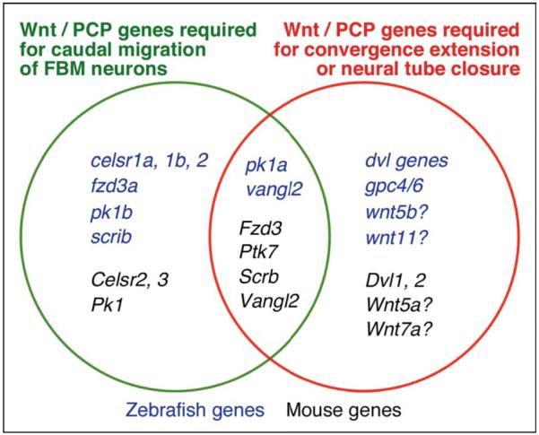
During development, facial branchiomotor (FBM) neurons, which innervate muscles in the vertebrate head, migrate caudally and radially within the brainstem to form a motor nucleus at the pial surface. Several components of the Wnt/planar cell polarity (PCP) pathway, including the transmembrane protein Vangl2, regulate caudal migration of FBM neurons in zebrafish, but their roles in neuronal migration in mouse have not been investigated in detail. Therefore, we analyzed FBM neuron migration in mouse looptail (Lp) mutants, in which Vangl2 is inactivated. In Vangl2(Lp/+) and Vangl2(Lp/Lp) embryos, FBM neurons failed to migrate caudally from rhombomere (r) 4 into r6. Although caudal migration was largely blocked, many FBM neurons underwent normal radial migration to the pial surface of the neural tube. In addition, hindbrain patterning and FBM progenitor specification were intact, and FBM neurons did not transfate into other non-migratory neuron types, indicating a specific effect on caudal migration. Since loss-of-function in some zebrafish Wnt/PCP genes does not affect caudal migration of FBM neurons, we tested whether this was also the case in mouse. Embryos null for Ptk7, a regulator of PCP signaling, had severe defects in caudal migration of FBM neurons. However, FBM neurons migrated normally in Dishevelled (Dvl) 1/2 double mutants, and in zebrafish embryos with disrupted Dvl signaling, suggesting that Dvl function is essentially dispensable for FBM neuron caudal migration. Consistent with this, loss of Dvl2 function in Vangl2(Lp/+) embryos did not exacerbate the Vangl2(Lp/+) neuronal migration phenotype. These data indicate that caudal migration of FBM neurons is regulated by multiple components of the Wnt/PCP pathway, but, importantly, may not require Dishevelled function. Interestingly, genetic-interaction experiments suggest that rostral FBM neuron migration, which is normally suppressed, depends upon Dvl function.


Figures

Figure 1
Figure 1
Vangl2 expression in the hindbrain. A-D, Ventricular views of flat-mounted hindbrains processed for in situ hybridization (ISH) with Tbx20 (A-B) and Vangl2 (C-D) probes. At E10.5 (A), Tbx20 is expressed in trigeminal branchiomotor neurons in r2-r3 and facial branchiomotor (FBM) neurons in r4. By E12.0 (B), Tbx20 expression is maintained in laterally-migrated trigeminal branchiomotor neurons in r2-r3 and caudally migrating FBM neurons spanning r4-r6. At the onset of FBM neuron migration, E10.5 (C) and E11.5 (D), Vangl2 mRNA is expressed at all axial levels of the hindbrain. Stronger expression is seen near the midline corresponding to the location of the FBM neurons. E-F, Coronal sections (r4 level) of embryos in C and D. At E10.5 (E), Vangl2 is expressed in the ventricular zone (white arrowhead) and in the FBM neuron domain (dotted circle), with low expression in the floorplate (black arrowhead). At E11.5 (F), Vangl2 expression is maintained in the ventricular zone (white arrowhead) and in the FBM neuron domain (dotted circle), and reduced in floorplate cells (black arrowhead).
Figure 2
Figure 2
FBM neurons fail to migrate caudally in looptail mutants and Vangl2 knockout embryos. The trigeminal motor nucleus (nV) is located in r2 in all embryos. A-C, Ventricular views of SE1::gfp hindbrains. In a WT embryo (A), FBM neurons (white arrowhead) migrate caudally from r4 into r6 and radially away from the midline. In Vangl2Lp/+ (B) and Vangl2Lp/Lp embryos (C), FBM neurons fail to migrate out of r4. GFP-expressing cells in r5 are abducens motor neurons (nVI; white arrowheads). D-F, Ventricular views of hindbrains processed for Tbx20 ISH. In a WT embryo (D), FBM neurons migrate caudally from r4 into r6 (white arrowhead), then migrate radially to form the facial motor nucleus (nVII) in r6. In Vangl2Lp/+ embryos (E), most Tbx20-expressing cells remain in r4 in medial and lateral positions, reflecting a near-failure of caudal migration. In Vangl2Lp/Lp embryos (F), caudal migration is completely abolished, with all FBM neurons located medially within r4. The apparent fusion of FBM populations across the midline may result from the defective floorplate and open neural tube in mutants. G-I, Pial views. In a WT embryo (G), FBM neurons have completed migration into r5/r6 to form the facial motor nucleus (nVII). In Vangl2Lp/+ embryos (H), the facial motor nucleus (nVII) is elongated within r4/r5, and is confined entirely to r4 in Vangl2Lp/Lp embryos (I), with some fused clusters (asterisk) at the midline. J-K, Ventricular views of hindbrains processed for Tbx20 ISH. In a WT embryo (J), FBM neurons (white arrowhead) migrate in characteristic fashion. In a Vangl2del/+ embryo (K), FBM neurons span r3 (black arrowhead) to r5 (white arrowhead), and do not migrate into r6, with most of the neurons confined to r4. In a Vangl2del/del embryo (L), FBM neurons do not migrate out of r4, with some cells migrating radially within r4 (white arrowheads).
Figure 3
Figure 3
Normal hindbrain development in looptail mutants. A-F, ventricular views; A’-C’, coronal sections. Math3 expression in r4 (white arrowhead) is predominantly in the motor neuron progenitor domain (pMN, dotted circle) in WT (A, A’) embryos, and is unaffected in Vangl2Lp/+ (B, B’) and Vangl2Lp/Lp (C, C’) embryos. Mash1 expression in medial aspect of r4 is also limited to the pMN domain in the WT embryo (D), and is unaffected in Vangl2Lp/+ (E), and Vangl2Lp/Lp (F) embryos. G-I, Lateral views, rostral to the left, of NF160-stained embryos. In a WT embryo (G), the trigeminal (V) and facial (VII, white arrowhead) nerves project into the first and second branchial arches, respectively, and the vagus (X) nerve exits from the caudal hindbrain. These projections are essentially normal in Vangl2Lp/+ (H) or Vangl2Lp/Lp (I) embryos.
Figure 4
Figure 4
Normal expression of Wnt/PCP genes in looptail mutants. In a WT embryo (A), Wnt5a is expressed in r5 and caudally, with a sharp r4/r5 boundary (black arrowhead). The rostral domain of Wnt5a expression (black arrow) extends into r3. Wnt5a expression is normal in Vangl2Lp/+ embryos (B). In a Vangl2Lp/Lp embryo (C), Wnt5a expression is reduced, although a boundary and the medial expression domain (arrow) are intact despite neural tube defects. In a WT embryo (D), Prickle1 is expressed in FBM neurons along their migratory route spanning r4 through r6. In Vangl2Lp/+ (E) and Vangl2Lp/Lp (F) embryos, Prickle1-expressing FBM neurons are confined to r4.
Figure 5
Figure 5
FBM neurons fail to migrate caudally in chuzhoi mutants. Ventricular (A-C) and pial (D-F) views of embryos processed for Tbx20 ISH. In E12.5 WT (A) and Ptk7chz/+ (B) embryos, FBM neurons migrate caudally from r4 into r6 (arrowheads) to form the facial motor nucleus (nVII). In Ptk7chz/chz mutants (C), FBM neurons completely or largely fail to migrate out of r4, with a few neurons migrating caudally into r5 (white arrowhead). By E14.5, FBM neurons in WT (D) and Ptk7chz/+ (E) embryos have completed their caudal and radial migrations to form facial motor nuclei (nVII) in r6. In Ptk7chz/chz mutants (F), the facial motor nuclei are rostrally displaced and located in r4 (8/8 embryos). The location of the trigeminal motor neurons (nV) in r2 is not affected in mutants.
Figure 6
Figure 6
Caudal migration of FBM neurons is not affected by loss of Dishevelled function. Ventricular (A-D) and pial (E-H) views of embryos processed for Tbx20 ISH. In E12.5 control Dvl1+/-;Dvl2+/- embryos (A), FBM neurons migrate caudally from r4 into r6 (arrowhead) to form the facial motor nucleus (nVII). This caudal migration occurs normally in Dvl1+/-;Dvl2-/- (B), Dvl1-/-;Dvl2+/- (C), and Dvl1-/-;Dvl2-/- mutants (D), inspite of neural tube closure defects in many of these embryos. By E14.5, FBM neurons in control embryos (E) have completed their caudal and radial migrations to form facial motor nuclei (nVII) in r6. Likewise, these nuclei are formed in r6 in Dvl-deficient embryos including Dvl1+/-;Dvl2-/- (F, 2/2 embryos), Dvl1-/-;Dvl2+/- (G, 6/6 embryos), and Dvl1-/-;Dvl2-/- mutants (H, 4/4 embryos). Neural tube defects were seen in Dvl1-/-;Dvl2-/- mutants. I-L, Dorsal views of 48 hpf Tg(isl1:gfp) zebrafish embryos processed for anti-GFP and zn5 immunohistochemistry. In control embryos (I), GFP-expressing FBM neurons migrate caudally from r4 into r6 (arrowhead) and r7 to form the facial motor nucleus (nVII). Zn5 staining labels rhombomere boundaries. The trigeminal (nV) and vagal (nX) motor neurons are located in r2 and r3, and the caudal hindbrain, respectively. Inset shows intact embryo with normal extension of the body axis. In embryos injected with Dvl-delPDZ (J), Dvl-delC (K), and N-Daam1 (L) RNA (200 pg/embryo), FBM neurons are able to migrate caudally (arrowheads) out of r4 despite convergence and extension defects resulting in a shortened body axis (insets). Positions of the nV and nX neurons are not affected by these treatments.
Figure 7
Figure 7
Genetic interactions between Vangl2Lp, Dvl2, and Celsr1Crsh for FBM neuron migration. The caudal migratory streams of FBM neurons are labeled with white arrowheads, while rostrally-migrating neurons in Celsr1-deficient embryos are labeled with black arrowheads. The location of the trigeminal (nV) motor nucleus (demarcating r2) is noted with an asterisk in every preparation except G. A-C, Removing Dvl2 genes from a wild-type background (A) did not affect FBM neuron migration (B, C). D-F, Removing Dvl2 genes from a Vangl2Lp/+ background (D), did not exacerbate the migration defect of Vangl2Lp/+ embryos, with thick streams of FBM neurons migrating caudally into r5 (E, F), compared to Vangl2Lp/Lp embryos (G). Fusion of FBM clusters across the midline may be a consequence of defects in neural tube and floorplate development in these embryos. H-J, While removing one copy of Dvl2 had no effect on the abnormal rostral migration of FBM neurons (black arrowhead, I), as seen in Celsr1Crsh/+ embryos (H), removing the second copy of Dvl2 completely blocked rostral migration (J), but not caudal migration. Rostral migration was also blocked in Vangl2Lp/+; Celsr1Crsh/+ embryos (K).
Figure 8
Figure 8
Differential roles for Wnt/PCP genes in regulating FBM neuron migration and neural tube morphogenesis. Mouse (black) and zebrafish (blue) Wnt/PCP genes are categorized based on their roles in regulating caudal migration of FBM neurons or hallmark PCP processes like convergence and extension movements during gastrulation (zebrafish) and neural tube closure (mouse). These data are based on the phenotypes of mouse mutants and zebrafish mutants or morphants, with the exception of zebrafish dvl genes, which were tested using dominant-negative reagents. The “?”s after the Wnt genes signify that double mutants have not been tested, so a putative role in FBM migration cannot be ruled out. Zebrafish data from Wada et al., 2006 (celsr1a, 1b, 2, fzd3a), Mapp et al., 2010, (pk1b), Wada et al., 2005 (scrib), Carreira-Barbosa et al., 2003 (pk1a), Jessen et al., 2002 (vangl2, wnt5b, wnt11), Bingham et al., 2002 (gpc4/6), and this report (dvl genes). Mouse data from Vivancos et al., 2009 (Vangl2, Fzd3, Scrb, Wnt5a, Wnt7a), Qu et al., 2010 (Celsr2, 3, Fzd3), and this report (Vangl2, Ptk7, Dvl1, 2). Mouse Pk1 knockout phenotype (B. Fritzsch, personal communication).

Similar articles

Cited by

References

    1. Bingham S, Higashijima S, Okamoto H, Chandrasekhar A. The Zebrafish trilobite gene is essential for tangential migration of branchiomotor neurons. Dev Biol. 2002;242:149–60. - PMC - PubMed
    1. Carpenter EM, Goddard JM, Chisaka O, Manley NR, Capecchi MR. Loss of Hox-A1 (Hox-1.6) function results in the reorganization of the murine hindbrain. Development. 1993;118:1063–75. - PubMed
    1. Carreira-Barbosa F, Concha ML, Takeuchi M, Ueno N, Wilson SW, Tada M. Prickle 1 regulates cell movements during gastrulation and neuronal migration in zebrafish. Development. 2003;130:4037–46. - PubMed
    1. Chandrasekhar A. Turning heads: development of vertebrate branchiomotor neurons. Dev Dyn. 2004;229:143–61. - PMC - PubMed
    1. Chandrasekhar A, Moens CB, Warren JT, Jr., Kimmel CB, Kuwada JY. Development of branchiomotor neurons in zebrafish. Development. 1997;124:2633–44. - PubMed

Publication types

MeSH terms

LinkOut - more resources