Review
. 2012 May;135(5):703-30.

Challenges in the diagnosis & treatment of miliary tuberculosis

Affiliations
Review

Challenges in the diagnosis & treatment of miliary tuberculosis

Surendra K Sharma et al. Indian J Med Res. 2012 May.

Abstract

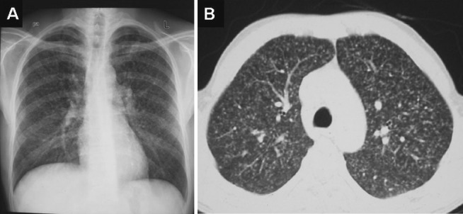
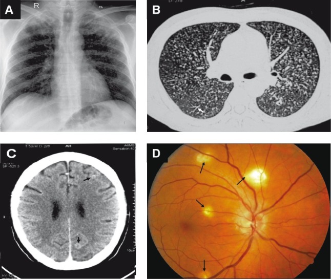
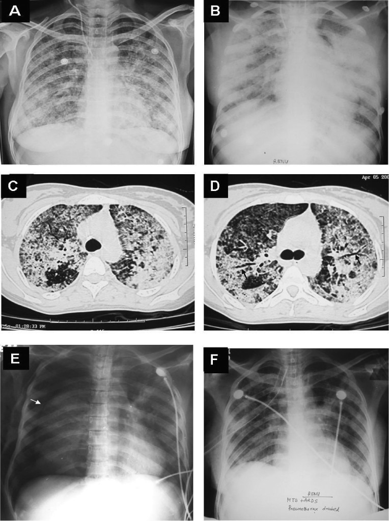
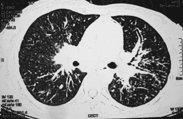
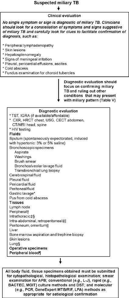
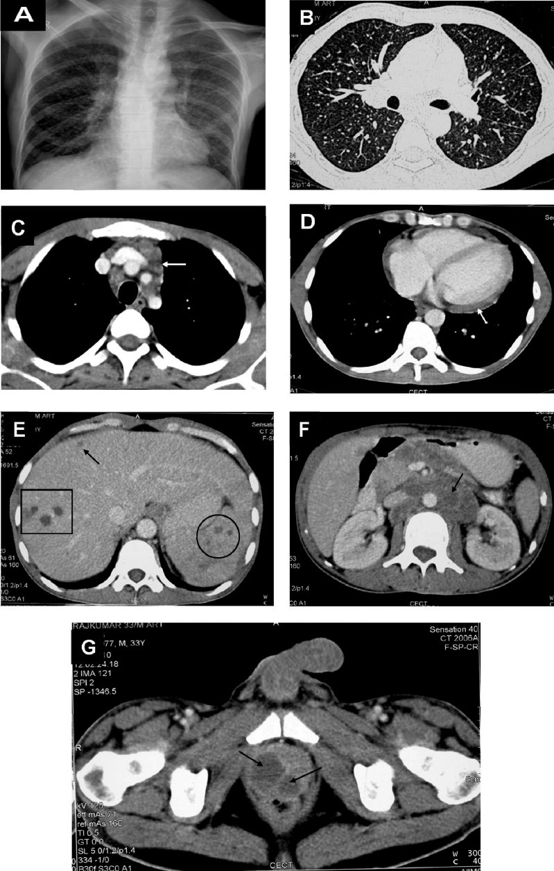
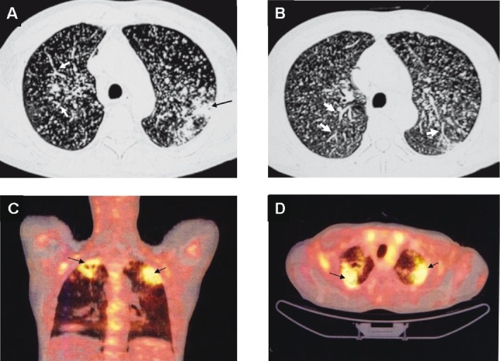
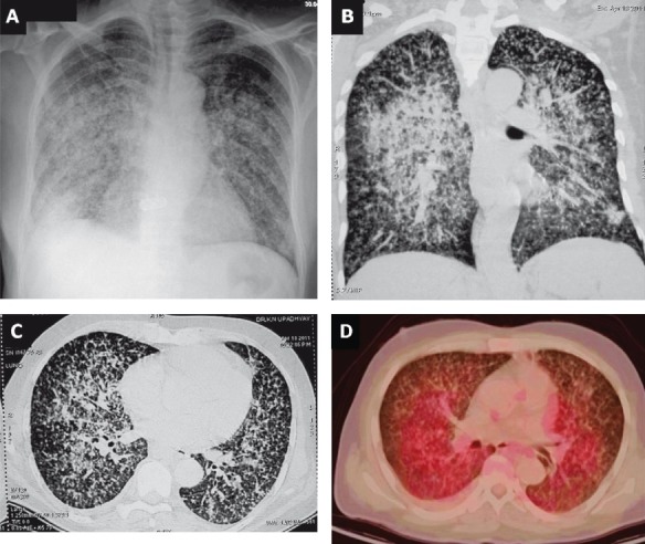
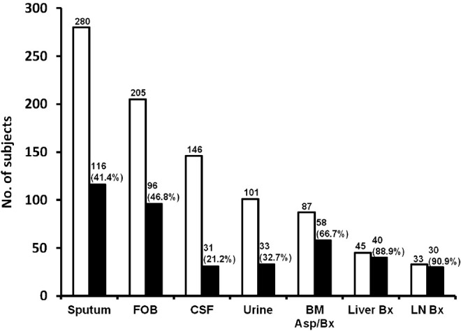
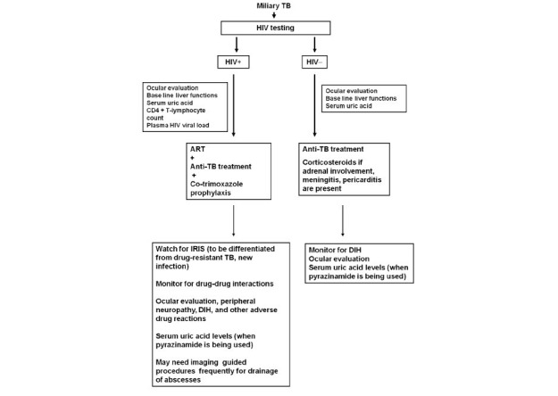
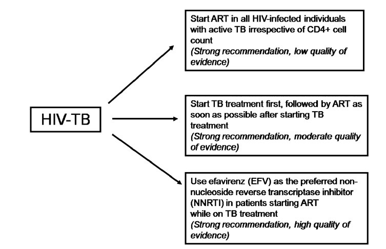
Miliary tuberculosis (TB) is a potentially lethal disease if not diagnosed and treated early. Diagnosing miliary TB can be a challenge that can perplex even the most experienced clinicians. Clinical manifestations are nonspecific, typical chest radiograph findings may not be evident till late in the disease, high resolution computed tomography (HRCT) shows randomly distributed miliary nodules and is relatively more sensitive. Ultrasonography, CT and magnetic resonance imaging (MRI) are useful in discerning the extent of organ involvement by lesions of miliary TB in extra-pulmonary locations. Fundus examination for choroid tubercles, histopathological examination of tissue biopsy specimens, conventional and rapid culture methods for isolation of Mycobacterium tuberculosis, drug-susceptibility testing, along with use of molecular biology tools in sputum, body fluids, other body tissues are useful in confirming the diagnosis. Although several prognostic markers have been described which predict mortality, yet untreated miliary TB has a fatal outcome within one year. A high index of clinical suspicion and early diagnosis and timely institution of anti-tuberculosis treatment can be life-saving. Response to first-line anti-tuberculosis drugs is good but drug-induced hepatotoxicity and drug-drug interactions in human immunodeficiency virus/acquired immunodeficiency syndrome (HIV/AIDS) patients pose significant problems during treatment. However, sparse data are available from randomized controlled trials to define the optimum regimen and duration of treatment in patients with drug-sensitive as well as drug-resistant miliary TB, including those with HIV/AIDS.


Figures

Fig. 1
Fig. 1
Chest radiograph (postero-anterior view) (A) and chest CT (lung window) (B) showing classical miliary pattern.
Fig. 2
Fig. 2
Median prevalence of symptoms and signs at initial presentation in adult patients with miliary tuberculosis. Data from references –,–,–,.
Fig. 3
Fig. 3
Clinical photograph of a child showing cutaneous lesions of miliary tuberculosis (Kind courtesy: Dr M. Ramam, Department of Dermatology, All India Institute of Medical Sciences, New Delhi, India).
Fig. 4
Fig. 4
Chest radiograph (postero-anterior view) (A) and chest CT (lung window) (B) showing classical miliary pattern, tree-in-bud appearance (B) (arrow). The patient also had cerebral tuberculomas (arrows) and TB meningitis (C). Choroid tubercles, located in the posterior pole of the orbit (D) (arrows) offered an early valuable clue to the diagnosis. The present case illustrates the importance of documenting the presence of neurological involvement, particularly, TB meningitis in patients with miliary TB and thereby ensuring adequate duration of anti-tuberculosis treatment and need for corticosteroid treatment.
Fig. 5
Fig. 5
Chest radiograph (postero-anterior view) of a pregnant woman who presented with prolonged pyrexia showing a classical miliary pattern (A). Fundus examination following mydriatic administration in both the eyes revealed choroid tubercles and had raised the suspicion of miliary TB. The patient developed ARDS during the course of her illness. Chest radiograph (antero-posterior view), obtained with a portable X-ray machine, bed-side showing bilateral frontal opacities suggestive of ARDS (B). CT chest obtained at the same time reveals air-space consolidation (C and D); air-bronchogram (arrow) (D). While assisted ventilation was being administered, the patient developed pneumothorax (asterisk) on the right side; collapsed lung border is also evident (arrow) (E). The patient required tube thoracostomy and underwater seal drainage. Eventually the patient was weaned off the ventilator and the intercostal tube was removed following resolution of the pneumothorax. The chest radiograph obtained thereafter shows significant improvement in the lesions (F). The patient survived the turbulent in-hospital course, went on to complete full-term of pregnancy and was successfully delivered a live baby. ARDS, acute respiratory distress syndrome; CT, computed tomography; TB, tuberculosis.
Fig. 6
Fig. 6
CT of the chest showing miliary sarcoidosis. While the lesions of miliary TB (Fig. 1B) are randomly distributed, the lesions of miliary sarcoidosis are distributed along the bronchovascular bundle (lymphangitic distribution). Thus transbronchial lung biopsy gives a higher diagnostic yield in miliary sarcoidosis.
Fig. 7
Fig. 7
Algorithm for the diagnostic work-up of a patient with suspected miliary TB. The clinical and imaging diagnostic work-up should also aim at accurately assessing the extent of extrapulmonary involvement to facilitate monitoring and ensure adequate duration of treatment. All laboratory testing, especially, antituberculosis drug-susceptibility testing must be carried out in quality assured, periodically accredited laboratories. *Often used in children; †FNAC/excision biopsy; ‡ Radiologically guided FNAC/biopsy; §Mediastinoscopic/video-assisted thoracoscopic surgery, biopsy; ||Laparoscopic biopsy; ¶Useful in advanced HIV infection. TB, tuberculosis; TST, tuberculin skin test; IGRA, interferon-γ release assays; HRCT, high resolution computed tomography; CECT, contrast enhanced computed tomography; MRI, magnetic resonance imaging; FNAC, fine needle aspiration cytology; HIV, human immunodeficiency virus; AFB, acid-fast bacilli; L-J, Lowenstein-Jensen medium; DST, drug-susceptibility testing; MGIT, mycobacterial growth inhibitor tube; BACTEC, radiometric culture method; PCR, polymerase chain reaction; GeneExpert MTB/RIF, GeneXpert MTB/RIF assay (Cepheid, Sunnyvale, CA); LPA, line probe assay.
Fig. 8
Fig. 8
Chest radiograph in a patient with HIV/AIDS (postero-anterior view) (A) and chest CT (lung window) (B) showing classical miliary pattern. The CECT chest (mediastinal window) also reveals intrathoracic lymphadenopathy (arrow) (C) and pericardial thickening and effusion (D). The CECT of the abdomen of the same patient reveals focal miliary lesions in the liver (square, arrow) and spleen (circle) (E) and retroperitoneal lymphadenopathy (arrow) (F); pelvic CECT shows a prostatic abscess (arrows) (G). Ultrasound guided trans-rectal prostatic aspirate smear and culture examination confirmed the diagnosis of miliary TB. The diagnostic evaluation of this patient illustrates the judicious use of imaging modalities to define the extent of organ system involvement and procuring tissue for diagnostic confirmation. Such extensive involvement usually occurs in HIV/AIDS with miliary TB. HIV, human immunodeficiency virus; AIDS, acquired immunodeficiency syndrome; CT, computed tomography; CECT, contrast enhanced computed tomography; TB, tuberculosis.
Fig. 9
Fig. 9
Chest CT (lung window) of the same patient as in Fig. 4 showing pulmonary parenchymal lesions (black arrow) (A). In addition to the miliary pattern, well-defined, linear, branching opacities (tree-in-bud appearance) (thick white arrows) (A and B) are also seen. This pattern is evident when centrilobular bronchioles are dilated, or, are filled with mucus, fluid or, pus and represents endobronchial spreading of infection. 18FDG-PET CT of the same patient showing increased activity in the pulmonary parenchymal lesions (arrows) but not in the miliary lesions (C and D). The 18FDG-PET has potential to further understanding the clinico-radiographic-functional correlation in miliary tuberculosis and merits further study. However, it may not be useful in intracranial TB. CT, computed tomography; 18FDG-PET CT, 18F labelled 2-deoxy-D-glucose positron emission tomography-computed tomography; TB, tuberculosis.
Fig. 10
Fig. 10
Chest radiograph (poster-anterior view) (A) and chest CT (lung window) (B and C) showing predominance of miliary lesions on the right side. 18FDG-PET CT of the same patient (D) showing increased activity in the coalesced pulmonary lesions, which is evident more prominently on the right side. CT, computed tomography; 18FDG-PET CT, 18F labelled 2-deoxy-D-glucose positron emission tomography-computed tomography.
Fig. 11
Fig. 11
Cumulative diagnostic yield of various body fluids and tissues in the diagnosis of miliary TB. Cumulative diagnostic yield is expressed as percentage. The data are pooled for various specimen categories and may not be comparable across various series because different criteria were employed; however, these can be appropriately used in the individual patient to ascertain the diagnosis of miliary TB. FOB, fibreoptic bronchoscopy; CSF, cerebrospinal fluid; LN, lymph node; BM, bone marrow; Bx, biopsy. Source: Refs ,–,–,–,
Fig. 12A
Fig. 12A
Algorithm for treatment of miliary TB patients with and without HIV co-infection.TB, tuberculosis; HIV, human immunodeficiency virus; +, seropositive; -, seronegative; ART, anti-retroviral treatment; IRIS, immune reconstitution inflammatory syndrome; DIH, anti-tuberculosis drug induced hepatotoxicity
Fig. 12B
Fig. 12B
Guidelines on timing of antiretroviral treatment in patients with HIV-tuberculosis co-infection. HIV, human immunodeficiency virus; TB, tuberculosis; ART, antiretroviral therapy Source: Ref.

Similar articles

Cited by

References

    1. Manget JJ. Sepulchretum sive anatomica practica. Vol. 1. London: Cramer and Perachon; 1700. Observatio XLVII (3 vols)
    1. Sahn SA, Neff TA. Miliary tuberculosis. Am J Med. 1974;56:494–505. - PubMed
    1. Sharma SK, Mohan A. Miliary tuberculosis. In: Schlossberg D, editor. Tuberculosis and nontuberculous mycobacterial infections. 6th ed. Washington: American Society for Microbiology Press; 2011. pp. 415–35.
    1. Sharma SK, Mohan A, Sharma A, Mitra DK. Miliary tuberculosis: new insights into an old disease. Lancet Infect Dis. 2005;5:415–30. - PubMed
    1. Alvarez S, McCabe WR. Extrapulmonary tuberculosis revisited: a review of experience at Boston City and other hospitals. Medicine (Baltimore) 1984;63:25–55. - PubMed

MeSH terms

Substances