Review
. 2012 Dec 3:1487:173-82.
doi: 10.1016/j.brainres.2012.05.059. Epub 2012 Jul 4.

Two independent forms of activity-dependent potentiation regulate electrical transmission at mixed synapses on the Mauthner cell

Affiliations
Review

Two independent forms of activity-dependent potentiation regulate electrical transmission at mixed synapses on the Mauthner cell

Roger Cachope et al. Brain Res. .

Abstract

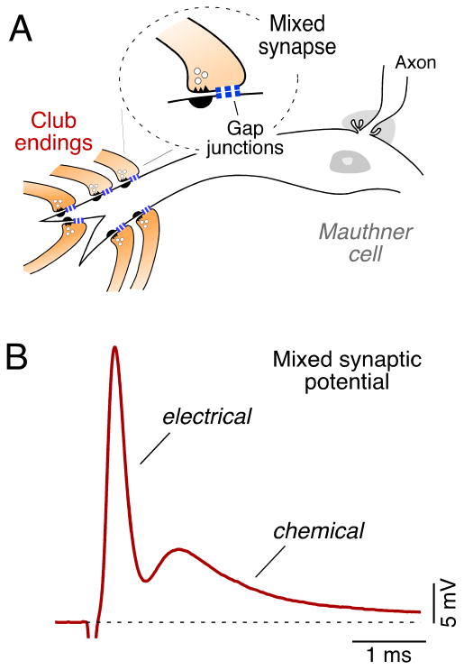
Mixed (electrical and chemical) synaptic contacts on the Mauthner cells, known as Club endings, constitute a valuable model for the study of vertebrate electrical transmission. While electrical synapses are still perceived by many as passive intercellular channels that lack modifiability, a wealth of experimental evidence shows that gap junctions at Club endings are subject to dynamic regulatory control by two independent activity-dependent mechanisms that lead to potentiation of electrical transmission. One of those mechanisms relies on activation of NMDA receptors and postsynaptic CaMKII. A second mechanism relies on mGluR activation and endocannabinoid production and is indirectly mediated via the release of dopamine from nearby varicosities, which in turn leads to potentiation of the synaptic response via a PKA-mediated postsynaptic mechanism. We review here these two forms of potentiation and their signaling mechanisms, which include the activation of two kinases with well-established roles as regulators of synaptic strength, as well as the functional implications of these two forms of potentiation. Special Issue entitled Electrical Synapses.


Figures

Figure 1
Figure 1. Club endings exhibit mixed synaptic transmission
(A) VIIIth afferents (Club endings) terminate as single mixed synaptic contact (Mixed synapse) on the lateral dendrite of the Mauthner cell (B) VIIIth nerve stimulation evokes a mixed electrical and chemical synaptic responses (trace represents the average of 20 individual responses).
Figure 2
Figure 2. Activity-dependent modulation of electrical coupling
(A) Discontinuous high-frequency stimulation (500 Hz Bursts protocol; indicated by HFS) of the VIIIth nerve can evoke persistent potentiation of both components of the mixed synaptic response. Plots here and in subsequent figures illustrate the amplitudes of the electrical (open circles) and chemical (filled circles) components versus time (each point represents the average of 20 traces) for one experiment. (B) Schematic representation of the proposed potentiating pathway. Ca++ entering through NMDA receptors activates CaMKII which phosphorylates either glutamate receptors and connexins or regulatory molecules. Modified from Pereda et al., 1998.
Figure 3
Figure 3. Endocannabinoid mediated activity-dependent modulation of electrical coupling
(A) Repetitive stimulation of the posterior VIIIth nerve (100 Hz protocol; indicated by HFS) evoked robust potentiation of both components of the mixed synaptic potential (n = 5). (B) Model for endocannabinoid-mediated potentiation of electrical and chemical synaptic transmission at Club endings. Synaptic activity leads to mGluR activation paired with postsynaptic membrane depolarization, triggering endocannabinoid (eCB) release from the postsynaptic Mauthner cell dendrite, which activates CB1Rs on dopaminergic fibers. CB1R activation leads to dopamine release that, by activating postsynaptic D1/5 receptors, increases PKA activity responsible for simultaneous potentiation of electrical (Cx35) and glutamatergic (GluR) synaptic transmission. Modified from Cachope et al., 2007.
Figure 4
Figure 4. Immunochemical evidence for the involvements of CaMKII and PKA in Club endings
Connexin35 and αCaMKII colocalize at Club endings and associate in goldfish brain. (A), Confocal z-section of a single terminal showing colocalization (yellow) of Cx35 (green) and αCaMKII (red). Labeling of Cx35 and αCaMKII colocalized at the periphery and the center of the synaptic contact. (B), Higher magnification of the labeled boxed region in B. αCaMKII and Cx35 colocalize at some Cx35 puncta (arrowheads) but is absent at others (asterisks). (C), Immunoblot detection of Cx35 in two different samples (lanes 2 and 3) with monoclonal Cx35/Cx36 antibody after IP of αCaMKII from goldfish hindbrain with G301 antibody. The Cx35 immunoblot exhibits three identifiable bands. Modified from Flores et al., 2010. (C) Polyclonal anti-Cx35/P110 antibody recognizes Cx35 as a pair of bands at 32–33 kDa (arrow, left lane, AP-) in membranes from goldfish hindbrain Labeling is lost in membranes digested with alkaline phosphatase (right lane, AP+). The high molecular weight phospho-proteins labeled nonspecifically by the antibody are not associated with Cx35 (Kothmann et al., 2007). 50 mg/lane crude membrane protein. (D) Laser scanning confocal immunofluorescence of Club endings with double labeling by polyclonal anti-Cx35/P110 (green, Alexa Fluor 488) and monoclonal anti-Cx35/36 (mCx35; red, Alexa Fluor 594) antibodies. The image shows extensive colocalization at individual Club endings in a section of the Mauthner cell lateral dendrite. A DIC image of this region is superimposed. Modified from Cachope et al., 2007.
Figure 5
Figure 5. Convergence of two mechanisms of activity-dependent potentiation in Club endings
Two different patterns of activity lead to potentiation of electrical (and chemical) transmission in Club endings. Discontinuous 500 Hz burst-like activity leads to activation of CaMKII via the involvement of NMDA receptors (“500 Hz Bursts”, red pathway). Sustained 100 Hz synaptic activation leads instead to activation of mGluR receptors and the release of endocannabinoids, which in turn lead to the local release of dopamine and the activation of postsynaptic PKA (“100 Hz”, black pathway). The involvement of PKA and CaMKII in regulating electrical transmission, which are also known to regulate the strength of chemical synapses, suggests a general role for these molecules in regulating interneuronal communication in general.

Similar articles

Cited by

References

    1. Alev C, Urschel S, Sonntag S, Zoidl G, Fort AG, Höher T, Matsubara M, Willecke K, Spray DC, Dermietzel R. The neuronal connexin36 interacts with and is phosphorylated by CaMKII in a way similar to CaMKII interaction with glutamate receptors. Proc Natl Acad Sci U S A. 2008;105:20964–9. - PMC - PubMed
    1. Alger BE. Retrograde signaling in the regulation of synaptic transmission: focus on endocannabinoids. Prog Neurobiol. 2002;68:247–86. - PubMed
    1. Asrican B, Lisman J, Otmakhov N. Synaptic strength of individual spines correlates with bound Ca2+-calmodulin-dependent kinase II. J Neurosci. 2007;27:14007–11. - PMC - PubMed
    1. Barria A, Muller D, Derkach V, Griffith LC, Soderling TR. Regulatory phosphorylation of AMPA-type glutamate receptors by CaM-KII during long-term potentiation. Science. 1997;276:2042–5. - PubMed
    1. Bartelmez GW. Mauthner's cell and the nucleus motorius tegmenti. The Journal of Comparative Neurology. 1915;25:87–128.

Publication types

MeSH terms

Substances