
TGFB2 mutations cause familial thoracic aortic aneurysms and dissections associated with mild systemic features of Marfan syndrome

Catherine Boileau et al. Nat Genet. .

Abstract

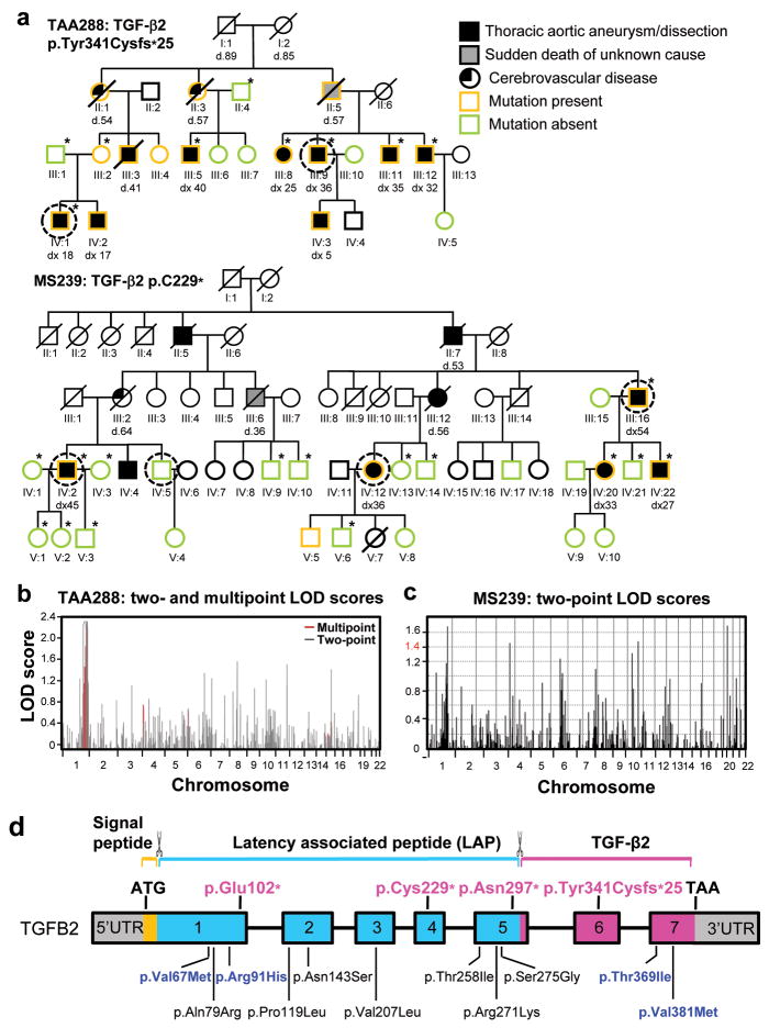
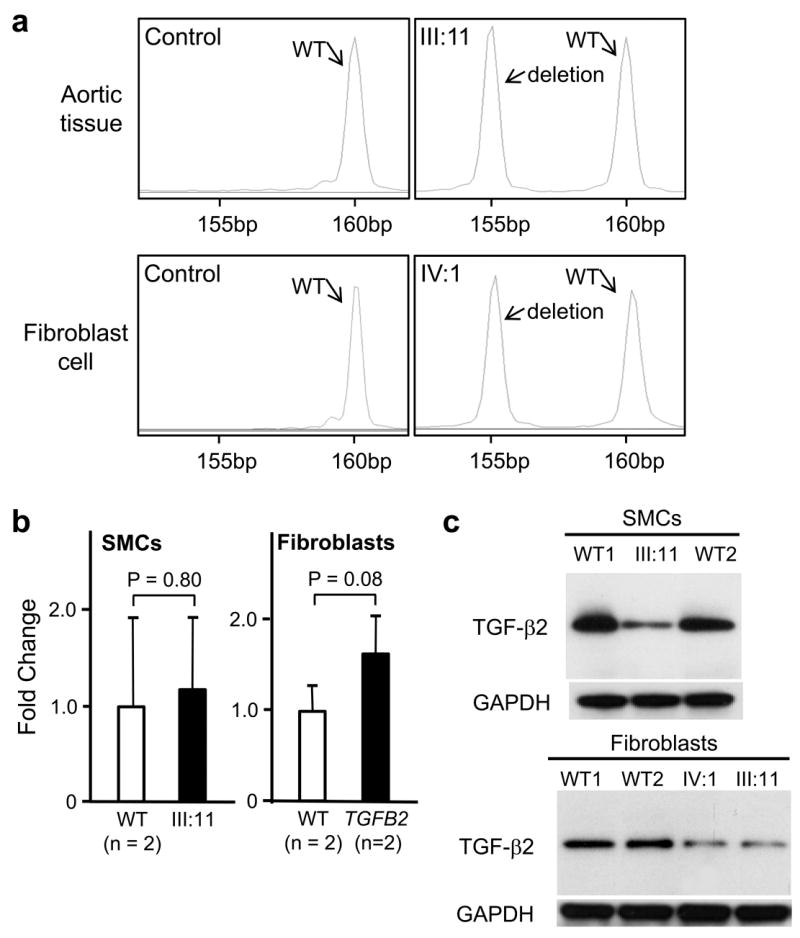
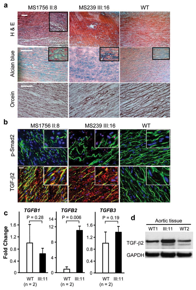
A predisposition for thoracic aortic aneurysms leading to acute aortic dissections can be inherited in families in an autosomal dominant manner. Genome-wide linkage analysis of two large unrelated families with thoracic aortic disease followed by whole-exome sequencing of affected relatives identified causative mutations in TGFB2. These mutations-a frameshift mutation in exon 6 and a nonsense mutation in exon 4-segregated with disease with a combined logarithm of odds (LOD) score of 7.7. Sanger sequencing of 276 probands from families with inherited thoracic aortic disease identified 2 additional TGFB2 mutations. TGFB2 encodes transforming growth factor (TGF)-β2, and the mutations are predicted to cause haploinsufficiency for TGFB2; however, aortic tissue from cases paradoxically shows increased TGF-β2 expression and immunostaining. Thus, haploinsufficiency for TGFB2 predisposes to thoracic aortic disease, suggesting that the initial pathway driving disease is decreased cellular TGF-β2 levels leading to a secondary increase in TGF-β2 production in the diseased aorta.


Conflict of interest statement

Competing financial interests

The authors declare no competing financial interests.

Figures

Figure 1
Figure 1
Identification of TGFB2 as the causative gene responsible for thoracic aortic disease in families TAA288 and MS239. (a) Pedigrees of family TAA288 and MS239 with the legend indicating the disease and mutation status of the family members. The age at diagnosis of aortic root enlargement and/or dissection (“dx”) is shown in years and “d” indicates age at death. A single asterisk indicates individuals whose DNA was used in genome-wide mapping. DNA from the circled individuals was used for exome sequencing. (b) Parametric two-point and multi-point LOD score profile for thoracic aortic aneurysms and dissections (TAAD) across the human genome in family TAA288 based on the Affymetrix 50K GeneChips Hind array data. The parametric two-point (grey) and multi-point (red) LOD scores are on the y-axis and are correlated to physical location of human chromosome on the x-axis. (c) Parametric two-point LOD score profile for TAAD across the human genome in family MS239. (d) Schematic representation of the TGFB2 gene and the protein domains and preproprotein proteolytic processing sites for mature TGF-β2. Boxes represent exons 1-7 with the untranslated regions (UTRs) and the open reading frame designated. The domains of the protein are designated using orange, blue and pink. The proteolytic sites of TGF-β2 preproprotein are marked with scissors symbol. Proteolytic cleavage sites remove the signal peptides from the amino-terminus and release the mature TGF-β2 from the latent associated peptide. The TGFB2 mutations identified in this study are indicated in pink type. Below the gene diagram are the rare variants found in the NHLBI exome sequencing variant server (http://evs.gs.washington.edu/EVS/); blue type designates variants predicted to be possibly or probably damaging by PolyPhen-2 analysis and black type designates variants predicted to be benign.
Figure 2
Figure 2
Transcript and protein analysis of the TGFB2 mutation p.Tyr341Cysfs*25 in exon 6. (a) RT-PCR of RNA extracted from normal ascending aortic specimens (n=2) or fibroblast cell cultures (n=2) showed only one PCR product at 160 bp. RT-PCR of RNA extracted from ascending aortic specimen of a patient (III:11) or fibroblast cell culture (IV:1) from family TAA288 demonstrated that the transcript from both normal and 5 bp deleted allele were present. (b) Expression of TGFB2 was quantified in aortic SMCs and dermal fibroblasts explanted from TGFB2 mutation carriers and matched controls using quantitative-PCR. TGFB2 expression levels between patients and controls were similar in both SMCs and fibroblasts. TGFB2 levels are standardized to GAPDH messages. The relative expression values were determined via the ΔΔCt method, and assays were performed in triplicate. Data are expressed as mean ± standard error of the mean for pooled experimental results. (c) Immunoblot analysis of TGF-β2 proprotein in cellular lysates of aortic SMCs and fibroblasts from patients (III:11 and IV:1, TAA288 family) and two normal controls. A protein band at 47kD, the molecular weight of the proprotein of TGF-β2, was identified with immunoblot analysis in both the patient and control lysates using a polyclonal antibody specific for TGF-β2. Decreased intensity of this band was found in patients’ cells, both SMCs and fibroblasts, when compared with control cells.
Figure 3
Figure 3
Aortic pathology and assessment of TGF-β signaling in patients with TGFB2 mutations. (a) Histology of aortic media from a dissection case (left column) with a TGFB2 mutation resulting in p.Glu102* (II:8, MS1756), an aneurysm case (middle column) with a TGFB2 mutation resulting in p.Cys229* (III:16, MS239 family), and a control (right column). Hematoxylin-eosin (H&E) staining displays disorganization of the aortic media with loss of SMCs and alcian blue reveals proteoglycan accumulation (blue). Note the loss, disarray and fragmentation of elastin fibers versus control with orcein staining. The white scale bars in the upper left pictures are 100μm. (b) Immunohistofluorescent staining for the same patients and control samples. Elastin fibers appear in green (autofluorescence), while nuclei are counterstained blue. Nuclear pSMAD2 staining (pink, upper panel) is present in the disease tissues and is absent in the control aorta. The lower panel shows the intense immunostaining for TGF-β2 using a polyclonal antibody specific for TGF-β2. Staining for pSMAD2 and TGF-β2 is located in the most disorganized areas of the aortic wall. The white scale bar in the upper left picture is 20μm. (c) The expression of TGFB1, TGFB2 and TGFB3 was assayed by Q-PCR using RNA isolated from the aorta of III:11 from TAA288. The expression of TGFB2 was increased over 10-fold when compared to aortic tissue isolated from controls aortas. Gene expression levels are standardized to GAPDH. The relative expression values were determined via the ΔΔCt method, and assays were performed in triplicate. Data are expressed as mean ± standard error of the mean for pooled experimental results. (d) Immunoblot analysis of the mutant and control aortas showed increased levels of the 47 kD TGF-β2 protein band in the patient’s aorta compared with two control aortas using a TGF-β2 specific antibody. Data are expressed as mean ± standard error of the mean for experimental results.
Figure 4
Figure 4
Clinical features associated with TGFB2 mutations. (a) CT scan imaging of the aorta of patient III:16 from family MS239 demonstrates dilatation predominating at the level of the sinuses of Valsalva (50 mm). (b) CT scan of a normal aorta with a diameter of 34 mm at the level of the sinuses of Valsalva. (c) Three dimensional CT scan from patient MS239, III:16 showing mild tortuosity (arrows) of cerebral arteries compared to normal control (d). Minimal arachnodactyly is evident in individual TAA288 III:8 based on positive wrist (Walker) sign (e) but negative thumb (Steinberg) sign (f).

Comment in

References

    1. Hiratzka LF, et al. 2010 ACCF/AHA/AATS/ACR/ASA/SCA/SCAI/SIR/STS/SVM Guidelines for the Diagnosis and Management of Patients With Thoracic Aortic Disease: Executive Summary. A Report of the American College of Cardiology Foundation/American Heart Association Task Force on Practice Guidelines, American Association for Thoracic Surgery, American College of Radiology, American Stroke Association, Society of Cardiovascular Anesthesiologists, Society for Cardiovascular Angiography and Interventions, Society of Interventional Radiology, Society of Thoracic Surgeons, and Society for Vascular Medicine. Circulation. 2010;121:e266–369. - PubMed
    1. Faivre L, et al. Pathogenic FBN1 mutations in 146 adults not meeting clinical diagnostic criteria for Marfan syndrome: further delineation of type 1 fibrillinopathies and focus on patients with an isolated major criterion. Am J Med Genet A. 2009;149A:854–860. - PubMed
    1. Loeys BL, et al. Aneurysm syndromes caused by mutations in the TGF-beta receptor. N Engl J Med. 2006;355:788–798. - PubMed
    1. van de Laar IM, et al. Mutations in SMAD3 cause a syndromic form of aortic aneurysms and dissections with early-onset osteoarthritis. Nat Genet. 2011;43:121–126. - PubMed
    1. Gomez D, et al. Syndromic and non-syndromic aneurysms of the human ascending aorta share activation of the Smad2 pathway. J Pathol. 2009;218:131–142. - PubMed

Publication types

MeSH terms