Comparative Study
. 2012 Aug 1;189(3):1418-30.
doi: 10.4049/jimmunol.1100431. Epub 2012 Jul 6.

Mutation at positively selected positions in the binding site for HLA-C shows that KIR2DL1 is a more refined but less adaptable NK cell receptor than KIR2DL3

Affiliations
Comparative Study

Mutation at positively selected positions in the binding site for HLA-C shows that KIR2DL1 is a more refined but less adaptable NK cell receptor than KIR2DL3

Hugo G Hilton et al. J Immunol. .

Abstract

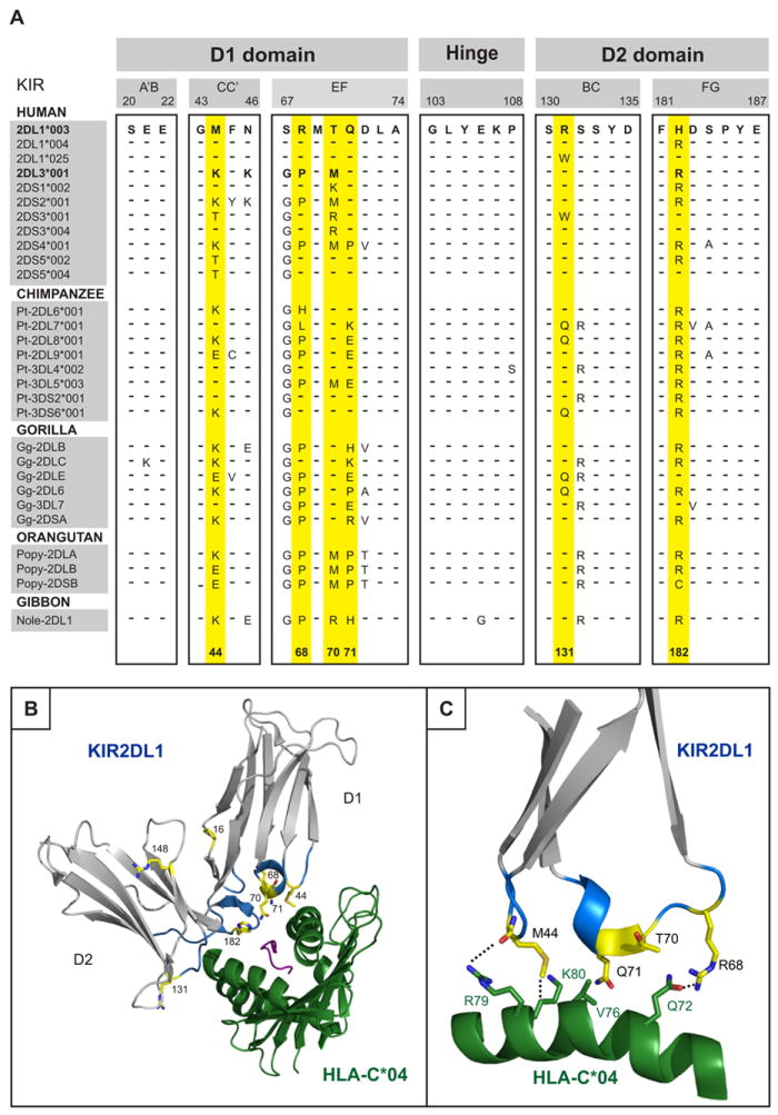
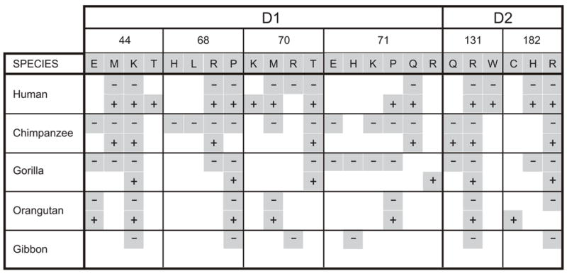
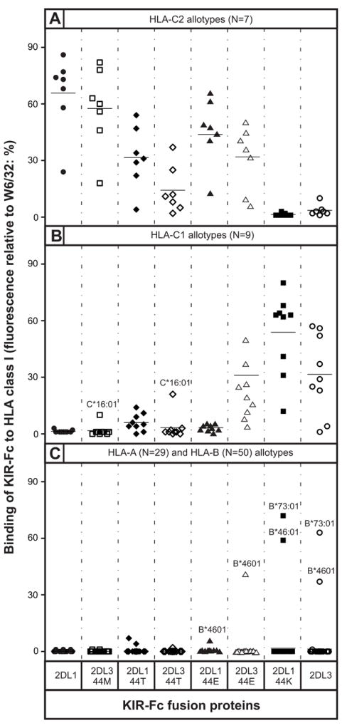
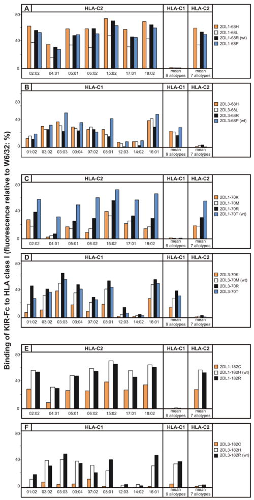
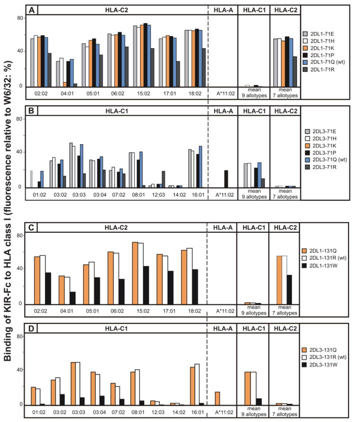
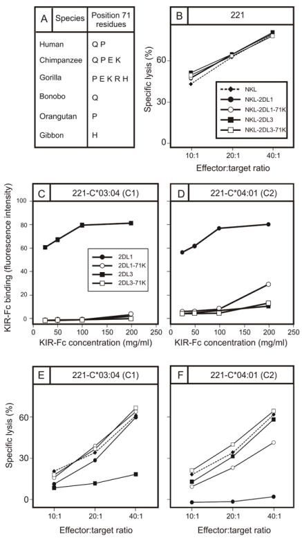
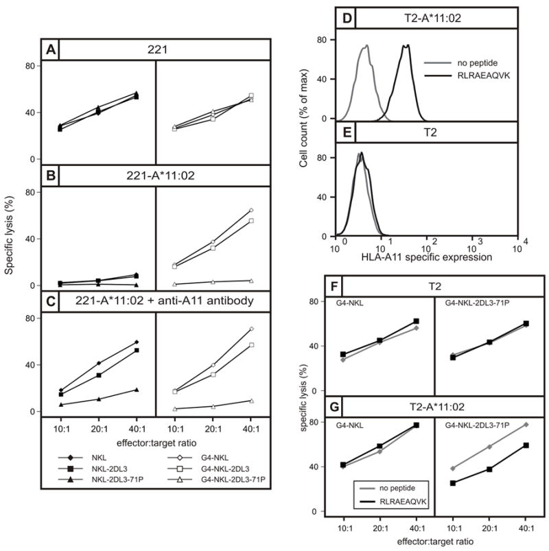
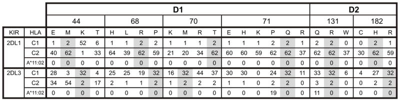
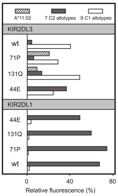
Through recognition of HLA class I, killer cell Ig-like receptors (KIR) modulate NK cell functions in human immunity and reproduction. Although a minority of HLA-A and -B allotypes are KIR ligands, HLA-C allotypes dominate this regulation, because they all carry either the C1 epitope recognized by KIR2DL2/3 or the C2 epitope recognized by KIR2DL1. The C1 epitope and C1-specific KIR evolved first, followed several million years later by the C2 epitope and C2-specific KIR. Strong, varying selection pressure on NK cell functions drove the diversification and divergence of hominid KIR, with six positions in the HLA class I binding site of KIR being targets for positive diversifying selection. Introducing each naturally occurring residue at these positions into KIR2DL1 and KIR2DL3 produced 38 point mutants that were tested for binding to 95 HLA- A, -B, and -C allotypes. Modulating specificity for HLA-C is position 44, whereas positions 71 and 131 control cross-reactivity with HLA-A*11:02. Dominating avidity modulation is position 70, with lesser contributions from positions 68 and 182. KIR2DL3 has lower avidity and broader specificity than KIR2DL1. Mutation could increase the avidity and change the specificity of KIR2DL3, whereas KIR2DL1 specificity was resistant to mutation, and its avidity could only be lowered. The contrasting inflexibility of KIR2DL1 and adaptability of KIR2DL3 fit with C2-specific KIR having evolved from C1-specific KIR, and not vice versa. Substitutions restricted to activating KIR all reduced the avidity of KIR2DL1 and KIR2DL3, further evidence that activating KIR function often becomes subject to selective attenuation.


Figures

Figure 1
Figure 1. Six positively selected residues in the binding site of hominoid lineage III KIR
(A) Alignment of partial amino acid sequences of hominoid lineage III KIR showing the loops of the D1 and D2 domains that contact HLA-C. Sequences were aligned to 2DL1*003, identities being indicated by dashes (–). The six positively selected residues in the binding site for HLA class I are colored yellow, as is also the case for panels B and C. (B) Ribbon diagram of KIR2DL1 (grey) bound to HLA-C*04:01 (green) (PDB1IM9) (56). The loops of the KIR molecule are colored blue and positively selected residues are colored yellow. (C) Details of the binding between the D1 domain contact loop of KIR2DL1*003 (in blue with positively selected residues in yellow) and the α1 domain helix of HLA-C*04:01 (green).
Figure 2
Figure 2. Sequence variation in human and ape lineage III KIR at each of the six positively selected positions located within the binding site for MHC-C
For each of the six positions (44, 68, 70, 71, 131, and 182) the variety of residues is shown. For each species, the presence of a given residue in their KIR is indicated by a grey-shaded box; a box with a dash (-) denotes its presence in inhibitory KIR, a box with a plus sign (+) denotes its presence in activating KIR.
Figure 3
Figure 3. The residue at position 44 determines the HLA-C specificity of lineage III KIR
Shown are the results of assays to measure the binding of wild-type and mutant KIR2DL1-Fc and KIR2DL3-Fc fusion proteins to beads coated with a representative range of HLA-A, -B, and -C allotypes. Mutations were restricted to position 44 where KIR2DL1 has methionine and KIR2DL3 has lysine. Fusion proteins tested were: 2DL1 (●), 2DL3-44M (□), 2DL1-44T (◆), 2DL3-44T (◇), 2DL1-44E (σ), 2DL3-44E (△), 2DL1-44K (■), and 2DL3-WT (○). Shown are representative results from (A) 7 C2-bearing HLA-C allotypes; (B) 9 C1-bearing HLA-C allotypes, and (C) 29 HLA-A and 50 HLA-B allotypes. The mean value for each allotype group is denoted by a horizontal line. Allotypes showing unusual patterns of binding are indicated by their official names (46).
Figure 4
Figure 4. The avidity of KIR2D for MHC-C has been modulated by positive selection at positions 68, 70, and 182
Shown is the binding of wild-type (wt) and mutant KIR2DL-Fc fusion proteins to beads coated with 16 HLA-C allotypes: 9 having the C1 epitope (HLA-C1) and 7 the C2 epitope (HLA-C2). Binding to each HLA-C allotype is given (left), as well as mean values for the HLA-C1 and HLA-C2 allotype groups (right). (A) Mutation at position 68 of KIR2DL1: 2DL1-68H (orange shading), 2DL1-68L (white shading), 2DL1-68R (wt) (black shading), 2DL1-68P (blue shading). (B) Mutation at position 68 of KIR2DL3: 2DL3-68H (orange shading), 2DL3-68L (white shading), 2DL3-68R (black shading) and 2DL3-68P (wt) (blue shading). (C) Mutation at position 70 of KIR2DL1: 2DL1-70K (orange shading), 2DL1-70M (white shading), 2DL1-70R (black shading) and 2DL1-70T (wt) (blue shading). (D) Mutation at position 182 of KIR2DL3: 2DL3-70K (orange shading), 2DL3-70M (wt) (white shading), 2DL3-70R (black shading) and 2DL1-70T (blue shading). (E) Mutation at position 182 of KIR2DL1: 2DL1-182C (orange shading), 2DL1-182H (wt) (white shading) and 2DL1-182R (black shading) and (F) Mutation at position 182 of KIR2DL3: 2DL3-182C (orange shading), 2DL3-182H (white shading) and 2DL3-182R (wt) (black shading). HLA-C*12:03 and C*14:02, routinely bind weakly compared to other C1-bearing allotypes.
Figure 5
Figure 5. Positive selection at positions 71 and 131 of KIR2D can introduce reactivity with HLA-A*11:02 and alter avidity for HLA-C
Shown is the binding of wild-type (WT) and mutant KIR2D-Fc fusion proteins to beads coated HLA-C (9 HLA-C1 and 7 HLA-C2) and A*11:02 allotypes. (A) Mutation at position 71 of KIR2DL1: 2DL1-71E (light-grey shading), 2DL1-71H (white shading), 2DL1-71K (orange shading), 2DL1-71P (black shading), 2DL1-71Q (wt) (blue shading), 2DL1-71R (dark-grey shading). (B) Mutation at position 71 of KIR2DL3: 2DL3-71E (light-grey shading), 2DL3-71H (white shading), 2DL3-71K (orange shading), 2DL3-71P (black shading), 2DL3-71Q (wt) (blue shading), 2DL3-71R (dark-grey shading). (C) Mutation at position 131 of KIR2DL1: 2DL1-131Q (orange shading), 2DL1-131R (wt) (white shading), 2DL1-131W (black shading). (D) Mutation at position 131 of KIR2DL3: 2DL3-131Q (orange shading), 2DL1-131R (wt) (white shading), 2DL1-131W (black shading). HLA-C*12:03 and C*1402, routinely bind weakly compared to other C1-bearing allotypes.
Figure 6
Figure 6. Lysine 71 abrogates interaction of KIR2DL3 with HLA-C and selectively impairs interaction of KIR2DL1 with HLA-C*04:01
(A) Shown are the residues present at position 71 in hominoid lineage III KIR. Notably lysine 71 is absent from humans but present in chimpanzees and gorillas. (B) Killing of class I-deficient 221 cells by NKL cells (◆, broken line), and by NKL cell transduced with KIR2DL1 (●), 2DL1-71K (○), KIR2DL3 (■), and 2DL3-71K (□). (C,D) Binding to 221 cells expressing C1-bearing HLA-C*03:04 (C) and C2-bearing HLA-C*04:01 (D) of KIR2DL-Fc fusion proteins: KIR2DL1 (●), 2DL1-71K (○), 2DL3 (■), and 2DL3-71K (□). (E, F) Killing of 221 target cells expressing HLA-C*03:04 (E) and HLA-C*04:01 (F) by NKL cells expressing KIR2DL1 (●), 2DL1-71K (○), 2DL3 (■), and 2DL3-71K (□).
Figure 7
Figure 7. Proline 71 present in KIR2DS4 and KIR3DL2 confers specificity for HLA-A*11:02 on KIR2DL3 but not on KIR2DL1
(A, B and C) Shows the results of cytotoxicity assays in which the target cells are (A) class I deficient 221 cells, (B) 221 cells expressing HLA-A*11:02, or (C) 221 cells expressing HLA-A*11:02 cells pre-incubated with antibody specific for HLA-A*11. In the panels on the left the effector cells are NKL cells (◆), NKL cells expressing 2DL3 (■), and NKL cells expressing 2DL3-71P (σ). In the panels on the right the effector cells are G4-NKL cells (◇), G4-NKL cells expressing 2DL3 (□), and G4-NKL cells expressing 2DL3-71P (△). NKL cells express the HLA class I receptor LILRB1, whereas G4-NKL is a NKL cell in which LILRB1 expression is suppressed. (D, E) Flow cytometric analysis of the binding of anti-A*11 antibody to (D) T2 cells transfected with HLA-A*11:02 (T2-A*11:02) and (E) untransfected T2 cells, following overnight incubation in the presence (black line) or absence (grey line) of the RLRAEAQVK peptide that binds HLA-A*11 (black histogram). (F, G) Shows the results of cytotoxicity assays in which the targets were (F) T2-A*11:02 cells and (G) T2 cells, following overnight incubation in the presence (black line) or absence (grey line) of peptide RLRAEAQVK. Effector cells were G4-NKL cells (left) and G4-NKL cells expressing 2DL3-71P.
Figure 8
Figure 8. Summary of the binding avidity of KIR2DL1 and KIR2DL3 mutants for the C1 and C2 epitopes of HLA-C and for HLA-A*1102
Each value is the binding of the KIR-Fc protein to the target HLA class I expressed as a percentage of the binding of the W6/32 monoclonal to the same HLA class I. Values for C1 and C2 are the means from 9 C1-bearing and 7 C2-bearing HLA-C allotypes, respectively. Grey shading indicates the residues in wild type KIR2DL1*003 and KIR2DL3*001.
Figure 9
Figure 9. KIR2DL1 has stronger avidity and narrower HLA class I specificity than KIR2DL3
Shown is the binding of wild-type (wt) and mutant KIR2DL3- (upper) and KIR2DL1-Fc (lower) fusion proteins to beads coated with HLA-A*11:02 (cross-hatch shading), HLA-C2 allotypes (dark grey shading, mean value for 7 allotypes) and 9 HLA-C1 allotypes (white shading, mean value for 9 allotypes). The specificity and avidity of KIR2DL1 are markedly more resistant to mutation at positions 44, 71 and 131 than KIR2DL3.

References

    1. Caligiuri MA. Human natural killer cells. Blood. 2008;112:461–469. - PMC - PubMed
    1. Campbell KS, Purdy AK. Structure/function of human killer cell immunoglobulin-like receptors: lessons from polymorphisms, evolution, crystal structures and mutations. Immunology. 2011;132:315–325. - PMC - PubMed
    1. Lanier LL. NK cell receptors. Annu Rev Immunol. 1998;16:359–393. - PubMed
    1. Moretta A, Bottino C, Vitale M, Pende D, Biassoni R, Mingari MC, Moretta L. Receptors for HLA class-I molecules in human natural killer cells. Annu Rev Immunol. 1996;14:619–648. - PubMed
    1. Braud VM, Allan DS, O’Callaghan CA, Soderstrom K, D’Andrea A, Ogg GS, Lazetic S, Young NT, Bell JI, Phillips JH, Lanier LL, McMichael AJ. HLA-E binds to natural killer cell receptors CD94/NKG2A, B and C. Nature. 1998;391:795–799. - PubMed

Publication types

MeSH terms