. 2012 Aug;18(8):1286-90.
doi: 10.1038/nm.2861. Epub 2012 Jul 8.

Ultraviolet radiation damages self noncoding RNA and is detected by TLR3

Affiliations

Ultraviolet radiation damages self noncoding RNA and is detected by TLR3

Jamie J Bernard et al. Nat Med. 2012 Aug.

Abstract

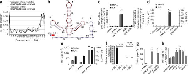
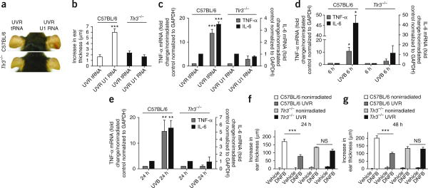
Exposure to ultraviolet B (UVB) radiation from the sun can result in sunburn, premature aging and carcinogenesis, but the mechanism responsible for acute inflammation of the skin is not well understood. Here we show that RNA is released from keratinocytes after UVB exposure and that this stimulates production of the inflammatory cytokines tumor necrosis factor α (TNF-α) and interleukin-6 (IL-6) from nonirradiated keratinocytes and peripheral blood mononuclear cells (PBMCs). Whole-transcriptome sequencing revealed that UVB irradiation of keratinocytes induced alterations in the double-stranded domains of some noncoding RNAs. We found that this UVB-damaged RNA was sufficient to induce cytokine production from nonirradiated cells, as UVB irradiation of a purified noncoding RNA (U1 RNA) reproduced the same response as the one we observed to UVB-damaged keratinocytes. The responses to both UVB-damaged self-RNAs and UVB-damaged keratinocytes were dependent on Toll-like receptor 3 (TLR3) and Toll-like receptor adaptor molecule 1 (TRIF). In response to UVB exposure, Tlr3(-/-) mice did not upregulate TNF-α in the skin. Moreover, TLR3 was also necessary for UVB-radiation-induced immune suppression. These findings establish that UVB damage is detected by TLR3 and that self-RNA is a damage-associated molecular pattern that serves as an endogenous signal of solar injury.


Figures

Figure 1
Figure 1
RNA from UVB-irradiated keratinocytes induces the production of inflammatory cytokines. (a) Quantitative PCR (qPCR) measurements of TNF-α and IL-6 mRNA (24 h) in human keratinocytes cultured for 24 h with control NHEK lysates (nonirradiated), lysates from UVB-irradiated (UVR) NHEKs or lysates from UVR NHEKs first treated with RNase. (b) The concentrations of TNF-α and IL-6 protein measured by ELISA from culture supernatants of NHEKs after the additions described in a. (c) The concentrations of TNF-α and IL-6 protein from PBMCs treated with lysates from UVR NHEKs as described in a. (d,e) qPCR measurements of TLR3 mRNA (24 h) (d) and of TNF-α and IL-6 mRNA (e) from NHEKs treated with TLR3 siRNA or control NHEKs treated with vehicle, transfection reagent (DF) or control oligonucleotides (ctrl siRNA) and then stimulated with lysates from UVR NHEKs. (f) Concentrations of TNF-α and IL-6 protein from culture supernatant of NHEKs measured by ELISA after the experiments described in e. (g) The ears of a wild-type C57BL/6 mouse and a Tlr3-/- mouse 24 h after intradermal ear injection of lysates from UVR NHEKs or equal amounts of lysates from nonirradiated NHEKs. (h) Thickness of the ear skin in mice treated as described in g. (i) qPCR measurements of TNF-α and IL-6 mRNA in tissue extracts of ear skin treated as described in g. To determine statistical significance between groups, comparisons were made using two-tailed t tests *P < 0.05, ***P < 0.001. Data are means ± s.e.m. and are representative of at least three independent experiments. n = 4–6 mice per group. GAPDH, glyceraldehyde-3-phosphate dehydrogenase.
Figure 2
Figure 2
UVB damage to U1 RNA generates products that induce the production of TNF-α and IL-6. (a) Base-read frequency of specific domains of U1 RNA from keratinocytes after exposure to 15 mJ cm−2 UVB, as determined by whole-transcriptome RNA sequencing (RNA-Seq). Data are shown with the per-base coverage as a proportion of the total sequencing coverage. (b) Representation of a sequencing analysis by base coverage showing that the loop domains a, b and c in U1 RNA increase in frequency after UVB exposure (red), whereas loop d and the liner domains decrease in frequency (blue). Numbers around the diagram indicate the base number in U1 RNA. (c) qPCR measurements of TNF- α and IL-6 mRNA in NHEKs 24 h after the addition of 100 ng of UVB irradiation (UVR) (15 mJ cm−2) U1 RNA. The addition of 100 ng of tRNA did not stimulate the NHEKs. (d) The amount of TNF-α and IL-6 protein released by NHEKs into the media 24 h after the addition of 100 ng of UVR (15 mJ cm−1) U1 RNA. (e) The amount of TNF-α and IL-6 protein released by PBMCs into the media 24 h after the addition of 100 ng of UVR (15 mJ cm−1) U1 RNA. (f) The relative abundance of U1 RNA detected at sizes greater than and less than 100 nt as determined by size exclusion gel purification before and after exposure to 15 mJ cm−2 UVB radiation. (g) The concentration of TNF-α protein in the NHEK media 24 h after the addition of UVB-generated fragments of U1 RNA less than 100 nt in length. (h) TNF-α mRNA (24 h) in NHEKs after treatment with synthetic oligonucleotides based on loops a–d of U1 RNA. To determine statistical significance between groups, comparisons were made using two-tailed t tests *P < 0.05, **P < 0.01, ***P < 0.001. Data are means ± s.e.m. and are representative of at least three independent experiments.
Figure 3
Figure 3
UVB damage to U1 RNA induces inflammatory cytokine release by activating TLR3. (a) TLR3 mRNA expression in NHEKs measured by qPCR after 24 h of culture with UVB-irradiated (UVR) U1 RNA. (b) TNF-α and IL-6 mRNA expression from NHEKs treated with siRNA to TLR3 and then stimulated with UVR U1 RNA for 24 h. (c) The concentrations of TNF-α and IL-6 in supernatants from NHEKs treated with siRNA to TLR3. (d) Intracellular colocalization of TLR3 (red fluorescence) and UVR U1 RNA labeled with Alexa Fluor 488 (green fluorescence). The blue staining is DAPI. Scale bars, 20× magnification, 50 μm; 100× magnification, 20 μm. (e) RelA and lamin B1 (loading control) detected by western blot of nuclear lysates of NHEKs treated with UVR U1 RNA for 1, 2 or 4 h. (f) Results from NHEKs cultured to 70% confluency on chamber slides, treated with siRNA to TLR3 and exposed to UVR U1 RNA for 4 h. Cells were fixed and stained with an antibody to RelA (red). Scale bar, 50 μm. To determine statistical significance between groups, comparisons were made using two-tailed t tests *P < 0.05, **P < 0.01, ***P < 0.001. Data are means ± s.e.m. and are representative of at least three independent experiments.
Figure 4
Figure 4
Recognition of UVB-irradiated RNA by TLR3 is necessary for the inflammatory response to UVB damage. (a) The ears of a wild-type C57BL/6 mouse and a Tlr3-/- mouse 24 h after intradermal ear injection of UVB-irradiated (UVR) U1 RNA or tRNA. (b) Thickness of the ear skin treated as described in a. (c) qPCR measurements of TNF-α and IL-6 mRNA in tissue extracts of ear skin treated as described in a.(d,e) TNF-α and IL-6 mRNA expression in the skin of wild-type C57BL/6 and Tlr3-/- mice 6 h (d) and 24 h (e) after exposure to 5 kJ m−2 UVB. (f,g) Ear swelling 24 h (f) and 48 h (g) after DNFB challenge (Online Methods). To determine statistical significance between groups, comparisons were made using two-tailed t tests, NS, not significant, *P < 0.05, **P < 0.01, ***P < 0.001. Data are means ± s.e.m. and are representative of at least three independent experiments. n = 4–6 mice per group.

References

    1. Armstrong BK, Kricker A. The epidemiology of UV induced skin cancer. J Photochem Photobiol B. 2001;63:8–18. - PubMed
    1. El Ghissassi F, et al. A review of human carcinogens–part D: radiation. Lancet Oncol. 2009;10:751–752. - PubMed
    1. De Fabo EC, Noonan FP. Mechanism of immune suppression by ultraviolet irradiation in vivo I Evidence for the existence of a unique photoreceptor in skin and its role in photoimmunology. J Exp Med. 1983;158:84–98. - PMC - PubMed
    1. Sontag Y, et al. Cells with UV-specifc DNA damage are present in murine lymph nodes after in vivo UV irradiation. J Invest Dermatol. 1995;104:734–738. - PubMed
    1. Setlow RB. The wavelengths in sunlight effective in producing skin cancer: a theoretical analysis. Proc Natl Acad Sci USA. 1974;71:3363–3366. - PMC - PubMed

Publication types

MeSH terms