. 2012 Aug;19(8):811-8.
doi: 10.1038/nsmb.2318. Epub 2012 Jul 8.

Cyclic AMP regulation of protein lysine acetylation in Mycobacterium tuberculosis

Affiliations

Cyclic AMP regulation of protein lysine acetylation in Mycobacterium tuberculosis

Ho Jun Lee et al. Nat Struct Mol Biol. 2012 Aug.

Abstract

Protein lysine acetylation networks can regulate central processes such as carbon metabolism and gene expression in bacteria. In Escherichia coli, cyclic AMP (cAMP) regulates protein lysine acetyltransferase (PAT) activity at the transcriptional level, but in Mycobacterium tuberculosis, fusion of a cyclic nucleotide-binding domain to a Gcn5-like PAT domain enables direct cAMP control of protein acetylation. Here we describe the allosteric activation mechanism of M. tuberculosis PAT. The crystal structures of the autoinhibited and cAMP-activated PAT reveal that cAMP binds to a cryptic site in the regulatory domain that is over 32 Å from the catalytic site. An extensive conformational rearrangement relieves this autoinhibition by means of a substrate-mimicking lid that covers the protein-substrate binding surface. A steric double latch couples the domains by harnessing a classic, cAMP-mediated conformational switch. The structures suggest general features that enable the evolution of long-range communication between linked domains.


Figures

Figure 1
Figure 1. A double steric latch auto-inhibits Mt-PatA
(a) Overall fold of Mt-PatA. The N-terminal regulatory domain (yellow) is joined to the catalytic PAT domain (magenta) by a short extended linker (red). A lid (cyan) inserted in the PAT domain buries the catalytic site, and a helix appended to the C-terminus of the PAT domain extends back to the regulatory domain and fills the cAMP-binding site. The bound acetyl-CoA substrate (green) is shown in stick representation. (b) The C-terminus occupies the cAMP-binding site in the regulatory domain. The surface of the cAMP-binding site (dashed arrow) engages the protein C-terminus. The terminal carboxylate of Gly333 and modeled cAMP are shown in stick representation. This figure is a view of the box in Fig. 1a. (c) Acetyl-CoA (spheres) binds in a shallow groove formed by conserved motifs of the PAT domain . (d) His173 (yellow) in the lid (cyan), contacts the catalytic base (Glu235, magenta), covers the acetyl donor and mimics a lysine substrate. Acetyl-CoA (green) and side chains are shown in stick model. The PAT domain is shown in magenta.
Figure 2
Figure 2. Crystal structure of the active form of Mt-PatA
(a) Difference-distance matrix between auto-inhibited and activated forms of Mt-PatA. Colors represent Cα differences of −40 to 40 Å. (b) Ribbon diagram of the auto-inhibited (left) and cAMP-activated (right) forms of Mt-PatA. The regulatory (yellow) and catalytic domains (magenta) are dramatically reoriented, and the lid (cyan) refolds. The C-terminal helix (blue) remains packed against the PAT domain. The hinge in the interdomain linker, Ser144, is shown as a gray sphere. Bound Ac-CoA (green) and cAMP (red) molecules are shown as sticks. (c) Sequence conservation displayed on the surface of the activated form of Mt-PatA shows that lid opening exposes a conserved surface (purple) around the acetyl donor site. Residues conserved in mycobacterial Mt-PatA orthologs include central β-strands of the catalytic domain and the residues binding the pantothenyl arm of acetyl-CoA. The view is the same as the activated form of Fig. 2b.
Figure 3
Figure 3. Mechanisms of interdomain communication
(a) Regulatory domains of auto-inhibited (blue) and active (red, ’) states superimposed using residues 37-113 reveal large shifts in the helical subdomain stabilized by cAMP (orange). (b) Cyclic-AMP binds at the N-terminus of a short helix and contacts conserved residues Glu89 and Arg98. Arg138 and Phe142 in helix F reorient to interact with the adenine ring. (c) Rotamer flips mediate the large conformational change in the regulatory domain upon binding cAMP (orange). Superimposed side chains (sticks) with Ringer correlation coefficients <0.2 between the electron density maps of the auto-inhibited (blue) and active (red) structures illustrate the extensive conformational remodeling. The backbone thickness is proportional to the Cα difference. (d) Residues on the active surface of Mt-PatA switch rotamers upon cAMP binding. Dihedral angles (balls) with Ringer correlation coefficients <0.2 (pink) or <0 (red) between the electron density maps of the active and auto-inhibited forms are displayed on the active form. Ac-CoA (yellow) marks the catalytic site. Over 37% of residues have Ringer correlation coefficients <0.2. Lid opening couples switches on the protein-substrate binding surface in the PAT domain (top) to changes in the regulatory domain (bottom). (e) The lid refolds to avoid a steric clash with the regulatory domain. The rotation of the regulatory domain exposes the cAMP-binding site and opens the lid. Helix F interacts with the helix I of the lid in the auto-inhibited state (blue), but changes stabilized by cAMP eliminate contacts between these helices (’) in the activated state (red).
Figure 4
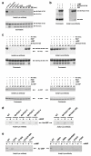
Figure 4. Mutants support a two-state model for Mt-PatA activation
(a) Auto-acetylation of His173Lys +/− cAMP. Purified His173Lys Mt-PatA (lane 1) was de-acetylated, dialyzed and de-acetylated again with Rv1151c (lanes 2-4). The His173Lys protein auto-acetylated efficiently in the absence but not presence of cAMP (lane 5-10). (b) Acetylation of USP by His173Lys +/− cAMP shows that cAMP activates the mutant enzyme to acetylate a heterologous substrate. USP alone (lane 1), or incubated with Mt-PatA His173Lys variant in the absence or presence of cAMP (lanes 2 and 3). (c) Deacetylation of Ac-His173Lys +/− cAMP and Ac-USP by Rv1151c +/− cAMP. Cyclic-AMP indirectly stimulates deacetylation of the Mt-PatA His173Lys by stabilizing the open conformation, but has no effect on deacetylation of USP. (d) Activity of C-terminal-truncations/additions. Deletion of four residues (-4) or addition of a Ser (+S) or Arg (+R) at the C-terminus activates Mt-PatA in the absence of cAMP. In contrast, Ala substitutions of conserved residues that contact cAMP block activation. These results emphasize the importance of the fit of the C-terminus into the regulatory site to form a “latch” that stabilizes the auto-inhibited state relative to the active state. (e) Changes in activity of mutants in the cAMP-binding-site (≤ residue 142) and lid (Arg184 and Phe185) reveal sites important to activate the enzyme.
Figure 5
Figure 5. The open lid in the active state uncovers a cavity for the substrate Lys
(a) Surface representation of the active conformation shows the access tunnel for the substrate Lys. Residues in the lid (cyan) and the conserved catalytic domain (magenta) form the surface surrounding the tunnel. Ac-CoA (sticks) projects the acetyl group (green C) and the sulfur of the leaving group (gold) into the active site. (b) Ribbon diagram of the binding surface for the substrate Lys in a same view and colors as Fig. 5a. The Arg184-Arg223 hydrogen bond is shown as a dotted line. (c) Catalytic site mutants reveal residues important for acetyl transfer. The pH dependence of the activity of wild-type (activity at pH 8 ≥ pH 9) and Arg223Ala (activity at pH 9 > pH 8) variants suggest the importance of Arg223 in modulating the substrate Lys pKa.
Figure 6
Figure 6. A steric double-latch regulates Mt-PatA
Release of the steric double-latch stabilized by cAMP binding. The N-terminal regulatory domain and the catalytic PAT domain are shown in yellow and magenta, respectively. The lid is shown in cyan, and the C-terminal helix is shown in blue. Cooperative interdomain communication is achieved indirectly, because the changes the regulatory domain stabilized by cAMP are incompatible with the closed lid.

References

    1. Frankel AD, Kim PS. Modular structure of transcription factors: implications for gene regulation. Cell. 1991;65:717–719. - PubMed
    1. Dorit RL, Gilbert W. The limited universe of exons. Curr. Opin. Genet. Dev. 1991;1:464–469. - PubMed
    1. Long M, de Souza SJ, Gilbert W. Evolution of the intron-exon structure of eukaryotic genes. Curr. Opin. Genet. Dev. 1995;5:774–778. - PubMed
    1. Geer LY, Domrachev M, Lipman DJ, Bryant SH. CDART: protein homology by domain architecture. Genome Res. 2002;12:1619–1623. - PMC - PubMed
    1. Marcotte EM, Pellegrini M, Thompson MJ, Yeates TO, Eisenberg D. A combined algorithm for genome-wide prediction of protein function. Nature. 1999;402:83–86. - PubMed

References for Online Methods

    1. Van Duyne GD, Standaert RF, Karplus PA, Schreiber SL, Clardy J. Atomic structures of the human immunophilin FKBP-12 complexes with FK506 and rapamycin. J. Mol. Biol. 1993;229:105–124. - PubMed
    1. Otwinowski Z, Minor W. Processing of X-ray diffraction data collected in oscillation mode. In: Carter Charles W., Jr., editor. Methods in Enzymology. Vol. 276. Academic Press; 1997. pp. 307–326. - PubMed
    1. Leslie AGW. Recent changes to the MOSFLM package for processing film and image plate data. Joint CCP4 + ESF-EAMCB Newsletter on Protein Crystallography. 1992:26.
    1. Collaborative Computational Project, N. The CCP4 Suite: Programs for Protein Crystallography. Acta Crystallographica. 1994;D50:760–763. - PubMed
    1. Adams PD, et al. PHENIX: a comprehensive Python-based system for macromolecular structure solution. Acta Crystallogr. D Biol. Crystallogr. 2010;66:213–221. - PMC - PubMed

Publication types

MeSH terms

LinkOut - more resources