. 2012 Oct;194(10):887-92.
doi: 10.1007/s00203-012-0830-1. Epub 2012 Jul 8.

Localization of new peptidoglycan at poles in Bacillus mycoides, a member of the Bacillus cereus group

Affiliations

Localization of new peptidoglycan at poles in Bacillus mycoides, a member of the Bacillus cereus group

Luana Turchi et al. Arch Microbiol. 2012 Oct.

Abstract

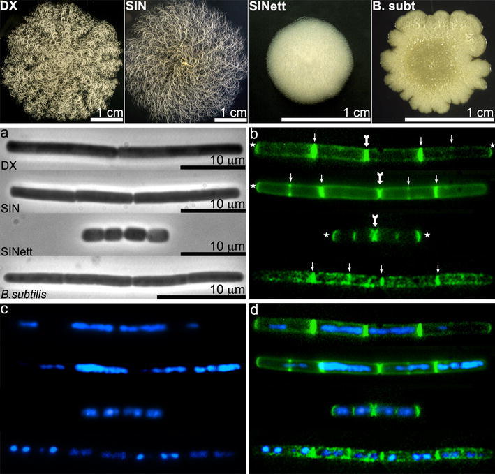
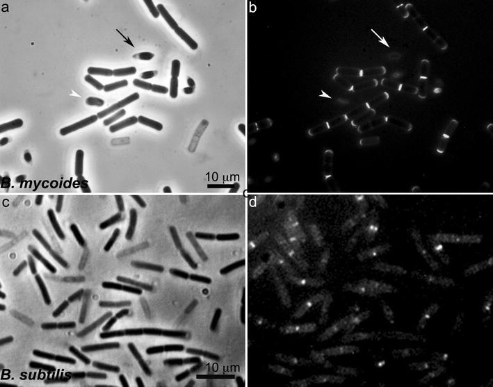
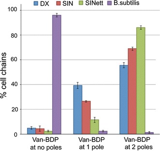
Bacillus mycoides is a sporogenic Gram-positive soil bacillus of the B. cereus group. This bacillus, which forms hyphal colonies, is composed of cells connected in filaments that make up bundles and turn clock- or counterclockwise depending on the strain. A thick peptidoglycan wall gives the rod cells of these bacilli strength and shape. One approach used to study peptidoglycan neoformation in Gram positives exploits the binding properties of antibiotics such as vancomycin and ramoplanin to nascent peptidoglycan, whose localization in the cell is monitored by means of a fluorescent tag. When we treated B. mycoides strains with BODIPY-vancomycin, we found the expected accumulation of fluorescence at the midcell septa and localization along the cell sidewall in small foci distributed quite uniformly. Intense fluorescence was also observed at the poles of many cells, more clearly visible at the outer edges of the cell chains. The unusual abundance of peptidoglycan intermediates at the cell poles after cell separation suggests that the construction process of this structure is different from that of B. subtilis, in which the free poles are rarely reactive to vancomycin.


Figures

Fig. 1
Fig. 1
Colony shape and cells of B. mycoides DX, SIN, SINett and B. subtilis 168. Top panel: colonies of B. mycoides DX, SIN and SINett were grown on TS agar for 72 h at 30 °C, while B. subtilis colonies were grown at 37 °C. Note the macroscopic hyphal pattern and the opposite direction of filament bundles in wild-type B. mycoides strains (EMBO Journal published this B. mycoides DX colony image on the cover of volume 28, 16 Sept. 2009). Bottom panel: B. mycoides DX, SIN, SINett and B. subtilis 168 cells in the exponential growth phase were treated with Van-BDP/vancomycin to stain new PG and with DAPI to visualize nucleoids as described in “Materials and methods”. a Phase contrast images. b Van-BDP fluorescence: arrows point to septa, thick arrows to internal newly formed poles, stars to fluorescent free poles. Note that fluorescence along the lateral cell wall is localized in small foci in B. mycoides and in large foci in B. subtilis. The external poles of the B. mycoides filaments are often brightly fluorescent, while they are rarely decorated by Van-BDP in B. subtilis. c DAPI staining of nucleoids. See multiple nucleoids in DX and SIN cells. d Overlay of Van-BDP (green) and DAPI (blue). Images were merged using Adobe Photoshop version 7.0
Fig. 2
Fig. 2
View of B. mycoides SIN and B. subtilis cells from germinated spores. The short filaments arose from spores seeded in TS broth. After 4 h at 30 °C (SIN) and at 37 °C (B. subtilis), Van-BDP/vancomycin at 2 μg/ml each was added for 10 min. a phase contrast and b Van-BDP fluorescence of B. mycoides SIN; c phase contrast and d Van-BDP fluorescence of B.subtilis 168. Note that the rod cells are mainly stained by Van-BDP at septa and poles in B. mycoides, and mainly at septa and along the lateral wall in large foci in B. subtilis. Some cells are “ghosts”, stained nonetheless by Van-BDP. The arrow points to a spore starting germination and the arrowhead to a spore in a more advanced germination stage
Fig. 3
Fig. 3
Van-BDP at the poles in B. mycoides strains and in B. subtilis. The histogram shows the percentage of the cell chains that show at the free external poles no Van-BDP binding, binding to only one pole or to both poles as the average of at least 3 independent experiments. Error bars show standard deviations. The poles of B. subtilis are very rarely bound by Van-BDP. Among the B. mycoides strains, Van-BDP is found more frequently at both poles in the mutant SINett

References

    1. Carballido-Lopez R. The bacterial actin-like cytoskeleton. Microbiol Mol Biol Rev. 2006;70:888–909. doi: 10.1128/MMBR.00014-06. - DOI - PMC - PubMed
    1. Clarke-Sturman AJ, Archibald AR, Hancock IC, Harwood CR, Merad T, Hobot JA. Cell wall assembly in Bacillus subtilis: partial conservation of polar wall material and the effect of growth conditions on the pattern of incorporation of new material at the polar caps. J Gen Microbiol. 1989;135:657–665. - PubMed
    1. Daniel RA, Errington J. Control of cell morphogenesis in bacteria: two distinct ways to make a rod-shaped cell. Cell. 2003;119:767–776. doi: 10.1016/S0092-8674(03)00421-5. - DOI - PubMed
    1. de Kruijff B, van Dam V, Breukink E. Lipid II: a central component in bacterial cell wall synthesis and a target for antibiotics. Prostaglandi Leukot Essent Fat Acids. 2008;79:117–121. doi: 10.1016/j.plefa.2008.09.020. - DOI - PubMed
    1. Di Franco C, Beccari E, Santini T, Pisaneschi G, Tecce G. Colony shape as a genetic trait in the pattern-forming Bacillus mycoides. BMC Microbiol. 2002;2:1–15. doi: 10.1186/1471-2180-2-1. - DOI - PMC - PubMed

Publication types

LinkOut - more resources