

Neural crest and olfactory system: new prospective
- PMID: 22773137
- PMCID: PMC3586243
- DOI: 10.1007/s12035-012-8286-5
Neural crest and olfactory system: new prospective
Abstract
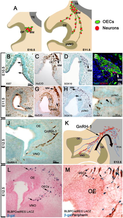
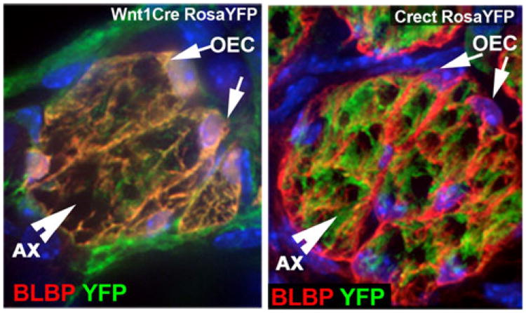
Sensory neurons in vertebrates are derived from two embryonic transient cell sources: neural crest (NC) and ectodermal placodes. The placodes are thickenings of ectodermal tissue that are responsible for the formation of cranial ganglia as well as complex sensory organs that include the lens, inner ear, and olfactory epithelium. The NC cells have been indicated to arise at the edges of the neural plate/dorsal neural tube, from both the neural plate and the epidermis in response to reciprocal interactions Moury and Jacobson (Dev Biol 141:243-253, 1990). NC cells migrate throughout the organism and give rise to a multitude of cell types that include melanocytes, cartilage and connective tissue of the head, components of the cranial nerves, the dorsal root ganglia, and Schwann cells. The embryonic definition of these two transient populations and their relative contribution to the formation of sensory organs has been investigated and debated for several decades (Basch and Bronner-Fraser, Adv Exp Med Biol 589:24-31, 2006; Basch et al., Nature 441:218-222, 2006) review (Baker and Bronner-Fraser, Dev Biol 232:1-61, 2001). Historically, all placodes have been described as exclusively derived from non-neural ectodermal progenitors. Recent genetic fate-mapping studies suggested a NC contribution to the olfactory placodes (OP) as well as the otic (auditory) placodes in rodents (Murdoch and Roskams, J Neurosci Off J Soc Neurosci 28:4271-4282, 2008; Murdoch et al., J Neurosci 30:9523-9532, 2010; Forni et al., J Neurosci Off J Soc Neurosci 31:6915-6927, 2011b; Freyer et al., Development 138:5403-5414, 2011; Katoh et al., Mol Brain 4:34, 2011). This review analyzes and discusses some recent developmental studies on the OP, placodal derivatives, and olfactory system.
Figures

References
-
- Bhattacharyya S, Bailey AP, Bronner-Fraser M, Streit A. Segregation of lens and olfactory precursors from a common territory: cell sorting and reciprocity of Dlx5 and Pax6 expression. Dev Biol. 2004;271:403–414. - PubMed
-
- Kozlowski DJ, Murakami T, Ho RK, Weinberg ES. Regional cell movement and tissue patterning in the zebrafish embryo revealed by fate mapping with caged fluorescein. Biochem Cell Biol. 1997;75:551–562. - PubMed
-
- Verwoerd CD, van Oostrom CG. Cephalic neural crest and placodes. Adv Anat Embryol Cell Biol. 1979;58:1–75. - PubMed
-
- Couly GF, Le Douarin NM. Mapping of the early neural primordium in quail-chick chimeras. I. Developmental relationships between placodes, facial ectoderm, and prosencephalon. Dev Biol. 1985;110:422–439. - PubMed
-
- Schlosser G. Do vertebrate neural crest and cranial placodes have a common evolutionary origin? Bioessays. 2008;30:659–672. - PubMed
Publication types
MeSH terms
Grants and funding
LinkOut - more resources
Full Text Sources
Miscellaneous
