. 2013 Sep;62(9):1270-9.
doi: 10.1136/gutjnl-2012-302401. Epub 2012 Jul 7.

Heterogeneity in mouse spasmolytic polypeptide-expressing metaplasia lineages identifies markers of metaplastic progression

Affiliations

Heterogeneity in mouse spasmolytic polypeptide-expressing metaplasia lineages identifies markers of metaplastic progression

Victoria G Weis et al. Gut. 2013 Sep.

Abstract

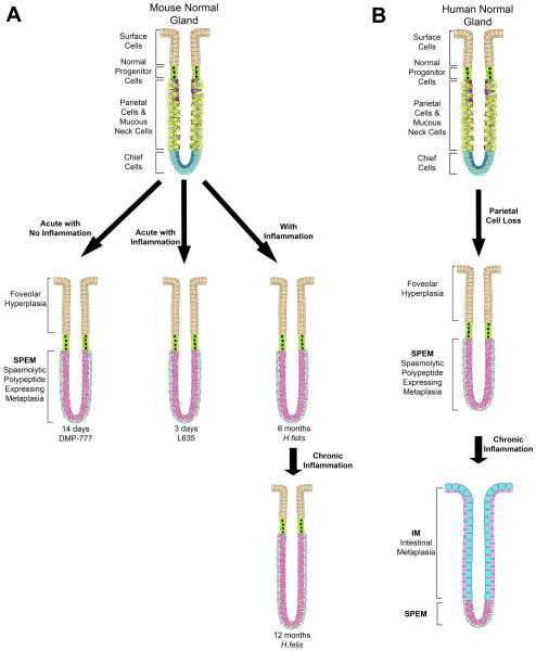
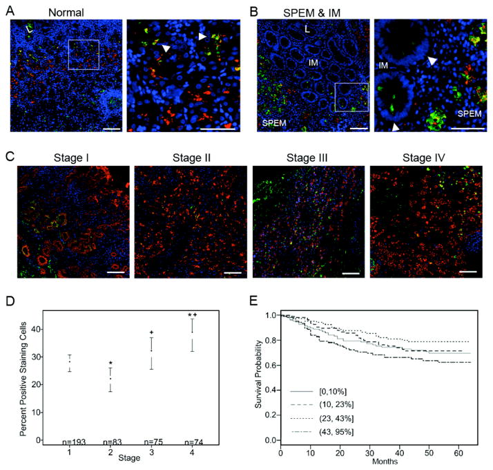
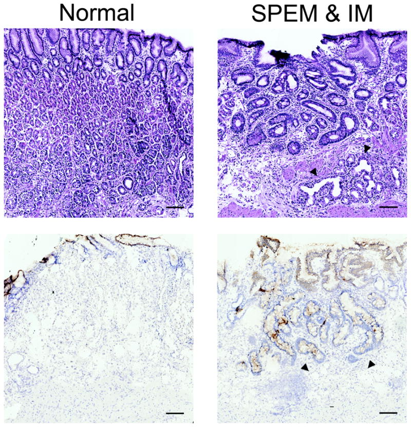
Objectives: Spasmolytic polypeptide-expressing metaplasia (SPEM) develops as a preneoplastic lesion in the stomachs of mice and humans after parietal cell loss. To identify the commonalities and differences between phenotypic SPEM lineages, SPEM were studied from three different mouse models of parietal cell loss: with chronic inflammation with Helicobacter felis infection; with acute inflammation with L635 treatment; and without inflammation following DMP-777 treatment.

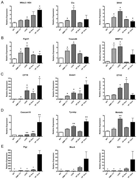
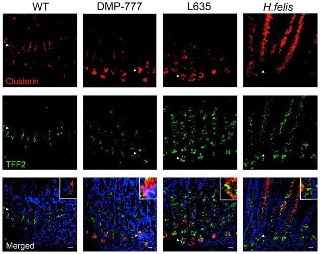
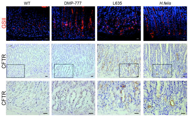
Design: RNA transcripts from laser capture microdissected normal chief cells and SPEM lineages were compared using gene microarray. Alterations in transcripts were validated by quantitative real-time PCR. Clusterin and cystic fibrosis transmembrane conductance regulator (CFTR) were selected for immunohistochemical analysis in all mouse models as well as in human SPEM, intestinal metaplasia and gastric cancer.

Results: Transcript expression patterns demonstrated differences among the phenotypic SPEM models. Clusterin expression was significantly upregulated in all three mouse SPEM models as well as in human SPEM. The highest clusterin expression in human gastric cancers correlated with poor survival. Conversely, CFTR expression was upregulated only in SPEM with inflammation in mice. In humans, intestinal metaplasia, but not SPEM, expressed CFTR.

Conclusions: While markers such as clusterin are expressed in all phenotypic SPEM lineages, distinct patterns of upregulated genes including CFTR are present in murine metaplasia associated with inflammation, indicative of progression of metaplasia towards a more intestinalised metaplastic phenotype.

Keywords: CFTR; DMP-777; H pylori-pathogenesis; Helicobacter felis; cholera; clusterin; cystic fibrosis; diarrhoeal disease; gastric cancer; gastrointestinal cancer; molecular pathology; spasmolytic polypeptide-expressing metaplasia; stem cells; trefoil peptides.


Conflict of interest statement

None of the authors have any conflicts of interest in the pursuit of this work.

Figures

Figure 1
Figure 1. Models of phenotypic SPEM in mice and of metaplastic progression in humans
(A) The three mouse models used in these investigations all display phenotypic SPEM. DMP-777 administration is an acute model of parietal cell loss that results in SPEM without inflammation. L635 administration is also an acute model of SPEM; however, L635-induced SPEM is accompanied by prominent inflammation. H. felis infection is a SPEM model phenotypcially similar to L635 but with chronic inflammation. H. felis infection induces SPEM at 6 months but progresses with chronic inflammation to acquire intestinal characteristics. (B) In humans, loss of parietal cells leads to the emergence of SPEM. Intestinal metaplasia arises from SPEM under the influences of chronic inflammation.
Figure 2
Figure 2. Quantitative Real Time PCR assay of selected upregulated transcripts from each comparison category
Expression of selected upregulated transcripts from each category was assessed by qRT-PCR in RNA isolated from the whole fundic stomach (3 mice per group). Pan-SPEM (A) and Acute SPEM (B) transcripts were associated with the emergence of SPEM, while SPEM with Inflammation (C), Specific to SPEM with Inflammation (D), and SPEM with Chronic Inflammation (E) relate to the progression of SPEM lineages. Results are shown as fold change as compared to the mean value of the control group (labeled WT). Single asterisks in A, B, and C signify significant upregulation as compared to WT. Double asterisks in D are significant as compared to DMP-777-induced SPEM. Significance in E (+) is noted as compared to 12 month H. felis infection. All values are shown as means +/− standard error of the mean, SE. (p < 0.05 by Mann-Whitney U test).
Figure 3
Figure 3. Expression of Clusterin in normal murine gastric mucosa and SPEM models
Sections of C57BL/6 mouse fundic mucosa were immunostained with antibodies against clusterin (top panels, red) and TFF2 (middle panels, green). In WT mice, clusterin was expressed in cells located in the isthmus and co-labeled with TFF2 in a small number of cells (arrowheads and higher magnification inset). Clusterin expression was detected throughout all SPEM lineages (14 day DMP-777 administration, 3 day L635 administration, and 12 month H. felis infection) (arrowheads and higher magnification insets). DAPI (blue). Bar = 20 μm.
Figure 4
Figure 4. Expression of CFTR in normal murine gastric mucosa and SPEM models
Frozen sections of C57BL/6 mouse fundic mucosa were immunostained with either GSII lectin (a mucous neck cell and SPEM marker) or an antibody against CFTR to investigate CFTR protein expression in SPEM. Top Panel: GSII (green) labels mucous neck cells in WT stomach mucosa and SPEM at the bases of glands in each of the SPEM models. Middle and Bottom Panels: Frozen sections immunohistochemical staining for CFTR showed no CFTR expression was detected in either WT mucosa or non-inflammatory SPEM (DMP-777-induced SPEM). The inset shows magnification of the bases of glands. CFTR expression was detected on the apical membranes of SPEM cells accompanied by inflammation (3 day L635 administration and 12 month H. felis infection). Scale bars = 20 μm.
Figure 5
Figure 5. Clusterin expression in human normal gastric mucosa, metaplasia of the stomach, and gastric cancer
(A) In normal glands, clusterin (green) was detected in the isthmus and in a few cells dual-labeled with TFF2 (red) as seen in the inset (arrowheads in top right panel). The sample shown was taken from the University of Tokyo metaplasia array. (B) Clusterin was prominently expressed throughout SPEM glands as marked by TFF2 in red. In intestinal metaplasia, clusterin was limited to a few cells at the base of glands (designated by arrowheads in bottom right panel). These staining patterns were found in 90% of SPEM samples and 45% of intestinal metaplasia samples on the University of Tokyo metaplasia tissue array. Left panel scale bar = 100 μm. Inset panel scale bar = 50 μm. (C) Representative images from a human gastric cancer TMA show clusterin (green) expression in gastric cancer cells in each stage. Pan-cytokeratin staining (red) marks epithelial cells. Clusterin expression was present in all stages of cancer (I–IV) with averages of 27.75%, 21.82%, 31.32%, and 37.94% clusterin positive epithelial cells, respectively. DAPI (blue). Bar = 100 μm. (D) The percentage of clusterin positive epithelial cells was statistically significantly related to tumor stage by analysis of variance (p = 0.05). There were significant differences in clusterin staining between stage 1 compared to stages 2 and 4 (labeled with *) and stage 2 versus stages 3 and 4 (labeled with +). (E) Estimated survival curves of clusterin by staining percentage categorized into 4 quartiles. The highest staining quartile (43–95% clusterin positive epithelial cells) correlated with the worst survival outcome.
Figure 6
Figure 6. CFTR expression in human normal gastric mucosa and in metaplasia of the human stomach
Frozen sections of human stomach were immunostained with an antibody against CFTR (bottom panels). Top panels are H&E staining of sections for orientation and morphology. CFTR was absent from normal mucosa (bottom left) and SPEM lineages (designated by arrowheads in right panels). The diffuse cytoplasmic immunoreactivity seen in surface cells is nonspecific as CFTR is localized to apical membranes. However, expression was observed in intestinal metaplasia (bottom right). Due to limitations of the CFTR antibody and the lack of availability of frozen adenocarcinoma samples, CFTR expression was not evaluated in cancers. Bar = 100 μm.

References

    1. Blaser M, Parsonnet J. Parasitism by the ‘slow’ bacterium Helicobacter pylori leads to altered gastric homeostasis and neoplasia. JClinInvest. 1994;94:4–8. - PMC - PubMed
    1. Yoshizawa N, Takenaka Y, Yamaguchi H, et al. Emergence of spasmolytic polypeptide-expressing metaplasia in Mongolian gerbils infected with Helicobacter pylori. Lab Invest. 2007;87:1265–76. - PubMed
    1. Nam KT, Lee HJ, Mok H, et al. Amphiregulin-deficient mice develop spasmolytic polypeptide expressing metaplasia and intestinal metaplasia. Gastroenterology. 2009;136:1288–96. - PMC - PubMed
    1. Goldenring JR, Nam KT, Wang TC, et al. Spasmolytic polypeptide-expressing metaplasia and intestinal metaplasia: time for reevaluation of metaplasias and the origins of gastric cancer. Gastroenterology. 2010;138:2207–10. 10e1. - PMC - PubMed
    1. Nam KT, Lee HJ, Sousa JF, et al. Mature chief cells are cryptic progenitors for metaplasia in the stomach. Gastroenterology. 2010;139:2028–37. e9. - PMC - PubMed

Publication types

MeSH terms