. 2012 Sep;303(5):L439-48.
doi: 10.1152/ajplung.00408.2011. Epub 2012 Jul 6.

Glycogen synthase kinase-3β/β-catenin signaling regulates neonatal lung mesenchymal stromal cell myofibroblastic differentiation

Affiliations

Glycogen synthase kinase-3β/β-catenin signaling regulates neonatal lung mesenchymal stromal cell myofibroblastic differentiation

Antonia P Popova et al. Am J Physiol Lung Cell Mol Physiol. 2012 Sep.

Abstract

In bronchopulmonary dysplasia (BPD), alveolar septa are thickened with collagen and α-smooth muscle actin-, transforming growth factor (TGF)-β-positive myofibroblasts. We examined the biochemical mechanisms underlying myofibroblastic differentiation, focusing on the role of glycogen synthase kinase-3β (GSK-3β)/β-catenin signaling pathway. In the cytoplasm, β-catenin is phosphorylated on the NH(2) terminus by constitutively active GSK-3β, favoring its degradation. Upon TGF-β stimulation, GSK-3β is phosphorylated and inactivated, allowing β-catenin to translocate to the nucleus, where it activates transcription of genes involved in myofibroblastic differentiation. We examined the role of β-catenin in TGF-β1-induced myofibroblastic differentiation of neonatal lung mesenchymal stromal cells (MSCs) isolated from tracheal aspirates of premature infants with respiratory distress. TGF-β1 increased β-catenin expression and nuclear translocation. Transduction of cells with GSK-3β S9A, a nonphosphorylatable, constitutively active mutant that favors β-catenin degradation, blocked TGF-β1-induced myofibroblastic differentiation. Furthermore, transduction of MSCs with ΔN-catenin, a truncation mutant that cannot be phosphorylated on the NH(2) terminus by GSK-3β and is not degraded, was sufficient for myofibroblastic differentiation. In vivo, hyperoxic exposure of neonatal mice increases expression of β-catenin in α-smooth muscle actin-positive myofibroblasts. Similar changes were found in lungs of infants with BPD. Finally, low-passage unstimulated MSCs from infants developing BPD showed higher phospho-GSK-3β, β-catenin, and α-actin content compared with MSCs from infants not developing this disease, and phospho-GSK-3β and β-catenin each correlated with α-actin content. We conclude that phospho-GSK-3β/β-catenin signaling regulates α-smooth muscle actin expression, a marker of myofibroblast differentiation, in vitro and in vivo. This pathway appears to be activated in lung mesenchymal cells from patients with BPD.


Figures

Fig. 1.
Fig. 1.
Treatment with TGF-β1 increases α-actin, p-glycogen synthase kinase (GSK)-3β, and β-catenin content in neonatal lung mesenchymal stromal cells (MSCs). Compared with unstimulated MSCs (A), MSCs treated with 10 ng/ml TGF-β1 for 48 h (B) show increases for α-actin (green), β-catenin (blue), and phospho-GSK-3β (red). β-Catenin and phospho-GSK-3β appear to be colocalized (purple) in the perinuclear space. Experiments are representative of 3 separate experiments. C: immunoblotting analysis for α-actin, β-catenin, phospho-GSK-3β, and GSK-3β in cells incubated with and without 10 ng/ml TGF-β1 for 48 h. D: group mean densitometry data for 4 experiments (*different from control, P < 0.05, 1-way ANOVA).
Fig. 2.
Fig. 2.
Transduction of MSCs with GSK-3β S9A, a nonphosphorylatable constitutively active mutant, blocks TGF-β1-induced β-catenin nuclear accumulation and myofibroblastic differentiation. Following TGF-β1 treatment (10 ng/ml for 48 h), cells transduced with empty vector (A and B) show increased phospho-GSK-3β content in the perinuclear space (green) and localization of β-catenin in the nucleus (blue). α-Actin staining shows increased expression in the cell cytoplasm and stress fibers indicative of incorporation of actin into contractile filaments. Cells transduced with nonphosphorylatable GSK-3β (D and E) fail to show increased α-actin expression or β-catenin nuclear translocation in response to TGF-β1. Following treatment with GSK-3β inhibitor, LiCl (10 mM for 48 h), cells transduced with empty vector (C) showed increased nuclear accumulation of β-catenin (blue) and expression of α-actin (green). In contrast, cells transduced with nonphosphorylatable GSK-3β failed to show β-catenin nuclear accumulation or increased α-actin expression following treatment with LiCl (F). G: immunoblotting of cells transduced with empty vector shows increased α-actin and β-catenin and no change in total GSK-3β content. In contrast, cells transduced with nonphosphorylatable GSK-3β fail to increase α-actin and β-catenin in response to TGF-β1 stimulation. Results are representative of 3 individual experiments.
Fig. 3.
Fig. 3.
Transduction of MSCs with ΔN-β-catenin, a nonphosphorylatable β-catenin, is sufficient for myofibroblastic differentiation. Compared with MSCs transduced with empty vector, (AD), MSCs transduced with ΔN-β-catenin (EH) show increases for α-actin (green) and β-catenin (red). α-Actin staining shows stress fibers indicative of incorporation of actin into contractile filaments. Anti-myc tag staining is blue (H). I: immunoblots for α-actin, β-catenin, myc, and β-actin are shown. MSCs were transduced with empty vector- or ΔN-β-catenin. These results are representative of 3 individual experiments.
Fig. 4.
Fig. 4.
Hyperoxic exposure of neonatal mice increases lung cell β-catenin and α-actin expression. Wild-type C57BL/6J mice (2–3 days old) were exposed to air or 75% oxygen for 14 days. Compared with air-exposed mice (AD), hyperoxic exposure (EH) caused the development of fewer and larger airspaces and thickened alveolar walls. Fluorescence microscopy showed basal phospho-GSK-3β content (red) in the epithelium of air-exposed mice (AD), and increased deposition of α-actin (green) and β-catenin (blue) in the lung interstitial of hyperoxia exposed lungs (E and F). α-Actin (green) and β-catenin colocalized with phospho-GSK-3β (white). DAPI staining of nuclei is depicted as black superimposed upon a visible image in the gray background. I: collagenase-digested erythrocyte-lysed lungs from 2–3-day-old wild-type C57BL/6J mice, exposed to air or 75% oxygen for 14 days, were fixed in ethanol and processed directly for flow cytometry. Hematopoietic cells were gated out with anti-CD45-AF750. Fluorescence for anti-pGSK-3β-AF488 (top), anti-α-actin-Cy3 (middle), and anti-β-catenin-AF633 (bottom) is shown (green, IgG immunoreactivity in the 3 channels; black, lung cells from normoxia-exposed animals; red, lung cells from hyperoxia exposed mice). These results are representative of 3 individual experiments. J: lung plasminogen activator inhibitor (PAI)-1 mRNA expression from 2-day-old mouse pups exposed to ambient air or 75% O2 for 14 days is shown (n = 9 for each group, *P < 0.01, unpaired t-test).
Fig. 5.
Fig. 5.
Lungs of infants with bronchopulmonary dysplasia (BPD) show increased expression of β-catenin and α-smooth muscle actin, which colocalizes with phospho-GSK-3β in the thickened alveolar interstitium. The lung of a full-term infant dying of a nonpulmonary cause is shown in (AE). Immunofluorescent staining shows α-actin positive (green) cells in the airway smooth muscle and at the tips of alveolar septa. Epithelial cells and red blood cells (arrows) stain positive for phospho-GSK-3β (red). The α-actin signal (green) does not colocalize with phospho-GSK-3β (red), and little or no β-catenin signal (blue) is present. Lung sections from infants dying with BPD (FJ) show abnormal architecture with widened alveolar spaces and thickened alveolar walls. Immunofluorescence staining shows increased expression of α-actin (green) and β-catenin (blue), which colocalize with phospho-GSK-3β in the thickened alveolar interstitium. Colocalization of α-actin, β-catenin, and phospho-GSK-3β appears white. DAPI staining of nuclei is depicted as black superimposed upon a visible image in the gray background. Phase contrast images depict differences in lung architecture between the normal (E) and BPD (J) lung.
Fig. 6.
Fig. 6.
Connective tissue growth factor (CTGF) induces phosphorylation of GSK-3β and accumulation of β-catenin in cultured neonatal lung MSCs and is increased in the lungs of hyperoxia-exposed neonatal mice and human infants with BPD. Compared with unstimulated MSCs (A), MSCs treated with 5 μg/ml CTGF for 48 h (B) show increases in α-actin (green), p-GSK-3β (red), and β-catenin (blue). C: lung CTGF mRNA expression from 2-day-old mouse pups exposed to ambient air or 75% O2 for 7 or 14 days is shown (n = 3, *different from air-exposed, P < 0.05). Lung sections from 14-day air-exposed (D and F) and hyperoxia-exposed (E and G) neonatal mice were immunostained for CTGF or isotype control and counterstained with hematoxylin (×200 original magnification). Lung sections from a normal-term infant (H and J) and terminal case of BPD (I and K) were immunostained for CTGF (×200 original magnification).
Fig. 7.
Fig. 7.
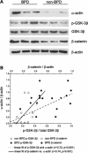
Phospho-GSK-3β, β-catenin, and α-actin content are increased in low-passage unstimulated neonatal lung MSCs from patients developing BPD. A: immunoblotting analysis shows higher levels of α-actin, phospho-GSK-3β, GSK-3β, and β-catenin in low-passage, unstimulated MSCs from infants developing BPD compared with MSCs from infants not developing this disease. B: phospho-GSK-3β and β-catenin content correlate with α-actin content in unstimulated neonatal lung MSCs.
Fig. 8.
Fig. 8.
TGF-β1 stimulation induces phosphatidylinositol (PI)3-kinase-dependent phosphorylation of Akt and GSK-3β. Immunoblotting analysis for phospho-Akt, Akt, phospho-GSK-3β, and GSK-3β in cells incubated with 10 ng/ml TGF-β1 and LY 294002.

References

    1. Abe M, Harpel JG, Metz CN, Nunes I, Loskutoff DJ, Rifkin DB. An assay for transforming growth factor-β using cells transfected with a plasminogen activator inhibitor-1 promoter-luciferase construct. Anal Biochem 216: 276–284, 1994. - PubMed
    1. Alapati D, Rong M, Chen S, Hehre D, Rodriguez MM, Lipson KE, Wu S. Connective tissue growth factor antibody therapy attenuates hyperoxia-induced lung injury in neonatal rats. Am J Respir Cell Mol Biol 45: 1169–1177, 2011. - PubMed
    1. Armstrong DD, Esser KA. Wnt/β-catenin signaling activates growth-control genes during overload-induced skeletal muscle hypertrophy. Am J Physiol Cell Physiol 289: C853–C859, 2005. - PubMed
    1. Ben-Ze'ev A, Shtutman M, Zhurinsky J. The integration of cell adhesion with gene expression: The role of β-catenin. Exp Cell Res 261: 75–82, 2000. - PubMed
    1. Bentley JK, Popova A, Bozyk P, Linn M, Baek A, Lei J, Goldsmith A, Hershenson M. Ovalbumin sensitization and challenge increases the number of lung cells possessing a mesenchymal stromal cell phenotype. Respir Res 11: 127, 2010. - PMC - PubMed

MeSH terms

LinkOut - more resources