. 2013 Jan;6(1):72-83.
doi: 10.1242/dmm.008946. Epub 2012 Jul 5.

Microtubule-targeting drugs rescue axonal swellings in cortical neurons from spastin knockout mice

Affiliations

Microtubule-targeting drugs rescue axonal swellings in cortical neurons from spastin knockout mice

Coralie Fassier et al. Dis Model Mech. 2013 Jan.

Abstract

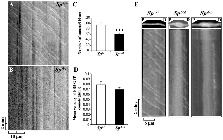
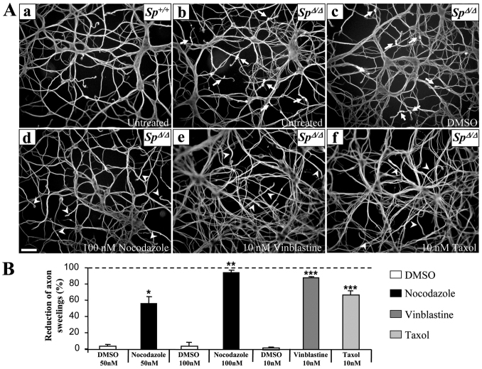
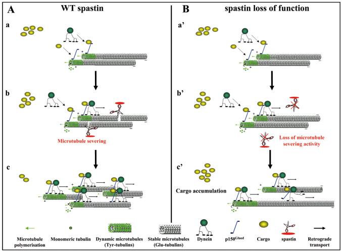
Mutations in SPG4, encoding the microtubule-severing protein spastin, are responsible for the most frequent form of hereditary spastic paraplegia (HSP), a heterogeneous group of genetic diseases characterized by degeneration of the corticospinal tracts. We previously reported that mice harboring a deletion in Spg4, generating a premature stop codon, develop progressive axonal degeneration characterized by focal axonal swellings associated with impaired axonal transport. To further characterize the molecular and cellular mechanisms underlying this mutant phenotype, we have assessed microtubule dynamics and axonal transport in primary cultures of cortical neurons from spastin-mutant mice. We show an early and marked impairment of microtubule dynamics all along the axons of spastin-deficient cortical neurons, which is likely to be responsible for the occurrence of axonal swellings and cargo stalling. Our analysis also reveals that a modulation of microtubule dynamics by microtubule-targeting drugs rescues the mutant phenotype of cortical neurons. Together, these results contribute to a better understanding of the pathogenesis of SPG4-linked HSP and ascertain the influence of microtubule-targeted drugs on the early axonal phenotype in a mouse model of the disease.


Figures

Fig. 1.
Fig. 1.
Absence of truncated spastin in SpΔ/Δ mutant mice. (A) Western blot analysis of protein extracted from non-transfected Cos-7 cells (NT) or transfected with constructs expressing GFP-Sp+ or GFP-SpΔ using S51 spastin polyclonal antibody (left panel) or GFP antibody (right panel). S51 antibody specifically recognizes the truncated spastin lacking its AAA domain (i.e. GFP-SpΔ), which is equally detected by the anti-GFP antibody. (B) Immunoblot analysis of spastin from 40 μg (left panel) or 10 μg (right panel) of 4-month-old Sp+/+, SpΔ/+ and SpΔ/Δ brain lysates using S51 spastin antibody. SpΔ/+ and SpΔ/Δ brain lysates do not show any traces of truncated spastin. Note the reduction or complete absence of wild-type spastin isoforms in SpΔ/+ and SpΔ/Δ mice, respectively. Actin was used as a loading control. (C) Quantification of spastin band density (including all spastin isoforms) normalized to actin values from three independent experiments. Spastin expression is reduced by 40% in SpΔ/+ mice compared with Sp+/+ mice, whereas no traces of wild-type spastin was detectable in SpΔ/Δ mutants. Vertical bars show s.e.m. *P<0.05, **P<0.001. A.U, arbitrary units.
Fig. 2.
Fig. 2.
Spastin deletion specifically affects the integrity of cortical axons. (A–H) Immunodetection of acetylated α-tubulin in primary cultures of DIV6 hippocampal (A,B,G,H) and cortical neurons (C–F) from Sp+/+ (A,C) and SpΔ/Δ (B,D–H) embryos. Unlike SpΔ/Δ cortical neurons that show obvious axonal swellings (D–F; arrows), SpΔ/Δ hippocampal neurons do not display any neurite swellings in the distal part of their axons (arrowheads) or in other regions of the axon (B,G,H). Neurite density was not affected in SpΔ/Δ hippocampal neurons (B) compared with Sp+/+ hippocampal neurons (A). Images E–H represent higher magnification of SpΔ/Δ cortical (E,F) and hippocampal (G,H) axon distal regions. Scale bars: 50 μm (A–D); 20 μm (E–H). (I) Western blot analysis of spastin isoforms in protein lysates (20 μg) from primary cultures of Sp+/+ cortical and hippocampal neurons using the S51 spastin antiserum. Actin was used as a loading control. H.N, hippocampal neurons; C.N, cortical neurons.
Fig. 3.
Fig. 3.
Ultrastructural analysis of SpΔ/Δ cortical neurons reveals a massive disorganization of the microtubule network within axonal swellings. (A–H) Ultrathin sections of DIV6 primary cultures of cortical neurons from SpΔ/Δ (A–F) and Sp+/+ mouse embryos (G–H). Images B and D are higher magnifications of axonal swellings indicated by arrows in images A and C, respectively. Images E, F and H are higher magnification of boxed regions in images B, D and G, respectively. Note the obvious disorganization and the tangled and bent aspect of microtubules within axonal swellings of SpΔ/Δ cortical neurons (B,D–F). This abnormal appearance of microtubules was never observed in Sp+/+ axons, in which microtubules are always organized in parallel arrays (G–H). White arrows indicate microtubules (E,F,H). Scale bars: 5 μm (A,C); 0.5 μm (B,D,G); 0.25 μm (E,F,H).
Fig. 4.
Fig. 4.
Impaired microtubule disassembly in SpΔ/Δ cultured cortical neurons. (A–L) Sp+/+ (A,B,E,F) and SpΔ/Δ (C,D,G–L) neurons at 4 days (A–H) or 6 days (I–L) post-plating. Neurons were incubated in the presence of 30 μM nocodazole for 40 minutes (E–H) or 40 μM nocodazole for 0 (I), 30 (J), 60 (K) or 90 minutes (L), then permeabilized in saponin-based buffer to extract free tubulin molecules and double-labeled for F-actin (red) and βIII-tubulin (green). After 40 minutes of exposure to 30 μM nocodazole, only a faint βIII-tubulin staining was still detectable in Sp+/+ cortical axons (E) whereas a persistent microtubule signal was observed in SpΔ/Δ axons (G). Note that the remaining microtubule signal was still present in SpΔ/Δ axonal swellings after 90-minute exposure to 40 μM nocodazole, whereas only a residual labeling was observed in the other regions of the axon shaft (L). Scale bars: 20 μm.
Fig. 5.
Fig. 5.
Spastin loss of function decreases the number of dynamic microtubule plus ends in cortical axon shaft. (A–E) Time-lapse recording of EB3-GFP comets in DIV6 Sp+/+ (n=20) and SpΔ/Δ (n=29) cortical axons. (A,B) Kymographs of a Sp+/+ (A) and SpΔ/Δ (B) axon from a 10-minute time-lapse recording of EB3-GFP comets. These kymographs revealed a striking decrease in EB3-GFP moving comets in SpΔ/Δ axon shaft, which is associated with a weak movement of diffuse EB3-GFP protein compared with Sp+/+ axon. (C) Quantification of the average number of EB3-GFP comets per 100 μm of axonal length. (D) Quantification of the mean EB3-GFP comet velocity. The average number of EB3-GFP comets is significantly reduced in SpΔ/Δ axons compared with Sp+/+ axons (***P<0.001), whereas the mean comet speed is not affected by spastin depletion. Vertical bars show s.e.m. (E) Kymograph analysis of microtubule plus ends (i.e. EB3-GFP comets) in a selected Sp+/+ axonal region (left panel; see supplementary material Movie 2) and two distinct SpΔ/Δ swollen axons (middle and right panels; supplementary material Movies 4 and 5). The analyzed regions are boxed in supplementary material Movies 2, 4 and 5 and are presented on the top of each kymograph. Kymographs showed that only very few EB3-GFP comets are moving within swellings and that the vast majority of these moving comets are located in the most distal end of the swelling. P, proximal; D, distal. (A,B,E) Anterograde comets are represented by diagonal lines leading to the bottom right, whereas stationary comets (i.e. Fig. 5E, right panel) are shown by vertical white lines.
Fig. 6.
Fig. 6.
Microtubule-targeting drugs rescue the pathological phenotype of SpΔ/Δ cortical neurons. (A) Immunolabeling of acetylated α-tubulin on DIV6 primary cultures of Sp+/+ (Aa) and SpΔ/Δ (Ab–Af) cortical neurons untreated (Aa,Ab) or treated 5 days post-plating with 100 nM of nocodazole (Ad), 10 nM of vinblastine (Ae), 10 nM of taxol (Af) or the equivalent volume of DMSO (vehicule; Ac). Note the absence of axonal swellings in the distal region of SpΔ/Δ cortical neurons treated with microtubule-targeting drugs (Ad–Af; arrowheads) compared with untreated or DMSO-treated SpΔ/Δ neurons (Ab,Ac; arrows). The neurite morphology of mutant neurons treated with microtubule-targeting drugs appears similar to that of Sp+/+ neurons (Aa). Scale bar: 50 μm. (B) The percentage of axonal swellings in SpΔ/Δ cortical neuron cultures was evaluated at DIV6. Note that the nanomolar concentration of microtubule targeting-drugs significantly decreases the proportion of neurite swellings in primary cultures of SpΔ/Δ neurons compared with DMSO-treated cultures. Asterisks indicate statistically different percentages between DMSO-treated neurons and 100 nM nocodazole-, 10 nM vinblastine- or 10 nM taxol-treated cells: *P<0.05, **P<0.001, ***P<0.0001. Vertical bars indicate s.e.m. More than 1000 neurons were analyzed per experimental condition.
Fig. 7.
Fig. 7.
Hypothetical model of spastin function. (A,B) Model of spastin function in the distal part of cortical axons in physiological (A) and pathological (B) conditions. Cargoes that should be retrogradely transported are recruited on microtubule plus ends via p150Glued, a member of the dynactin complex. Then, the recruitment of the molecular motor dynein via p150Glued allows retrograde transport of cargoes to the soma along microtubules (Aa; Ba′). In the distal part of the axon, spastin severs microtubules to generate new microtubules plus ends (Ab) that might serve as local nucleation points for the synthesis of dynamic microtubules (Ac) and also improve retrograde axonal transport efficiency by increasing the number of microtubules plus ends, and thereby the capacity of cargo loading onto microtubules via p150Glued (Ac). In this region of the axon, loss of spastin microtubule-severing activity (Bb′) might reduce the number of microtubule plus ends, which will subsequently affect both microtubule dynamics and cargo loading efficiency (Bc′).

Similar articles

Cited by

References

    1. Baas P. W., Qiang L. (2005). Neuronal microtubules: when the MAP is the roadblock. Trends Cell Biol. 15, 183–187 - PubMed
    1. Baas P. W., Karabay A., Qiang L. (2005). Microtubules cut and run. Trends Cell Biol. 15, 518–524 - PubMed
    1. Blackstone C., O’Kane C. J., Reid E. (2011). Hereditary spastic paraplegias: membrane traffic and the motor pathway. Nat. Rev. Neurosci. 12, 31–42 - PMC - PubMed
    1. Butler R., Wood J. D., Landers J. A., Cunliffe V. T. (2010). Genetic and chemical modulation of spastin-dependent axon outgrowth in zebrafish embryos indicates a role for impaired microtubule dynamics in hereditary spastic paraplegia. Dis. Model. Mech. 3, 743–751 - PMC - PubMed
    1. Charvin D., Cifuentes-Diaz C., Fonknechten N., Joshi V., Hazan J., Melki J., Betuing S. (2003). Mutations of SPG4 are responsible for a loss of function of spastin, an abundant neuronal protein localized in the nucleus. Hum. Mol. Genet. 12, 71–78 - PubMed

Publication types

MeSH terms