. 2012 Sep 1;113(5):766-74.
doi: 10.1152/japplphysiol.01424.2011. Epub 2012 Jul 5.

Impact of hydroxyl radical-induced injury on calcium handling and myofilament sensitivity in isolated myocardium

Affiliations

Impact of hydroxyl radical-induced injury on calcium handling and myofilament sensitivity in isolated myocardium

Kaylan M Haizlip et al. J Appl Physiol (1985). .

Abstract

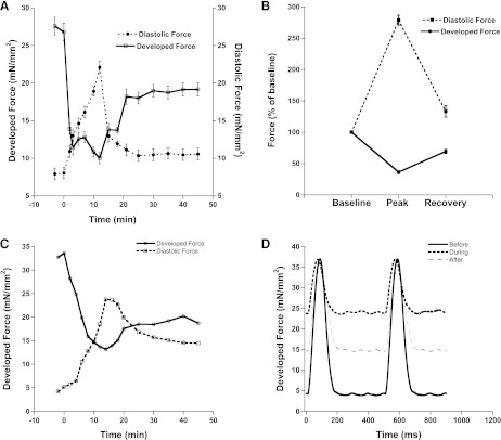
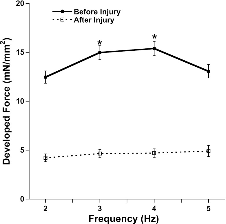
Hydroxyl radicals (OH) are involved in the pathogenesis of reperfusion injury and are observed in acute heart failure, stroke, and myocardial infarction. Two different subcellular defects are involved in the pathogenesis of OH injury, deranged calcium handling, and alterations of myofilament responsiveness, but their temporal impact on contractile function is not resolved. Initially, after brief OH exposure, there is a corresponding marked increase in diastolic calcium and diastolic force. We followed these parameters until a new steady-state level was reached at ~45 min post-OH exposure. At this new baseline, diastolic calcium had returned to near-normal, pre-OH levels, whereas diastolic force remained markedly elevated. An increased calcium sensitivity was observed at the new baseline after OH-induced injury compared with the pre-OH state. The acute injury that occurs after OH exposure is mainly due to calcium overload, while the later sustained myocardial dysfunction is mainly due to the altered/increased myofilament responsiveness.


Figures

Fig. 1.
Fig. 1.
A: effect of 2-min exposure of hydroxyl radicals (OH˙) on the contractile parameters of cardiac trabeculae (n = 10). Values are means ± SE. B: percentage effect of OH˙ on the contractile parameters of cardiac trabeculae (n = 10). At peak of contracture (occurring at ∼12 min after OH˙ exposure), there is significant increase in diastolic force and a decrease in developed force. New steady state was marked by significant elevated diastolic force and reduced developed force. Values are means ± SE. C: representative tracing developed and diastolic force of one muscle (101013_3) before and after treatment show similar effects to average data. D: representative twitch tracings for one muscle taken before, during, and after treatment with OH˙. Stimulation frequency was 2 Hz and temperature was 37°C throughout the experiment.
Fig. 2.
Fig. 2.
Time from peak tension to 50% relaxation (RT50; A) and time from 50% relaxation to 90% relaxation (RT90-RT50; B) before and after OH˙-induced injury are significantly different. At baseline frequency of 2 Hz, RT50 and RT90-RT50 are significantly longer after injury compared with before frequency. Values are means ± SE. *Difference of P < 0.05 between conditions in the same group. C: representative recording of relaxation kinetics from individual muscle (101013_3).
Fig. 3.
Fig. 3.
Force-frequency relationship before (following 45 min of 2-Hz stabilization) and after 2 min of OH˙ exposure on RV trabeculae (n = 8). The new steady state was marked by the flat force-frequency relationship compared with positive force-frequency relationship (*P < 0.05) before application of OH˙. Values are means ± SE.
Fig. 4.
Fig. 4.
A: relationship between intracellular calcium and diastolic force after the exposure of OH˙ in right ventricular rabbit trabeculae. Raw record of diastolic force and intracellular calcium before, during, and after exposure to OH˙ (from one experiment) are shown. B: percentage effect of OH˙ on the intracellular calcium and diastolic force (n = 8). At peak of contracture (occurring at ∼12 min after OH˙ exposure), there is a significant increase in diastolic force and a decrease in diastolic calcium. New steady state was marked by a significant elevated diastolic force, but diastolic calcium returned to near pre-interventional values. Values are means ± SE.
Fig. 5.
Fig. 5.
A: assessment of myofilament responsiveness before (following 45 min of 2-Hz stabilization) and after the OH˙-induced injury. Using the K+ contracture protocol, a steady-state force-Ca2+ concentration relationship was measured before and after OH˙-induced injury. This shows that the curve shifts up and left after the injury. B: EC50 decreased after OH˙-induced injury. *Difference at P < 0.05 between conditions in the same group. On average, neither maximal force nor cooperativity (Hill coefficient) was significantly different (not shown). Values are means ± SE.
Fig. 6.
Fig. 6.
Myofilament fraction protein phosphorylation was determined by comparing the protein band density of the Sypro Ruby protein gel stain (total protein) on the same gel stained with Pro-Q diamond (phosphor-protein). Molecular weight (MW) markers include lane 1 BioRad dual stain ladder and lane 2 peppermint stick (Molecular Probes) phosphor-protein marker positive control. None of the indicated bands was significantly different between control and OH˙-exposed muscles (n = 5 per group). A: densitometric measurements of phosphorylated protein compared with total actin. Values are means ± SE. B: representative blot of Sypro Ruby Gel stain (Invitrogen). C: representative blot of ProQ Diamond phospho-protien stain (Invitrogen). MyBP-C, myosin-binding protein C; TnT, troponin T; TnI, troponin I; MLC-2, myosin light chain 2; AU, arbitrary units.

Similar articles

Cited by

References

    1. Avner BS, Hinken AC, Yuan C, Solaro RJ. H2O2 alters rat cardiac sarcomere function and protein phosphorylation through redox signaling. Am J Physiol Heart Circ Physiol 299: H723–H730, 2010 - PMC - PubMed
    1. Backx PH, Ter Keurs HE. Fluorescent properties of rat cardiac trabeculae microinjected with fura-2 salt. Am J Physiol Heart Circ Physiol 264: H1098–H1110, 1993 - PubMed
    1. Biesiadecki BJ, Tachampa K, Yuan C, Jin JP, de Tombe PP, Solaro RJ. Removal of the cardiac troponin I N-terminal extension improves cardiac function in aged mice. J Biol Chem 285: 19688–19698, 2010 - PMC - PubMed
    1. Bolli R, Marban E. Molecular and cellular mechanisms of myocardial stunning. Physiol Rev 79: 609–634, 1999 - PubMed
    1. Bupha-Intr T, Haizlip KM, Janssen PM. Temporal changes in expression of connexin 43 after load-induced hypertrophy in vitro. Am J Physiol Heart Circ Physiol 296: H806–H814, 2009 - PMC - PubMed

Publication types

MeSH terms

LinkOut - more resources