. 2012 Sep;194(18):4920-32.
doi: 10.1128/JB.00793-12. Epub 2012 Jul 6.

TolC-dependent secretion of an ankyrin repeat-containing protein of Rickettsia typhi

Affiliations

TolC-dependent secretion of an ankyrin repeat-containing protein of Rickettsia typhi

Simran J Kaur et al. J Bacteriol. 2012 Sep.

Abstract

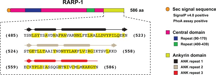
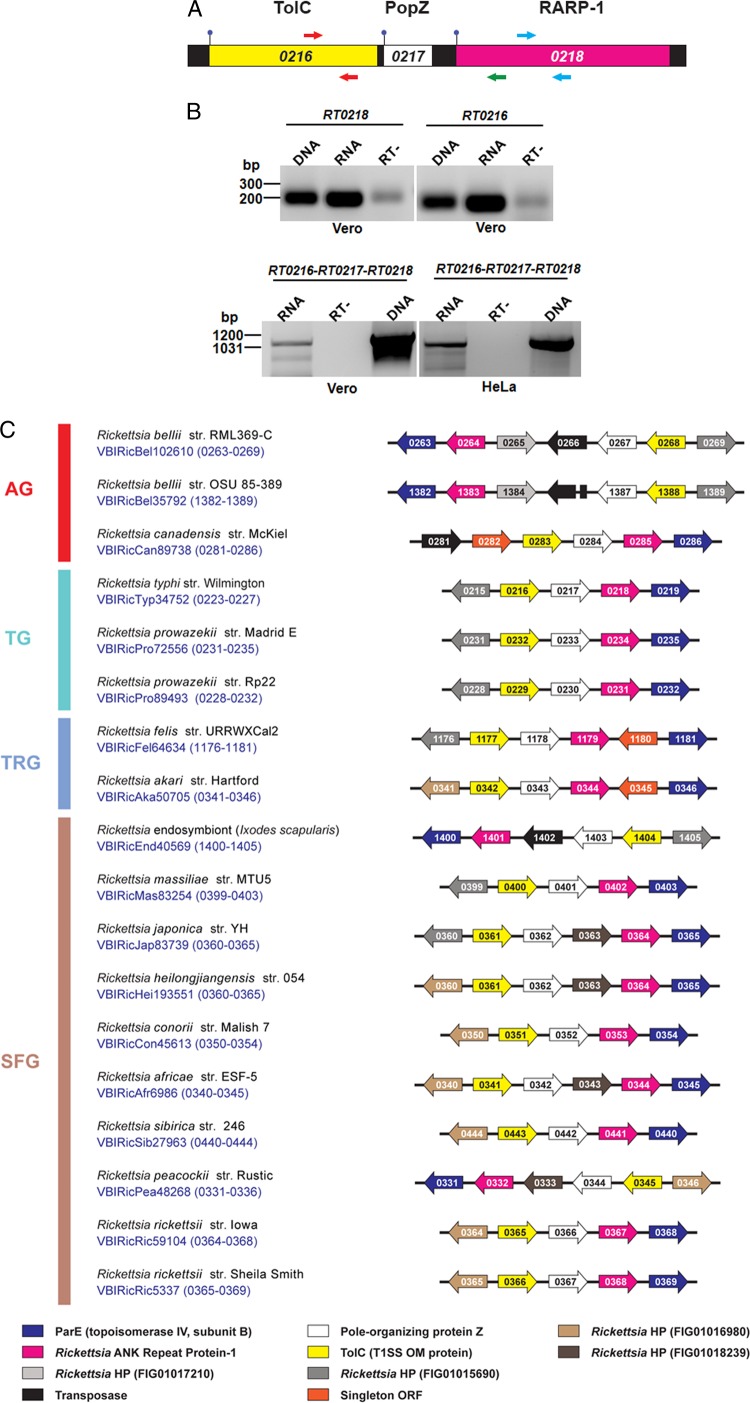
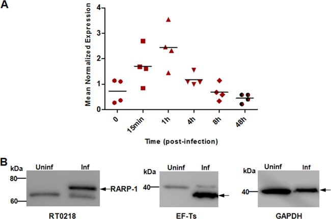
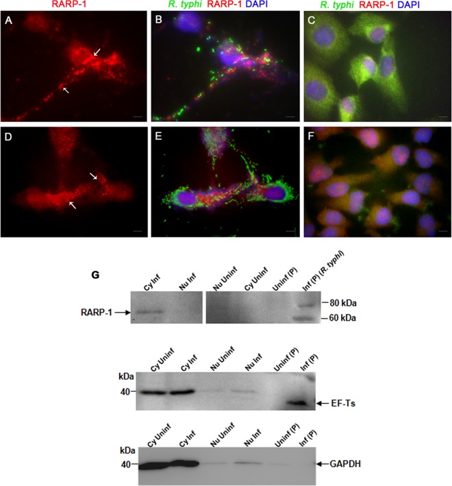
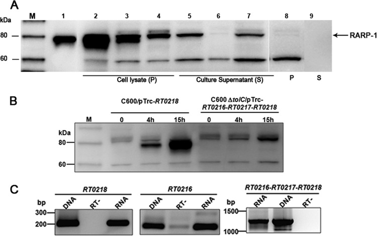
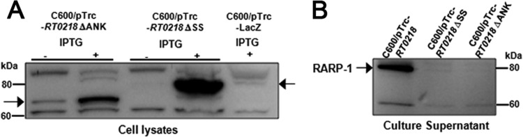
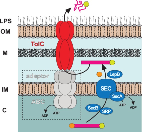
Rickettsia typhi, the causative agent of murine (endemic) typhus, is an obligate intracellular pathogen with a life cycle involving both vertebrate and invertebrate hosts. In this study, we characterized a gene (RT0218) encoding a C-terminal ankyrin repeat domain-containing protein, named Rickettsia ankyrin repeat protein 1 (RARP-1), and identified it as a secreted effector protein of R. typhi. RT0218 showed differential transcript abundance at various phases of R. typhi intracellular growth. RARP-1 was secreted by R. typhi into the host cytoplasm during in vitro infection of mammalian cells. Transcriptional analysis revealed that RT0218 was cotranscribed with adjacent genes RT0217 (hypothetical protein) and RT0216 (TolC) as a single polycistronic mRNA. Given one of its functions as a facilitator of extracellular protein secretion in some Gram-negative bacterial pathogens, we tested the possible role of TolC in the secretion of RARP-1. Using Escherichia coli C600 and an isogenic tolC insertion mutant as surrogate hosts, our data demonstrate that RARP-1 is secreted in a TolC-dependent manner. Deletion of either the N-terminal signal peptide or the C-terminal ankyrin repeats abolished RARP-1 secretion by wild-type E. coli. Importantly, expression of R. typhi tolC in the E. coli tolC mutant restored the secretion of RARP-1, suggesting that TolC has a role in RARP-1 translocation across the outer membrane. This work implies that the TolC component of the putative type 1 secretion system of R. typhi is involved in the secretion process of RARP-1.


Figures

Fig 1
Fig 1
Domain organization of R. typhi RARP-1. The N-terminal signal peptide (orange; residues 1 to 23) was predicted with SignalP, version 4.0 (45). The large central domain (magenta; residues 24 to 484) contains two repeat regions (blue and green), predicted using HHrepID, version 2.16.1 (9), with no significant matches to any previously defined conserved domain. The three ankyrin (ANK) repeats at the C terminus (light green; residues 485 to 586) were identified by PSI-BLAST (3) matches to ANK domains from other bacterial and eukaryotic proteins, with structural features within each repeat determined using a conserved ANK model (40). Residues within ANK repeats highlighted in yellow depict invariant residues in RARP-1 homologs encoded within 17 other Rickettsia genomes (see Fig. S1 in the supplemental material for more details).
Fig 2
Fig 2
Cotranscription of RT0216-RT0217-RT0218. (A) Structure of three genes, RT0216-RT0217-RT0218, in the operon. Genes encoding TolC (genome coordinates 278261 to 279725), HP RT0217 (279757 to 280140) (see Fig. S3 in the supplemental material), and RARP-1 (280273 to 282033) are diagrammed at the top according to open reading frame lengths. DNA schema depicts coding sequences for RT0216 (yellow), RT0217 (white), and RT0218 (magenta) and intergenic regions (black). Arrows depict RT0216-specific (red) and RT0218-specific (blue) primers, with an additional reverse primer (green) used in conjunction with the RT0216 forward primer to amplify a fragment across all three genes. The ball-and-stick symbol indicates the predicted start codon for each open reading frame. (B) RT-PCR analysis of total RNA isolated from R. typhi-infected Vero76 and HeLa cells at 48 h postinfection using primers designed for targeted genes (as shown in panel A). The anticipated size (1,069 bp) of the RT0216-RT0217-RT0218 amplicon was identified. Reverse transcriptase was omitted (RT−) in one of the reaction mixtures to confirm the absence of DNA. As a positive control, PCR was performed with R. typhi DNA using Taq polymerase. The molecular mass standards (in kDa) are shown on the left. (C) RT0216-RT0217-RT0218 map across Rickettsia genomes. Gene structure within each genome was drawn with reference to 18 genomes displayed in the Compare Region Viewer tool at PATRIC (29). Locus tags refer to the PATRIC annotation, which can be translated to NCBI Refseq numbers using the ID Mapping tool at PATRIC. Genes pointing right are carried by the forward strand; genes pointing left are carried by the reverse strand. Colors for all genes are described at bottom. At right, the genomes are classified into four rickettsial groups based on phylogenomic analysis (30), with rickettsial groups as follows: red, ancestral group (AG); aquamarine, typhus group (TG); blue, transitional group (TRG); brown, spotted fever group (SFG). The Refseq protein numbers (RT0215 to RT0219) are shown for the R. typhi operon for consistency. str, strain.
Fig 3
Fig 3
RT0218 transcriptional profile and protein expression during infection. (A) Vero76 cells were infected with R. typhi, and total RNA was isolated at the indicated time points. Real-time quantitative RT-PCR was performed using total RNA for each time point, and RT0218 mean normalized expression was calculated relative to the rickettsial housekeeping gene (rpsl) transcript abundance. The dots represent data from four independent experiments. (B) Cell lysate was prepared from R. typhi-infected ([Inf] MOI of ∼100) or uninfected (Uninf) Vero76 cells, and proteins were electrophoresed by SDS-PAGE. The membranes were probed either with RT0218-specific antibodies preadsorbed with Vero76 cell lysate or with anti-EF-Ts antibodies to detect rickettsial housekeeping cytoplasmic EF-Ts protein (expected molecular mass of ∼34 kDa). The antibody against the host cell cytoplasmic GAPDH protein (∼37 kDa) was used as the host cell protein loading control. The arrows on the right indicate the expected positions of the proteins.
Fig 4
Fig 4
Translocation of RARP-1 into host cells. Vero76 cells (A and B) and HeLa cells (D and E) were fixed with 4% paraformaldehyde at 24 h postinfection and immunolabeled using anti-RT0218 antibodies and anti-R. typhi rat serum as primary antibodies. The anti-rat-Alexa Fluor-488 (green) and anti-rabbit-Alexa Fluor-594 (red) were used as secondary antibodies. The cell nuclei are stained with DAPI (blue). The immunolabeled uninfected Vero76 and HeLa cells are shown in panels C and F, respectively. The white arrows show punctate structures indicating RARP-1 in the host cell cytoplasm. Scale bar, 5 μm. (G) Immunoblotting of the subcellular protein fractions (cytoplasmic [Cy], nuclear [Nu], and pellet [P]) of infected (MOI of ∼100) or uninfected Vero76 cells using anti-RT0218 antibodies. Controls included a blot probed with anti-EF-Ts (rickettsial housekeeping cytoplasmic protein) antibodies or monoclonal anti-GAPDH antibodies. The arrow indicates the secreted RARP-1 band with a molecular mass slightly smaller than the band detected in the pellet fraction containing rickettsial cell lysate, due to either nonspecific degradation or proteolytic processing of the protein. The faint band of EF-Ts (∼34 kDa) in the nuclear protein fraction indicates a minor carryover from the pellet fraction containing rickettsiae. The presence of GAPDH (∼36 kDa) in the nuclear fractions may be due to the presence of residual cytoplasmic proteins.
Fig 5
Fig 5
RARP-1 is secreted into extracellular milieu by E. coli in a TolC-dependent manner, and R. typhi TolC restores RARP-1 secretion in tolC deficient E. coli strain. (A) Wild-type E. coli C600 and an isogenic tolC mutant harboring the indicated plasmids were grown to mid-log phase, and protein expression was induced by the addition of IPTG to a final concentration of 1 mM at 16°C. Following centrifugation of culture aliquots, proteins in the supernatants were precipitated with trichloroacetic acid. The total cell lysate equivalent to approximately 1.0 OD600 unit and concentrated (100-fold) culture supernatant equivalent to 2 ml of culture supernatant prior to concentration were loaded on each lane and subjected to SDS-PAGE. RARP-1 was detected by Western blotting using anti-RT0218 antibodies. The recombinant RARP-1 with a C-terminal His6 tag was purified from the total cell lysate of C600/pTrc-RT0218 using Ni-NTA agarose and used as a positive control. Lane 1, recombinant purified RARP-1; lane 2, cell lysate of C600/pTrc-RT0218; lane 3, cell lysate of C600 ΔtolC/pTrc-RT0218; lane 4, cell lysate of C600 ΔtolC/pTrc-RT0216-RT0217-RT0218; lane 5, supernatant of C600/pTrc-RT0218; lane 6, supernatant of C600 ΔtolC/pTrc-RT0218; lane 7, supernatant of C600 ΔtolC/pTrc-RT0216-RT0217-RT0218; lane 8, cell lysate of C600/pTrc-lacZ (for control plasmid); lane 9, supernatant of C600/pTrc-lacZ. Results are representative of three independent experiments. The arrow indicates the expected position of RARP-1. The molecular mass markers (lane M) are shown in kDa on the left. (B) C600/pTrc-RT0218 or C600 ΔtolC/pTrc-RT0216-RT0217-RT0218 was grown to mid-log phase and induced by the addition of IPTG for expression of a full-length RARP-1 fusion protein with His6 tag at the C terminus. The bacterial cell lysates were prepared by collecting culture at indicated time points, and the protein was detected by Western blotting using anti-RT0218 antibodies. The molecular mass markers (M) are shown in kDa on the left. (C) Total RNA was isolated from the tolC-complemented strain after protein expression was induced overnight at 16°C. One-step RT-PCR was performed using primers designed for targeted genes (as described in the legend of Fig. 2A). Controls included the PCR analysis of the plasmid DNA isolated from the complemented strain and a no-RT reaction. The anticipated sizes of the RT0216, RT0218, and RT0216-RT0217-RT0218 amplicons were identified, indicating the expression of RT0218 and RT0216 individually and the cotranscription of all three genes of the operon.
Fig 6
Fig 6
RARP-1 is not secreted in the absence of the N-terminal signal peptide or the C-terminal ankyrin repeat. The WT E. coli C600 strain harboring pTrc-RT0218 ΔSS or pTrc-RT0218 ΔANK was grown to mid-log phase and induced with IPTG for recombinant protein expression. (A) The whole-cell lysate of uninduced and induced cultures equivalent to an OD600 of 1.0 was loaded in each lane and subjected to SDS-PAGE for subsequent analysis by immunoblotting using anti-RT0218 antibodies. The cell lysate of WT E. coli containing pTrc-lacZ was used as a negative control. (B) The culture supernatants were precipitated with trichloroacetic acid, and a concentrated supernatant sample equivalent to 1.5 ml of culture medium was loaded in each lane and analyzed for the presence of RARP-1 by immunoblot analysis using anti-RT0218 antibodies. The supernatant from the C600 strain carrying pTrc-RT0218 was used as a positive control. Numbers indicate molecular masses (in kDa) of the ladder. The arrows indicate the expected positions of the proteins.
Fig 7
Fig 7
Schematic model depicting the proposed mechanism of RARP-1 secretion. The model proposed here shows the translocation of RARP-1 from the bacterial cytoplasm to the periplasm via the Sec translocon (blue), with subsequent engagement to the TolC channel (red) for secretion through the outer membrane to the cell exterior. In a canonical T1SS mechanism, TolC interacts with the IM ABC and periplasmic adaptor proteins (gray) to secrete substrates directly from the cytoplasm to the cell exterior. The role of these proteins (dashed box) in the secretion of RARP-1 is currently unknown. LPS, lipopolysaccharide; OM, outer membrane; M, murein layer; IM, inner membrane; C, cytoplasm.

Similar articles

Cited by

References

    1. Al-Khodor S, Price CT, Habyarimana F, Kalia A, Abu Kwaik Y. 2008. A Dot/Icm-translocated ankyrin protein of Legionella pneumophila is required for intracellular proliferation within human macrophages and protozoa. Mol. Microbiol. 70:908–923 - PMC - PubMed
    1. Al-Khodor S, Price CT, Kalia A, Abu Kwaik Y. 2010. Functional diversity of ankyrin repeats in microbial proteins. Trends Microbiol. 18:132–139 - PMC - PubMed
    1. Altschul SF, et al. 1997. Gapped BLAST and PSI-BLAST: a new generation of protein database search programs. Nucleic Acids Res. 25:3389–3402 - PMC - PubMed
    1. Ammerman NC, Gillespie JJ, Neuwald AF, Sobral BW, Azad AF. 2009. A typhus group-specific protease defies reductive evolution in rickettsiae. J. Bacteriol. 191:7609–7613 - PMC - PubMed
    1. Ammerman NC, Rahman MS, Azad AF. 2008. Characterization of Sec-translocon-dependent extracytoplasmic proteins of Rickettsia typhi. J. Bacteriol. 190:6234–6242 - PMC - PubMed

Publication types

MeSH terms

Substances

LinkOut - more resources