. 2012 Sep;194(18):4941-50.
doi: 10.1128/JB.00914-12. Epub 2012 Jul 6.

Physical interaction between coat morphogenetic proteins SpoVID and CotE is necessary for spore encasement in Bacillus subtilis

Affiliations

Physical interaction between coat morphogenetic proteins SpoVID and CotE is necessary for spore encasement in Bacillus subtilis

Melissa de Francesco et al. J Bacteriol. 2012 Sep.

Abstract

Endospore formation by Bacillus subtilis is a complex and dynamic process. One of the major challenges of sporulation is the assembly of a protective, multilayered, proteinaceous spore coat, composed of at least 70 different proteins. Spore coat formation can be divided into two distinct stages. The first is the recruitment of proteins to the spore surface, dependent on the morphogenetic protein SpoIVA. The second step, known as encasement, involves the migration of the coat proteins around the circumference of the spore in successive waves, a process dependent on the morphogenetic protein SpoVID and the transcriptional regulation of individual coat genes. We provide genetic and biochemical evidence supporting the hypothesis that SpoVID promotes encasement of the spore by establishing direct protein-protein interactions with other coat morphogenetic proteins. It was previously demonstrated that SpoVID directly interacts with SpoIVA and the inner coat morphogenetic protein, SafA. Here, we show by yeast two-hybrid and pulldown assays that SpoVID also interacts directly with the outer coat morphogenetic protein, CotE. Furthermore, by mutational analysis, we identified a specific residue in the N-terminal domain of SpoVID that is essential for the interaction with CotE but dispensable for the interaction with SafA. We propose an updated model of coat assembly and spore encasement that incorporates several physical interactions between the principal coat morphogenetic proteins.


Figures
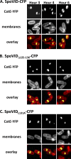
Fig 1
Fig 1
Leucine 131 in the N-terminal domain of SpoVID is required for encasement. Cells were sporulated at 37°C and imaged at the indicated times after resuspension in SM medium. Cells were stained with membrane stain (FM4-64) and analyzed by fluorescence microscopy. A few representative cells are shown for each strain. (First rows) CotE-YFP fluorescence images; (second rows) fluorescence images of membranes; (third rows) overlays of CotE-YFP (yellow) and membranes (red). (A) The CotE-YFP fusion in a spoVID::kan strain carrying amyE::spoVID-cfp (KW363). (B) CotE-YFP fusion in a spoVID::kan strain carrying amyE::spoVIDΔ125–136-cfp (PE2094). (C) CotE-YFP fusion in a spoVID::kan strain carrying amyE::spoVIDL131A-cfp (PE2276).
Fig 2
Fig 2
CotE interaction with the N-terminal domain of SpoVID in yeast two-hybrid assays. Saccharomyces cerevisiae strains bearing plasmids producing the N-terminal domain of SpoVID (amino acids 1 to 144) fused to the BD of the Gal4p transcription factor or an empty vector producing the unfused Gal4BD and either CotE (amino acids 1 to 158) fused to the AD of Gal4 (first two columns) or an empty vector producing the Gal4AD with no fusion (third and fourth columns) were plated on SC -Leu -Trp -His selection plates with or without 5 mM 3AT. The first, second, and third rows contain strains expressing the SpoVID(1–144)-BD fragment in its wild-type, L131A, and I133A mutated forms, respectively. The fourth row corresponds to a strain producing the Gal4BD from an empty vector. Intersections of the rows and columns indicate strains expressing the indicated protein fusions. Strains containing wild-type SpoVID(1–144)-BD and CotE(1–158)-AD truncation fragments or SpoVIDI133A-BD and CotE(1–158)-AD were able to grow on SC -Leu -Trp -His plates (first column) as well as on medium supplemented with 5 mM 3AT (second column). Strains containing SpoVIDL131A-BD and CotE(1–158)-AD truncated fusion proteins were unable to grow on the selection plate. All SpoVID(1–144)-BD isoforms were unable to grow in the presence of an empty Gal4AD vector. A strain containing empty AD and BD vectors was unable to grow on SC -Leu -Trp -His medium. A positive-control strain, containing AD and BD fusions to the known binding partners, Fos and Jun, respectively, was able to grow on SC -Leu -Trp -His medium.
Fig 3
Fig 3
SpoVID physically interacts with CotE in vitro. Results of His tag pulldown assays. (Top panel) Protein lysates from E. coli strains containing expression plasmids fused to spoVID-his6, preincubated with Ni2+ beads, and untagged cotE were mixed together, incubated with shaking for an hour at room temperature, and applied to a gravity column. After washing, elution fractions were collected and tested for retention of CotE. The blot was analyzed with anti-CotE antiserum. The three lanes containing the elution fractions (E1, E2, and E3) exhibited retention of CotE. Washing and elution were performed with the following imidazole concentrations: wash 1 (W1), 40 mM; W2, 100 mM; W3, 250 mM; elution 1 (E1) and E2, 500 mM; E3, 1 M. (Bottom panel) As a negative control, the second half of the protein lysate from the E. coli strain producing untagged cotE was incubated with Ni2+ beads for an hour at room temperature and applied to a gravity column. After washing, elution fractions were collected and tested for retention of CotE. The blot was analyzed with anti-CotE antiserum. The three lanes containing the elution fractions (E1, E2, and E3) were negative for retention of CotE.
Fig 4
Fig 4
Specific residues in the N-terminal domain of SpoVID are essential for interaction with CotE in vitro, as shown by these results of His tag pulldown assays. Protein lysates from separate E. coli strains containing expression plasmids fused to spoVID-his (Δ125–136, L125A, I127A, L131A, or I133A), preincubated with Ni2+ beads, and untagged cotE were mixed together, incubated with shaking for an hour at room temperature, and applied to a gravity column. After washing, elution fractions were collected and tested for retention of CotE. The blots were analyzed with anti-CotE antiserum. Rows 1 (SpoVIDΔ125–136), 3 (SpoVIDL127A), and 4 (SpoVIDL131A) showed no or very limited retention of CotE. Rows 2 (SpoVIDL125A) and 5 (SpoVIDI133A) exhibited retention of CotE.
Fig 5
Fig 5
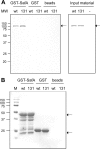
Residue L131 is dispensable for direct interaction with SafA. A fusion of the first 162 residues of SafA to GST (GST-SafA162) was bound to glutathione-Sepharose beads and incubated with equal amounts of extracts from E. coli BL21(DE3) overproducing wild-type SpoVID-His6 or a version of SpoVID-His6 carrying the L131A substitution. Immobilized GST and beads alone were also incubated with the two forms of SpoVID-His6 as negative controls. (A) Pulled down proteins were eluted and resolved by SDS-PAGE and immunoblotted with an anti-SpoVID antibody (left). The amount of SpoVID-His6 or of SpoVID-His6 (L131A) present in the extracts is also shown (right). (B) The nitrocellulose membrane used for immunoblotting was then stained with Ponceau Red as a control for the amount of GST-SafA or GST bound to the glutathione beads.
Fig 6
Fig 6
A model for spore encasement mediated by SpoVID. (A) Proteins of the basement layer of the spore coat localize to the surface of the spore coat, according to previously characterized interactions. The small peptide SpoVM (black) binds to curved membranes in vitro (46) and interacts directly with SpoIVA (white triangles) (45). SpoIVA has been shown to multimerize in vitro (47), restrict SpoVM to the forespore in vivo (45), and to interact with the C-terminal domain of SpoVID (gray circles) (35, 58). Additional interactions have been reported between the SpoVID N-terminal domain (gray triangles) and SafA N-terminal domain (yellow triangles) (5), as well as between SpoIVA, SafA, and SpoVID (35). Evidence for a direct interaction between SpoIVA and CotE (blue ovals) is lacking, but subcellular localization of a CotE-GFP fusion is dependent on spoIVA (59). (B) Encasement is favored by the SpoVID N-terminal domain and its interactions with SafA and CotE. An interaction between SafA full-length protein and SafA-C30 (short form of SafA [yellow ovals]) has been described by Ozin et al. (38). Evidence for an interaction between the SpoVID N-terminal domain and CotE was obtained in the current study. (C) Completion of encasement. The multimerization of CotE is in agreement with the data of Little and Driks (30) and Costa et al. (6). A possible increase in CotE levels in the late stages of sporulation (i.e., postengulfment) has been suggested by Zheng and Losick (62) and McKenney and Eichenberger (34).

Similar articles

Cited by

References

    1. Boland FM, Atrih A, Chirakkal H, Foster SJ, Moir A. 2000. Complete spore-cortex hydrolysis during germination of Bacillus subtilis 168 requires SleB and YpeB. Microbiology 146:57–64 - PubMed
    1. Boxem M, et al. 2008. A protein domain-based interactome network for C. elegans early embryogenesis. Cell 134:534–545 - PMC - PubMed
    1. Cangiano G, et al. 2010. Direct and indirect control of late sporulation genes by GerR of Bacillus subtilis. J. Bacteriol. 192:3406–3413 - PMC - PubMed
    1. Chen I, Provvedi R, Dubnau D. 2006. A macromolecular complex formed by a pilin-like protein in competent Bacillus subtilis. J. Biol. Chem. 281:21720–21727 - PMC - PubMed
    1. Costa T, Isidro AL, Moran CP, Jr, Henriques AO. 2006. Interaction between coat morphogenetic proteins SafA and SpoVID. J. Bacteriol. 188:7731–7741 - PMC - PubMed

Publication types

MeSH terms

Substances

LinkOut - more resources