Comparative Study
. 2012 Jul 24;109(30):12099-104.
doi: 10.1073/pnas.1204948109. Epub 2012 Jul 6.

Critical function for the Ras-GTPase activating protein RASA3 in vertebrate erythropoiesis and megakaryopoiesis

Affiliations
Comparative Study

Critical function for the Ras-GTPase activating protein RASA3 in vertebrate erythropoiesis and megakaryopoiesis

Lionel Blanc et al. Proc Natl Acad Sci U S A. .

Abstract

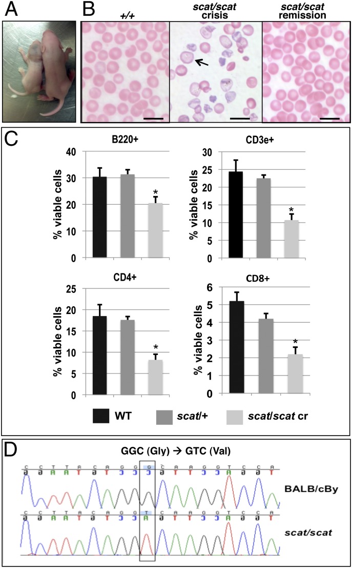
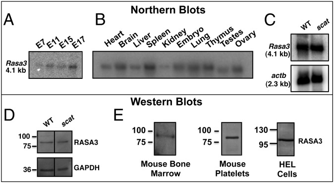
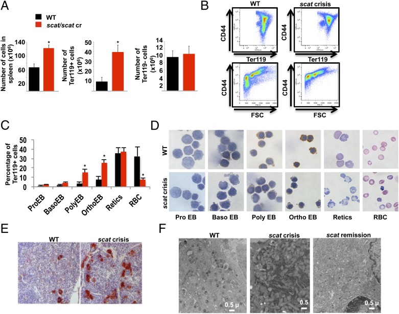
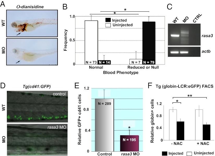
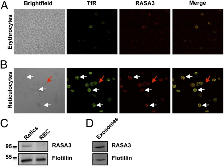
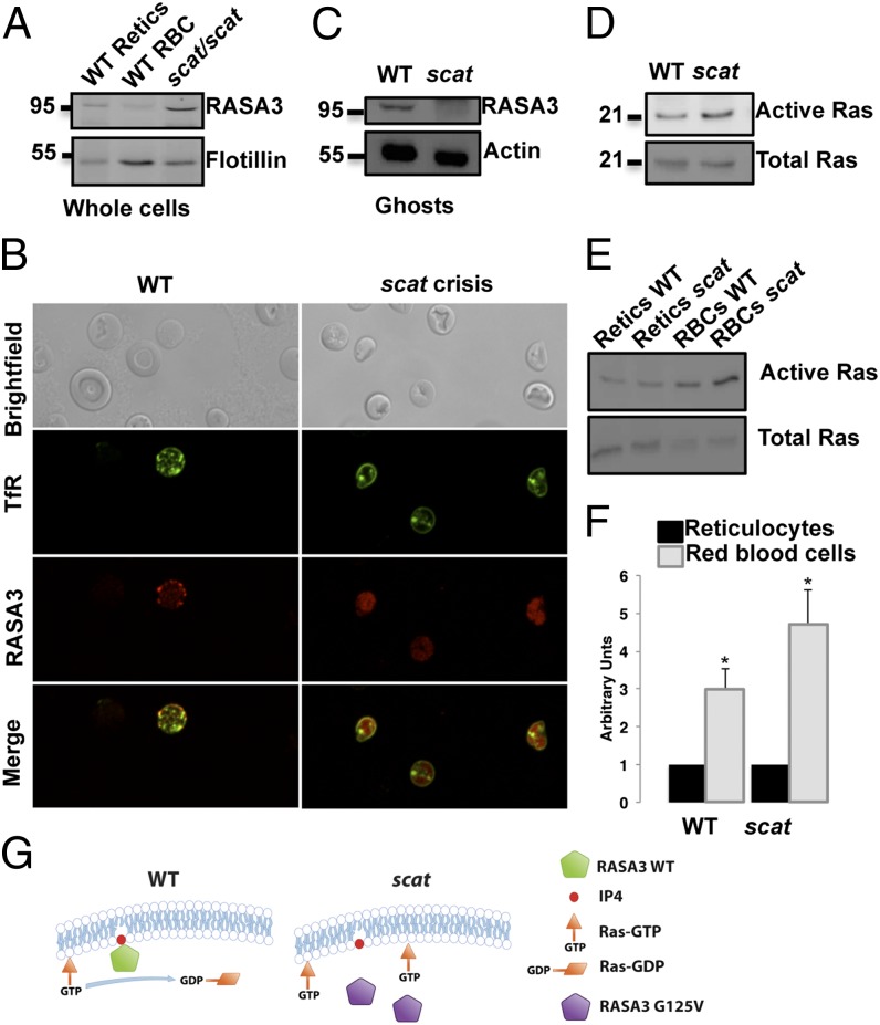
Phenotype-driven approaches to gene discovery using inbred mice have been instrumental in identifying genetic determinants of inherited blood dyscrasias. The recessive mutant scat (severe combined anemia and thrombocytopenia) alternates between crisis and remission episodes, indicating an aberrant regulatory feedback mechanism common to erythrocyte and platelet formation. Here, we identify a missense mutation (G125V) in the scat Rasa3 gene, encoding a Ras GTPase activating protein (RasGAP), and elucidate the mechanism producing crisis episodes. The mutation causes mislocalization of RASA3 to the cytosol in scat red cells where it is inactive, leading to increased GTP-bound Ras. Erythropoiesis is severely blocked in scat crisis mice, and ~94% succumb during the second crisis (~30 d of age) from catastrophic hematopoietic failure in the spleen and bone marrow. Megakaryopoiesis is also defective during crisis. Notably, the scat phenotype is recapitulated in zebrafish when rasa3 is silenced. These results highlight a critical, conserved, and nonredundant role for RASA3 in vertebrate hematopoiesis.


Conflict of interest statement

The authors declare no conflict of interest.

Figures

Fig. 1.
Fig. 1.
The scat phenotype is due to a mutation in Rasa3. (A) Scat homozygous mice in crisis (Left) are small, pale, and show extensive bruising compared with their normal littermates (Right). (B) Wright-Giemsa stained peripheral blood smears demonstrate severe poikilocytosis, anisocytosis, and polychromatophilia with frequent hypochromic cells (arrow) during crisis compared with WT (+/+); red cell morphology reverts to normal during remission. (Scale bar, 10 μM.) (C) Leukopenia in scat homozygotes during crisis primarily reflects a striking loss of lymphocytes. X ± SEM *P < 0.05, Tukey HSD test. (D) Fine mapping and sequencing reveals a G-to-T transversion in the Rasa3 gene in scat that creates a missense mutation (G125V) in a highly conserved residue.
Fig. 2.
Fig. 2.
Rasa3 appears early in development and is widely expressed in adult tissues. (A) Northern blot analysis of WT embryos shows that Rasa3 is expressed early in development, from at least embryonic day 7. (B) Northern blot analysis of adult WT tissues demonstrates widespread expression of Rasa3. (C) Normal levels of Rasa3 mRNA are found in scat spleen. (D) Western blot analyses demonstrate normal levels of RASA3 protein in scat fetal liver cells. (E) Abundant levels of RASA3 are present in WT bone marrow cell membranes and platelets and in human erythroleukemia (HEL) cells.
Fig. 3.
Fig. 3.
Delayed erythropoiesis and megakaryopoiesis in scat spleen. (A) An increase in total spleen cell number (Left) attributable to a dramatic increase in Ter119+ erythroid cells (Center) is seen in the scat crisis spleen. All values X ± SEM; *P < 0.05, t test. (B) By flow cytometry using CD44 and Ter119 or CD44 and forward scatter (FSC) as markers of differentiation, a significant block in erythropoiesis is observed in scat during crisis. (C and D) Quantitation of the different spleen populations demonstrates an accumulation of polychromatophilic and orthochromatic erythroblasts (C) associated with a decrease in hemoglobinization within these cells (D). X ± SEM; *P < 0.05, t test. (E) An increased number of megakaryocytes is seen during crisis as evidenced by acetylcholinesterase staining. (F) By transmission electron microscopy, scat crisis megakaryocytes display features characteristic of a significant developmental delay. See text for details. During remission, megakaryocyte characteristics normalize.
Fig. 4.
Fig. 4.
rasa3 knockdown in zebrafish recapitulates the scat anemia and thrombocytopenia phenotype. (A) In WT embryos, abundant ο-dianisidine positive hemoglobinized cells are seen in the heart and vessels (arrow) at 72 h postfertilization, whereas positive cells are absent in rasa3 morphants (MO). (B) Quantitation of the range of embryos containing normal and reduced or null number of hemoglobinized cells at 72 h postfertilization from control (uninjected) and rasa3 MO (injected). X ± SEM; *P < 0.05, t test. (C) By RT-PCR, abnormally processed rasa3 mRNA splice forms are found in MO compared with −RNA control (CTRL) and uninjected embryos (WT). actb serves as a control of off-target effects. (D) Silencing of rasa3 results in loss of GFP+ cd41-thrombocytes (green) in the transgenic Tg(cd41:GFP) line (MO) compared with control (WT). (E) Quantitation of GFP+ cd41-thrombocytes by flow cytometry in control and rasa3 morphant (MO) embryos. X ± SEM; *P < 0.05, t test. (F) Treatment of WT zebrafish (uninjected) and rasa3 MO (injected) from the Tg(globin LCR:eGFP) transgenic line with the potent antioxidant N-acetyl cysteine (+NAC) fails to improve anemia in the rasa3 MO embryos, indicating that generation of ROS per se is not a contributing factor in the anemia. −NAC, exposed to vehicle carrier only. X ± SEM; *P < 0.05; **P < 0.01, t test.
Fig. 5.
Fig. 5.
RASA3 is lost during reticulocyte maturation through the exosomal pathway. (A) In WT red cells, RASA3 is found in very few cells that are also positive for the TfR by immunofluorescence. (B) After erythropoietic stress by phlebotomy and isolation of reticulocytes on a Percoll gradient, a strong staining for RASA3 is observed on the plasma membrane and in some internal compartments where it colocalizes with the TfR (white arrows). Mature red cells, assessed by negative staining of the TfR (red arrow) are also negative for RASA3. (C) Western blot analyses of reticulocytes and mature erythrocytes (RBC) show that RASA3 is lost during reticulocyte maturation, and (D) is associated with exosomes secreted during maturation.
Fig. 6.
Fig. 6.
RASA3 is mislocalized in scat leading to its loss of function. (A) Western blot analyses on scat peripheral blood show that RASA3 is present in whole red cells (predominantly reticulocytes). (B) By immunofluorescence, RASA3 is mislocalized to the cytosol in scat. (C) Western blot analysis of the red cell membrane ghost fractions confirms the presence of RASA3 on the membrane in WT mice and loss of membrane-bound RASA3 in scat. (D) As a consequence of mislocalization to the cytosol, active GTP-bound active Ras is increased in scat. (E) Consistent with the loss of RASA3 during maturation, active Ras levels are increased in mature red blood cells (RBCs), both in WT and scat as shown by Western blot. (F) Quantitation of the bands demonstrates a higher degree of increase in scat RBCs. X ± SEM; *P < 0.05, t test. (G) Proposed model in which the scat disease during crisis episodes results from RASA3 protein mislocalization leading to loss of its GAP activity.

References

    1. Mikkola HK, Orkin SH. Gene targeting and transgenic strategies for the analysis of hematopoietic development in the mouse. Methods Mol Med. 2005;105:3–22. - PubMed
    1. Orkin SH. Transcription factors and hematopoietic development. J Biol Chem. 1995;270:4955–4958. - PubMed
    1. Orkin SH, Zon LI. Genetics of erythropoiesis: Induced mutations in mice and zebrafish. Annu Rev Genet. 1997;31:33–60. - PubMed
    1. Kerenyi MA, Orkin SH. Networking erythropoiesis. J Exp Med. 2010;207:2537–2541. - PMC - PubMed
    1. Peters LL, McFarland-Starr EC, Wood BG, Barker JE. Heritable severe combined anemia and thrombocytopenia in the mouse: Description of the disease and successful therapy. Blood. 1990;76:745–754. - PubMed

Publication types

MeSH terms

Substances