. 2012 Aug 24;287(35):29312-23.
doi: 10.1074/jbc.M112.362681. Epub 2012 Jul 6.

The Wnt receptor Ryk plays a role in mammalian planar cell polarity signaling

Affiliations

The Wnt receptor Ryk plays a role in mammalian planar cell polarity signaling

Maria L Macheda et al. J Biol Chem. .

Abstract

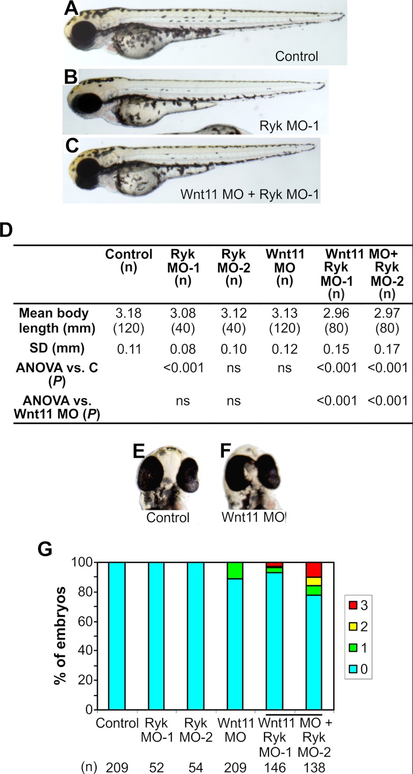
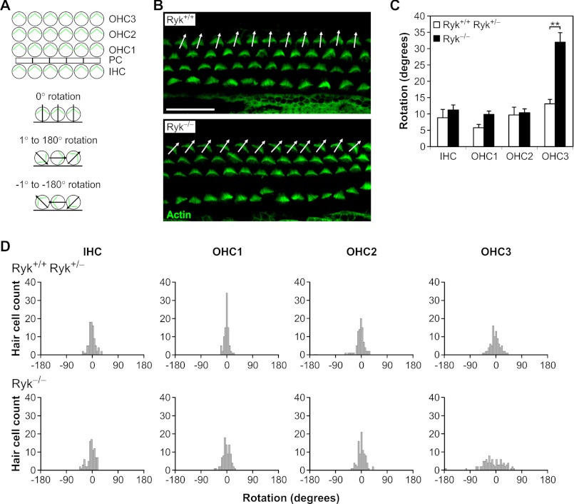
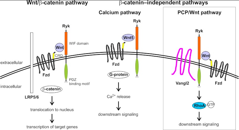
Wnts are essential for a wide range of developmental processes, including cell growth, division, and differentiation. Some of these processes signal via the planar cell polarity (PCP) pathway, which is a β-catenin-independent Wnt signaling pathway. Previous studies have shown that Ryk, a member of the receptor tyrosine kinase family, can bind to Wnts. Ryk is required for normal axon guidance and neuronal differentiation during development. Here, we demonstrate that mammalian Ryk interacts with the Wnt/PCP pathway. In vitro analysis showed that the Wnt inhibitory factor domain of Ryk was necessary for Wnt binding. Detailed analysis of two vertebrate model organisms showed Ryk phenotypes consistent with PCP signaling. In zebrafish, gene knockdown using morpholinos revealed a genetic interaction between Ryk and Wnt11 during the PCP pathway-regulated process of embryo convergent extension. Ryk-deficient mouse embryos displayed disrupted polarity of stereociliary hair cells in the cochlea, a characteristic of disturbed PCP signaling. This PCP defect was also observed in mouse embryos that were double heterozygotes for Ryk and Looptail (containing a mutation in the core Wnt/PCP pathway gene Vangl2) but not in either of the single heterozygotes, suggesting a genetic interaction between Ryk and Vangl2. Co-immunoprecipitation studies demonstrated that RYK and VANGL2 proteins form a complex, whereas RYK also activated RhoA, a downstream effector of PCP signaling. Overall, our data suggest an important role for Ryk in Wnt/planar cell polarity signaling during vertebrate development via the Vangl2 signaling pathway, as demonstrated in the mouse cochlea.


Figures

FIGURE 1.
FIGURE 1.
Interaction of Ryk with Wnts and Wnt/β-catenin signaling assays. A, Ryk and WIF-1 constructs used in co-immunoprecipitation experiments and TCF-luciferase reporter assays. B, immunoprecipitation of 400 μg of lysate from HEK293T cells transfected with mouse Ryk.Fc constructs and Wnt5a.Myc5. IP, immunoprecipitation; IB, immunoblot. C, co-immunoprecipitation of 200 μg of lysate from HEK293T cells transfected with empty vector (V), Wnt1.Myc5, Wnt3a.Myc5, or Wnt5a.Myc5, and 500 ng of purified RYK.Fc protein. D, TCF-luciferase reporter assays of HEK293T cells transfected with RYK constructs, superTOP-FLASH and Renilla, and treated with diluent (C) or 25 ng/ml purified Wnt3a for 24 h. Data are normalized to empty vector-transfected cells. The graph shows the mean ± S.D. of two to four independent experiments of triplicate determinations. No statistically significant differences were observed. E, TCF-luciferase reporter assay of HEK293T cells transfected with superTOP-FLASH and superFOP-FLASH reporters and treated with purified Wnt3a at various concentrations for 24 h. The graph shows the mean ± S.D. of one experiment of triplicate determinations. F, TCF-luciferase reporter assays of MEFs from Ryk+/+ or Ryk−/− embryos transfected with TOP-FLASH (containing three TCF sites in promoter) or FOP-FLASH (containing mutated TCF sites in promoter) and treated with 20 ng/ml purified Wnt3a for 24 h. The graph shows the mean ± S.E. of three independent experiments of triplicate determinations. No statistically significant differences were observed. RLU, relative luciferase units. G, TCF-luciferase reporter assays of HEK293T cells transfected with WIF-1.Fc constructs, superTOP-FLASH and Renilla, and treated with 25 ng/ml purified Wnt3a for 24 h. Data are normalized to empty vector-transfected cells. The graph shows the mean ± S.D. of two independent experiments of triplicate determinations. *, p < 0.05; ***, p < 0.001. H, co-immunoprecipitation of 200 μg of lysate from HEK293T cells transfected with empty vector (V), Wnt1.Myc5, Wnt3a.Myc5, Wnt5a.Myc5, or Wnt11.Myc5 and 1 μg of purified WIF-1.RYKWD.Fc or WIF-1.Fc protein.
FIGURE 2.
FIGURE 2.
Effects of Ryk and Wnt11 knockdown in zebrafish embryos at 54 hpf. A, lateral view of an uninjected control embryo. B, example of an embryo injected with Ryk MO-1, which had reduced body length. C, example of an embryo injected with Wnt11 MO + Ryk MO-1, showing significantly reduced body length. D, quantitation of body length in embryos injected with Ryk MO-1, Ryk MO-2, and Wnt11 MO. (n), number of embryos analyzed in each treatment; SD, standard deviation of body length; C, control embryos. E, frontal view of a control embryo, showing normal eye spacing. F, frontal view of an embryo injected with Wnt11 MO, showing eyes touching. G, quantitation of eye phenotype in zebrafish embryos injected with Ryk MOs and/or Wnt11 MO. Eye phenotype was defined as follows: 0 = wild-type; 1 = eyes close together or just touching; 2 = eyes partly fused; 3 = eyes fused. (n), number of embryos analyzed in each treatment. ANOVA, analysis of variance.
FIGURE 3.
FIGURE 3.
Ryk-deficient mice demonstrate a role for Ryk in the PCP pathway. A, schematic representations of stereociliary bundle orientation in cochlear hair cells of wild-type mice and showing the 0° base line of cell rotation, rotation to the left of center (negative degrees), and rotation to the right of center (positive degrees). Abbreviations used are as follows: IHC, inner hair cell; OHC, outer hair cell; PC, pillar cell. B, orientation of stereociliary bundles in E18.5 Ryk+/+ and Ryk−/− mouse embryos, as observed at the mid-point of the sensory epithelia by staining with phalloidin (to detect actin). Arrows show the orientation of the third row outer hair cells (OHC3). Many cells in Ryk−/− embryos have a misoriented bundle orientation. Scale bar, 20 μm. C, analysis of the orientation of hair cell stereociliary bundles. Results represent the mean ± S.E. for three embryos. **, p < 0.01. D, distribution histograms of stereociliary bundle orientation in E18.5 wild-type and Ryk-deficient cochleae. Bundle orientations are confined to a 45° segment centered on a line parallel to the medial-lateral axis in wild-type embryos. However, the distribution of OHC3 orientation is broader in the Ryk−/− mice. Stereociliary bundle orientation data were collected from three embryos of each genotype.
FIGURE 4.
FIGURE 4.
Ryk is expressed in the mouse cochlea. A, cross-section of an E18.5 Ryk-deficient (RykLacZ/LacZ) cochlea showing β-gal staining (blue). Ryk mRNA was distributed throughout the cochlear duct, with strong expression in the OHCs and pillar cells (PC). In the bottom panel, the location of the hair cells is indicated with myosin 6 (red). A single IHC row (arrowhead) and three OHC rows (arrows 1–3) can be identified in the organ of Corti. Scale bar, 50 μm. B, surface view of a P0 wild-type mouse cochlear whole mount, showing Ryk protein expression in outer hair cells (OHC1–3) and pillar cells (PC) using an anti-Ryk antibody (top panel). The bottom panel shows co-staining with phalloidin (to detect actin). Scale bar, 20 μm.
FIGURE 5.
FIGURE 5.
Ryk-deficient mouse embryos display neural tube defects in specific genetic backgrounds. A, lateral and dorsal views of a wild-type and a Ryk × Vangl2 double-heterozygous (Ryk+/−;Vangl2Lp/+) E18.5 embryo from the same litter. The neural tube is open along its whole length in the double heterozygote. Embryo heads were removed in dorsal view images. B, Ryk+/+/Ryk+/− and Ryk−/− E18.5 embryo heads from mice on a 129T2/Sv or a mixed C57BL/6 × 129T2/Sv genetic background; embryos sharing a genetic background were from the same litter. The 129T2/Sv knock-out has an open neural tube at the head only (exencephaly); this phenotype was observed in 13% of Ryk−/− embryos on this genetic background. No exencephaly was observed in Ryk−/− embryos on a mixed background.
FIGURE 6.
FIGURE 6.
Crosses of Ryk-deficient mice with Vangl2 mutant mice reveal that Ryk interacts with core PCP proteins in the cochlea. A, cochleae from E18.5 embryos with genotypes Ryk+/−, Vangl2Lp/+, and double heterozygotes Ryk+/−;Vangl2Lp/+ were stained with G. simplicifolia lectin and examined at the mid-point of the sensory epithelia. Arrows show the orientation of some misoriented hair cells (OHC3). Scale bar, 10 μm. B, analysis of the orientation of hair cell stereociliary bundles. Results represent the mean ± S.E. for three embryos. *, p < 0.05; **, p < 0.01. C, comparison of distribution histograms for stereociliary bundle orientation in E18.5 mouse cochleae. Bundle orientations in Ryk+/− and Vangl2Lp/+ cochleae are confined to a 60° segment centered on a line parallel to the medial-lateral axis. However, in the Ryk+/−;Vangl2Lp/+ double-heterozygous cochleae the distribution of OHC3 orientation is markedly broader. Stereociliary bundle orientation data were collected from three embryos of each genotype.
FIGURE 7.
FIGURE 7.
RYK forms a complex with VANGL2 and activates RhoA. A, immunoprecipitation (IP) of 200 μg of lysate from HEK293T cells transfected with empty vector (V), Myc2.RYK, Myc2.RYKΔPDZBM (RYKΔP), or HA2.VANGL2. Arrows indicate the band of interest (RYK constructs on the left or VANGL2 on the right). B, quantitation of body length in zebrafish embryos injected with Ryk MO-1 and VANGL2 RNA. (n), number of embryos analyzed in each treatment; SD, standard deviation of body length; C, control embryos; ANOVA, analysis of variance. C, active RhoA pulldowns using 1 ml of cell lysate from CHO-K1 cells transfected with empty vector (V) or RYKFCT. Western blot was performed with pulldowns or 10 μl of lysate using anti-RhoA antibody. D, quantitation of CHO-K1 cell lysate active RhoA pulldowns. Results represent the mean ± S.D. of three independent experiments. **, p < 0.01. E, active RhoA pulldowns using 1 ml of cell lysate from CHO-K1 cells mock transfected (no siRNA) or transfected with RYK siRNA and then transfected with empty vector (V) or Wnt3a.Myc5 (Wnt3a). Western blot was performed with pulldowns or 10 μl of lysate using anti-RhoA antibody. IB, immunoblot.
FIGURE 8.
FIGURE 8.
Schematic diagram of the interaction between Ryk and Wnt signaling pathways. Ryk binds to Wnts via its WIF domain and modulates both Wnt/β-catenin and β-catenin-independent signaling (14, 18, 22, 24). As was shown in this study, Ryk interacts with the PCP pathway protein Vangl2 and can activate downstream RhoA signaling (boxed). CRD, cysteine-rich domain; Fzd, Frizzled; LRP5/6, low density lipoprotein-related protein 5 or 6; PDZ, PSD-95/Dlg/ZO-1 homology; WIF, Wnt inhibitory factor.

References

    1. Seifert J. R., Mlodzik M. (2007) Frizzled/PCP signaling. A conserved mechanism regulating cell polarity and directed motility. Nat. Rev. Genet. 8, 126–138 - PubMed
    1. Keller R. (2002) Shaping the vertebrate body plan by polarized embryonic cell movements. Science 298, 1950–1954 - PubMed
    1. Tada M., Smith J. C. (2000) Xwnt11 is a target of Xenopus Brachyury; Regulation of gastrulation movements via Dishevelled but not through the canonical Wnt pathway. Development 127, 2227–2238 - PubMed
    1. Wallingford J. B., Rowning B. A., Vogeli K. M., Rothbächer U., Fraser S. E., Harland R. M. (2000) Dishevelled controls cell polarity during Xenopus gastrulation. Nature 405, 81–85 - PubMed
    1. Heisenberg C. P., Tada M., Rauch G. J., Saúde L., Concha M. L., Geisler R., Stemple D. L., Smith J. C., Wilson S. W. (2000) Silberblick/Wnt11 mediates convergent extension movements during zebrafish gastrulation. Nature 405, 76–81 - PubMed

Publication types

MeSH terms

Substances