. 2012 Aug 24;287(35):29554-67.
doi: 10.1074/jbc.M112.390047. Epub 2012 Jul 7.

Oxidation of dihydrotestosterone by human cytochromes P450 19A1 and 3A4

Affiliations

Oxidation of dihydrotestosterone by human cytochromes P450 19A1 and 3A4

Qian Cheng et al. J Biol Chem. .

Abstract

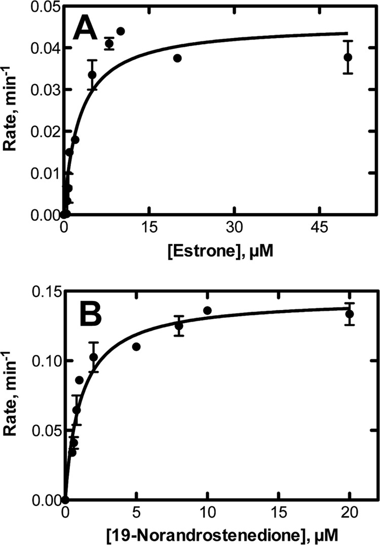
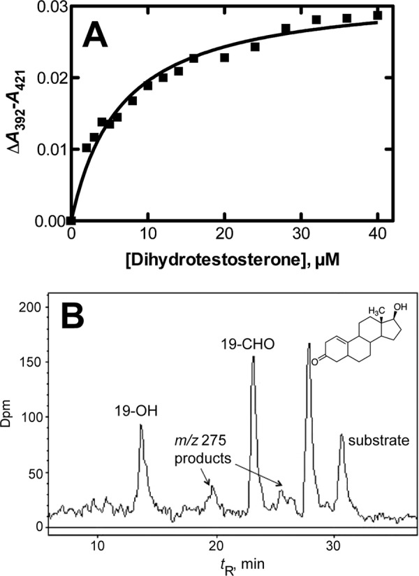
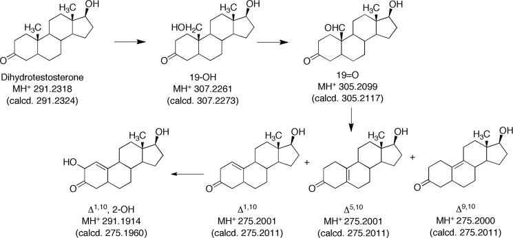
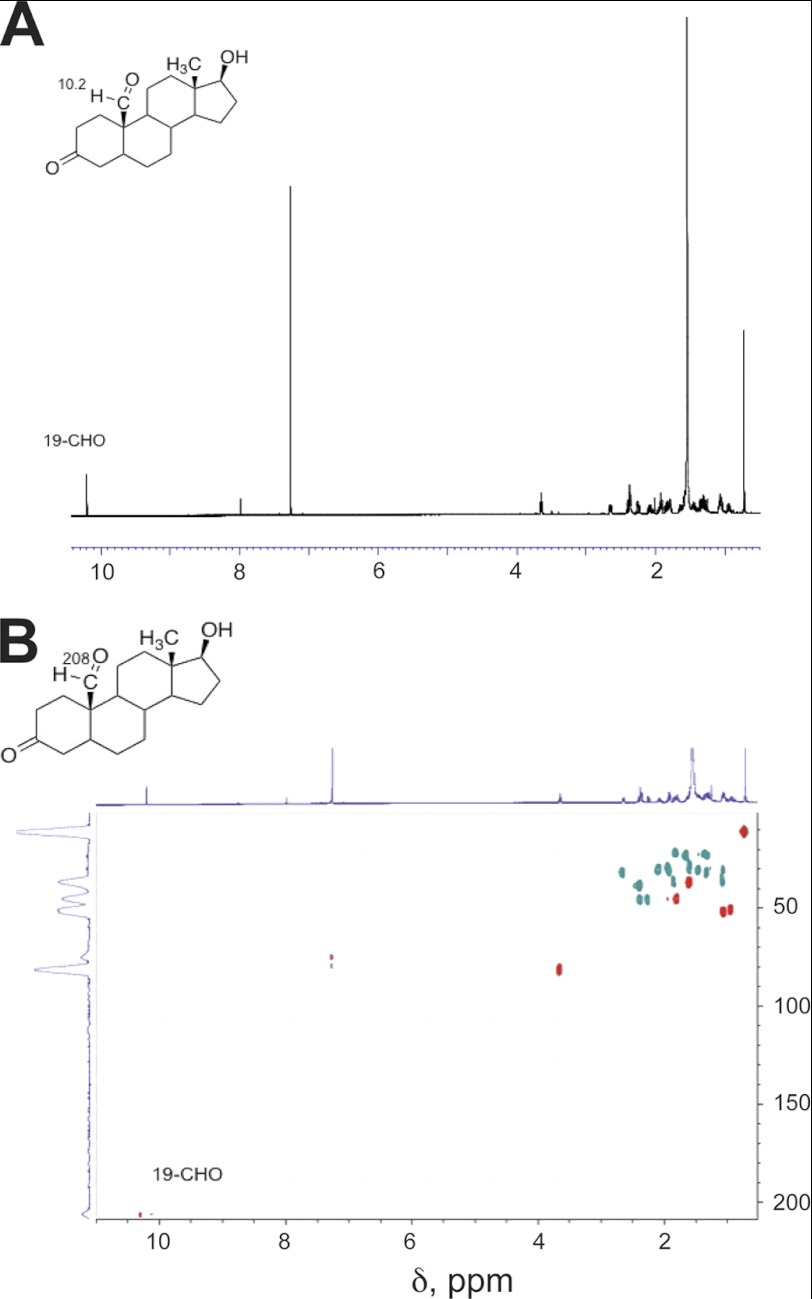
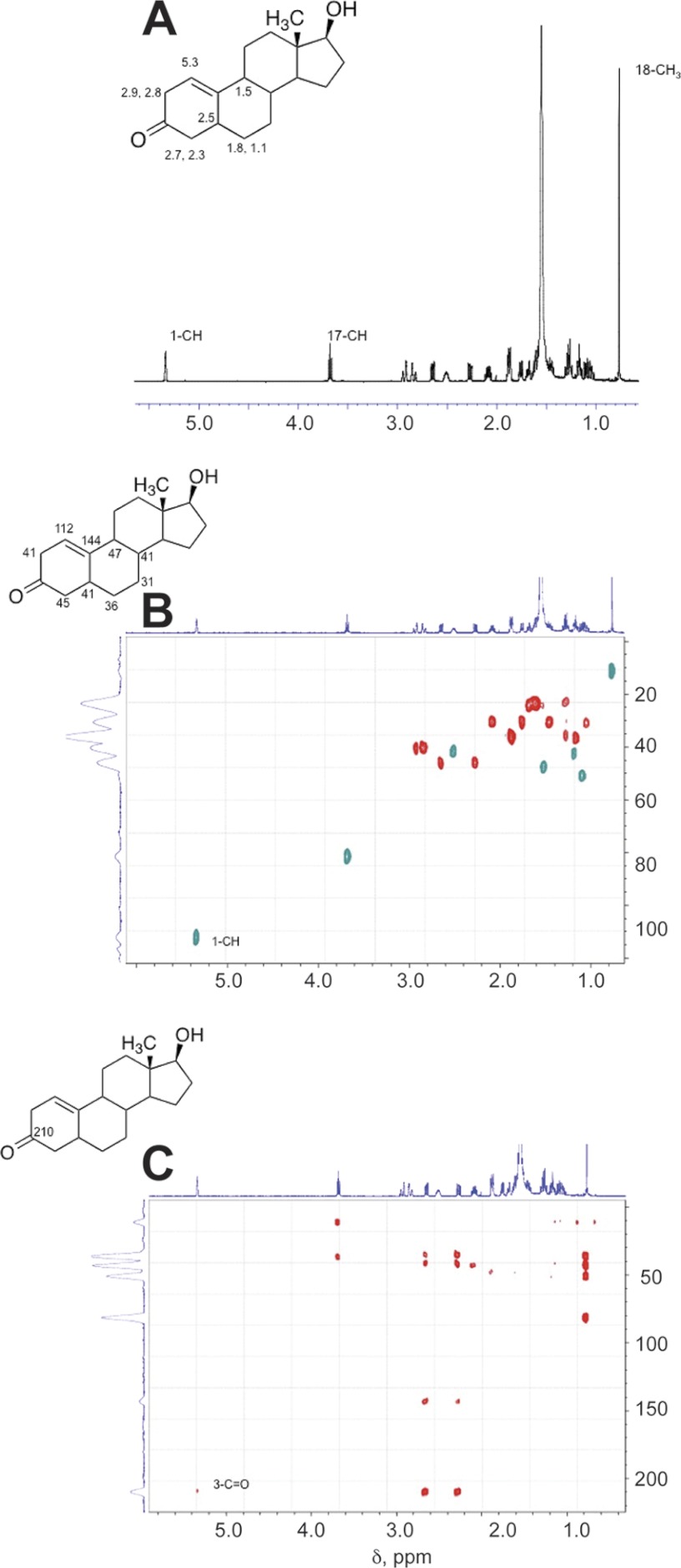
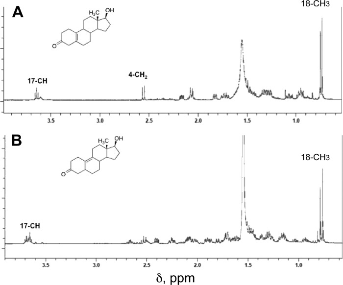
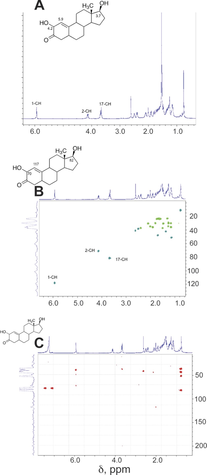
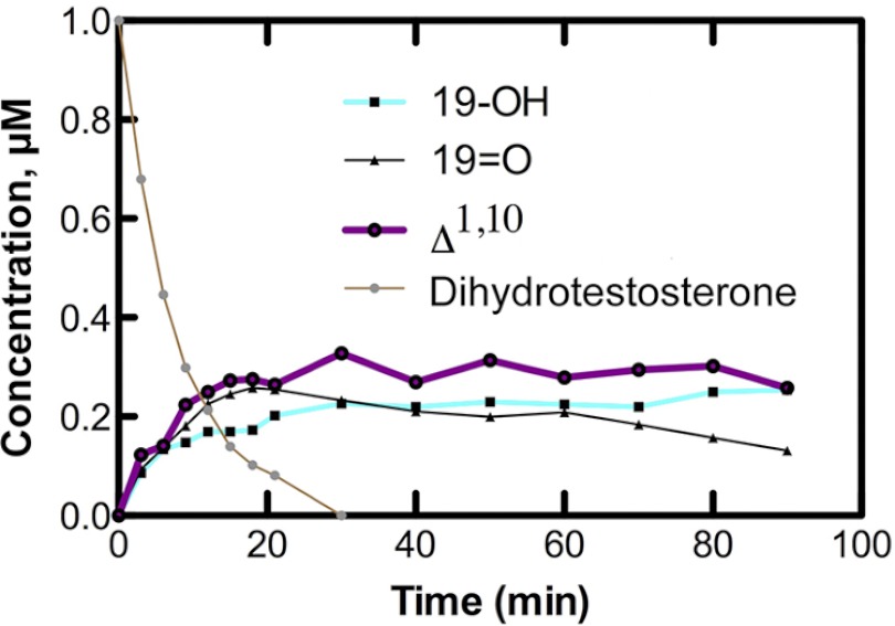
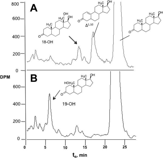
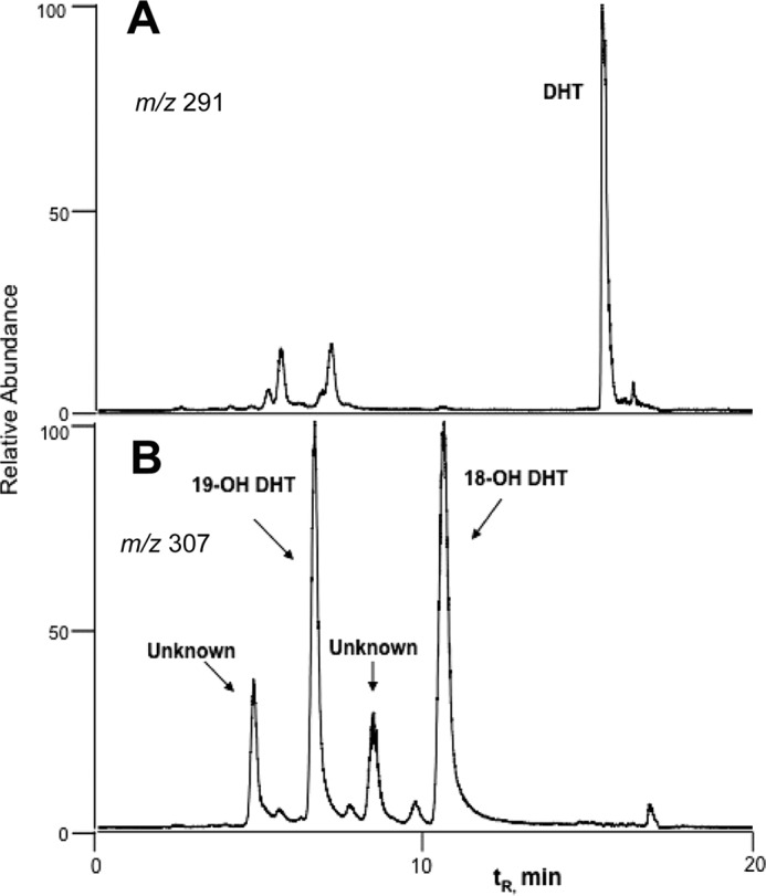
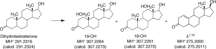
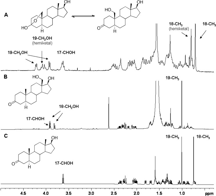
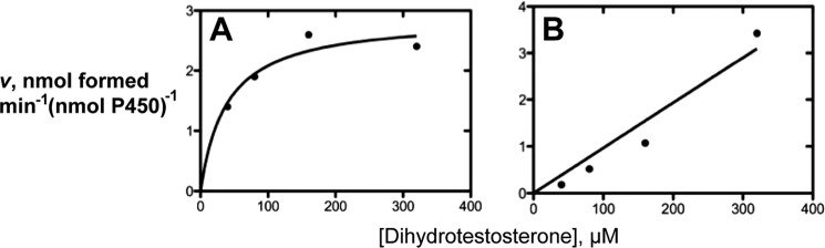
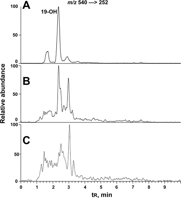
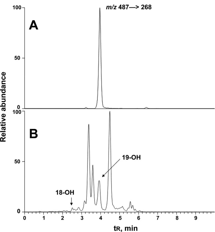
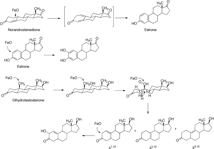
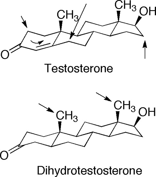
Dihydrotestosterone is a more potent androgen than testosterone and plays an important role in endocrine function. We demonstrated that, like testosterone, dihydrotestosterone can be oxidized by human cytochrome P450 (P450) 19A1, the steroid aromatase. The products identified include the 19-hydroxy- and 19-oxo derivatives and the resulting Δ(1,10)-, Δ(5,10)-, and Δ(9,10)-dehydro 19-norsteroid products (loss of 19-methyl group). The overall catalytic efficiency of oxidation was ~10-fold higher than reported for 3α-reduction by 3α-hydroxysteroid dehydrogenase, the major enzyme known to deactivate dihydrotestosterone. These and other studies demonstrate the flexibility of P450 19A1 in removing the 1- and 2-hydrogens from 19-norsteroids, the 2-hydrogen from estrone, and (in this case) the 1-, 5β-, and 9β-hydrogens of dihydrotestosterone. Incubation of dihydrotestosterone with human liver microsomes and NADPH yielded the 18- and 19-hydroxy products plus the Δ(1,10)-dehydro 19-nor product identified in the P450 19A1 reaction. The 18- and 19-hydroxylation reactions were attributed to P450 3A4, and 18- and 19-hydroxydihydrotestosterone were identified in human plasma and urine samples. The change in the pucker of the A ring caused by reduction of the Δ(4,5) bond is remarkable in shifting the course of hydroxylation from the 6β-, 2β-, 1β-, and 15β-methylene carbons (testosterone) to the axial methyl groups (18, 19) in dihydrotestosterone and demonstrates the sensitivity of P450 3A4, even with its large active site, to small changes in substrate structure.


Figures

FIGURE 1.
FIGURE 1.
Chemical synthesis of 19-hydroxydihydrotestosterone. PPTS, pyridinium p-toluenesulfonate; PTSA, p-toluenesulfonic acid. Yields are shown in the scheme. 19-Hydroxydihydrotestosterone exists in equilibrium with its hemiketal form (31). See “Experimental Procedures” for details.
FIGURE 2.
FIGURE 2.
Steady-state kinetics of alternate oxidations catalyzed by P450 19A1. A, 2-hydroxylation of estrone; B, oxidation of 19-norandrostenedione to estrone.
FIGURE 3.
FIGURE 3.
Binding and oxidation of dihydrotestosterone by P450 19A1. A, binding as observed by perturbation of the heme Soret spectra. B, radio-HPLC of oxidation products of dihydrotestosterone formed by P450 19A1. The m/z 275 products, including the Δ1,10 compound whose structure is shown, were identified by MS and NMR (Figs. 4, 5, and 7).
FIGURE 4.
FIGURE 4.
HRMS of oxidation products of dihydrotestosterone formed by P450 19A1. HRMS parent ions (m/z values) are those determined, with the calculated values (calcd.) are listed below each. All measured values were within 5 ppm of the calculated values.
FIGURE 5.
FIGURE 5.
NMR spectra of oxidation products of dihydrotestosterone formed by P450 19A1: dihydrotestosterone 19-aldehyde (see Fig. 3). A, 1H NMR spectrum; B, HSQC spectrum (CDCl3). Key assignments are shown.
FIGURE 6.
FIGURE 6.
NMR spectra of oxidation products of dihydrotestosterone formed by P450 19A1: Δ1,10-dehydro 19-nordihydrotestosterone (see Fig. 3). A, 1H NMR spectrum; B, HSQC spectrum; C, HMBC spectrum (CDCl3). Key assignments are shown.
FIGURE 7.
FIGURE 7.
1H NMR spectra of oxidation products of dihydrotestosterone formed by P450 19A1 (see Fig. 3). A, Δ5,10-dehydro 19-nordihydrotestosterone; B, Δ9,10-dehydro 19-nordihydrotestosterone. The solvent was CDCl3. Key assignments are shown.
FIGURE 8.
FIGURE 8.
NMR spectra of oxidation products of dihydrotestosterone formed by P450 19A1: 2-hydroxy-Δ1,10-dehydro 19-nordihydrotestosterone (see Fig. 3). A, 1H NMR spectrum; B, HSQC spectrum; C, HMBC spectrum (CDCl3). Key assignments are shown.
FIGURE 9.
FIGURE 9.
Time course of conversion of dihydrotestosterone to products by P450 19A1. The initial concentration of [3H]dihydrotestosterone was 1.0 μm and the P450 19A1 concentration was 0.5 μm (the NADPH-P450 reductase concentration was 1.0 μm).
FIGURE 10.
FIGURE 10.
Radio-HPLC traces of [3H]dihydrotestosterone oxidation products. A, human liver microsomes; B, P450 3A4 bicistronic membranes. The reaction time was 60 min in both cases.
FIGURE 11.
FIGURE 11.
LC-MS of metabolites of dihydrotestosterone formed by P450 3A4. Four hydroxy metabolites (m/z 307) were detected. The two major products were identified as 19-OH dihydrotestosterone (DHT) by co-chromatography and 18-OH DHT by 1H NMR (see Fig. 13), respectively. A, m/z 291; B, m/z 307.
FIGURE 12.
FIGURE 12.
HRMS of oxidation products of dihydrotestosterone formed in human liver microsomes. HRMS parent ions (m/z values) are those determined, with the calculated values (calcd.) listed below each.
FIGURE 13.
FIGURE 13.
One-dimensional 1H NMR spectra of dihydrotestosterone and 18-OH and 19-OH dihydrotestosterone recovered from P450 3A4 incubations. A, 19-hydroxydihydrotestosterone; B, 18-hydroxydihydrotestosterone; C, dihydrotestosterone. The 18-methyl signal of dihydrotestosterone is missing in the 18-OH dihydrotestosterone spectrum and appears (as -CH2OH) at δ 3.82.
FIGURE 14.
FIGURE 14.
Steady-state kinetics of P450 3A4-catalyzed 18- and 19-hydroxylations of dihydrotestosterone. A, 19-hydroxylation by P450 3A4; B, 18-hydroxylation by P450 3A4. The fits shown were done using a hyperbolic fit (A) and linear regression (B) (only slope estimates are considered in the text).
FIGURE 15.
FIGURE 15.
LC-MS of dansylated 19-hydroxydihydrotestosterone in human urine and plasma. The transition m/z 540 [rarrrow] 252 was monitored. A, derivatization of synthetic 19-hydroxydihydrotestosterone led to one major product (identified as a mono-dansyl derivative) and two minor products (unidentified); B, dansylated 19-hydroxydihydrotestosterone in a human urine sample; C, dansylated 19-hydroxydihydrotestosterone in human plasma.
FIGURE 16.
FIGURE 16.
LC-MS of 2,4-DNPH derivatives of 19-hydroxydihydrotestosterone in human urine. The transition m/z 487 → 268 was monitored. A, synthetic 19-hydroxydihydrotestosterone derivatized with 2,4-DNPH; B, a human urine sample extracted and derivatized with 2,4-DNPH.
FIGURE 17.
FIGURE 17.
Some oxidations of steroids catalyzed by P450 19A1. Site of oxidation by P450 FeO complexes are shown. The losses of individual protons of dihydrotestosterone 19-aldehyde are shown with arrows.
FIGURE 18.
FIGURE 18.
Oxidations of testosterone and dihydrotestosterone catalyzed by P450 3A4. Sites of hydroxylation are shown with arrows. All testosterone hydroxylations occur at the β face (18).

References

    1. Conney A. H. (1967) Pharmacological implications of microsomal enzyme induction. Pharmacol. Rev. 19, 317–366 - PubMed
    1. Miller J. A. (1970) Carcinogenesis by chemicals. An overview. G. H. A. Clowes Memorial Lecture. Cancer Res. 30, 559–576 - PubMed
    1. Ortiz de Montellano P. R. (ed) (2005) Cytochrome P450: Structure, Mechanism, and Biochemistry, 3rd ed., Kluwer Academic/Plenum Publishers, New York
    1. Nebert D. W., Russell D. W. (2002) Clinical importance of the cytochromes P450. Lancet 360, 1155–1162 - PubMed
    1. Zhao B., Lei L., Kagawa N., Sundaramoorthy M., Banerjee S., Nagy L. D., Guengerich F. P., Waterman M. R. (2012) A three-dimensional structure of steroid 21-hydroxylase (cytochrome P450 21A2) with binary substrate occupancy reveals locations of disease-associated variants. J. Biol. Chem. 287, 10613–10622 - PMC - PubMed

Publication types

LinkOut - more resources