. 2012:18:1640-8.
Epub 2012 Jun 20.

Effects of melatonin and its receptor antagonist on retinal pigment epithelial cells against hydrogen peroxide damage

Affiliations

Effects of melatonin and its receptor antagonist on retinal pigment epithelial cells against hydrogen peroxide damage

Richard B Rosen et al. Mol Vis. 2012.

Abstract

Purpose: Recently, we reported finding that circulating melatonin levels in age-related macular degeneration patients were significantly lower than those in age-matched controls. The purpose of this study was to investigate the hypothesis that melatonin deficiency may play a role in the oxidative damage of the retinal pigment epithelium (RPE) by testing the protective effect of melatonin and its receptor antagonist on RPE cells exposed to H(2)O(2) damage.

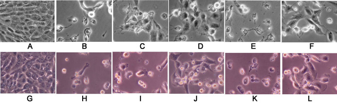
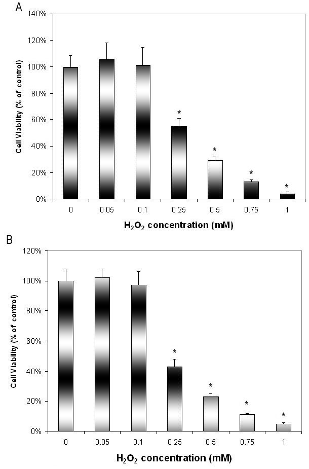
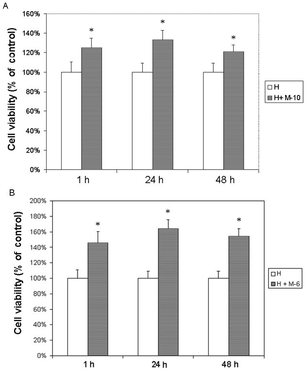
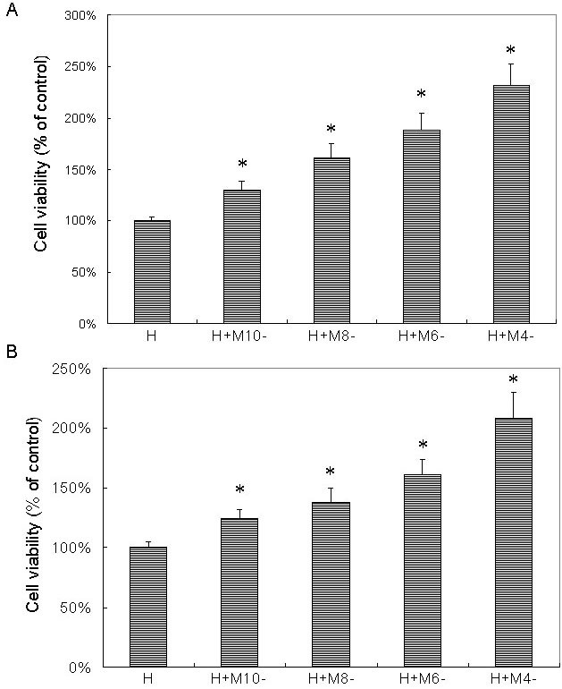
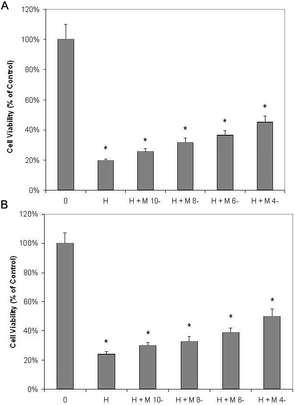
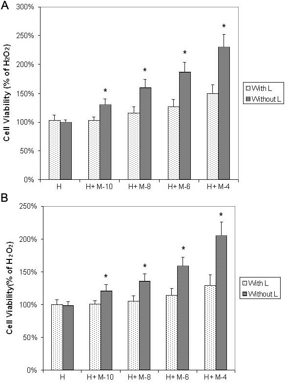
Methods: Cultured human RPE cells were subjected to oxidative stress induced by 0.5 mM H(2)O(2). Cell viability was measured using the microculture tetrazoline test (MTT) assay. Cells were pretreated with or without melatonin for 24 h. Luzindole (50 μM), a melatonin membrane-receptor antagonist, was added to the culture 1 h before melatonin to distinguish direct antioxidant effects from indirect receptor-dependent effects. All tests were performed in triplicate.

Results: H(2)O(2) at 0.5 mM decreased cell viability to 20% of control levels. Melatonin showed dose-dependent protective effects on RPE cells against H(2)O(2). Cell viability of RPE cells pretreated with 10(-10), 10(-8), 10(-6), and 10(-4) M melatonin for 24 h was 130%, 160%, 187%, and 230% of cells treated with H(2)O(2) alone (all p<0.05). Using cells cultured without H(2)O(2) as the control, cell viability of cells treated with H(2)O(2) after pretreatment with 10(-10)-10(-4) M melatonin was still significantly lower than that of the controls, suggesting that melatonin significantly decreased but did not completely abolish the in vitro cytotoxic effects of H(2)O(2). Luzindole completely blocked melatonin's protective effects at low concentrations of melatonin (10(-10)-10(-8) M) but not at high concentrations (10(-6)-10(-4) M).

Conclusions: Melatonin has a partial protective effect on RPE cells against H(2)O(2) damage across a wide range of concentrations (10(-10)-10(-4) M). This protective effect occurs through the activation of melatonin membrane receptors at low concentrations (10(-10)-10(-8) M) and through both the direct antioxidant and indirect receptor activation effects at high concentrations (10(-6)-10(-4) M).


Figures

Figure 1
Figure 1
Melatonin protected the retinal pigment epithelium (RPE) cells against H2O2 damage, especially in high concentrations (10−4 M) and luzindole decreased the protective effects of melatonin. Phase-contrast microscopic images of the effects of melatonin and its membrane-receptor antagonist (luzindole) on retinal pigment epithelial cells against H2O2 damage. A-F: The RPE cells are from the ARPE-19 cell line (an immortal RPE cell line from a 19-year-old donor). G-L: The RPE cells are from the primary culture (PC) from the donor eye. ARPE-19 and PC cells were cultured with or without H2O2, melatonin, and luzindole, as follows: without H2O2, melatonin, and luzindole(A and G); with H2O2 at 0.5 mM concentrations for 24 h (B and H); with H2O2 and a pretreatment of melatonin at 10−10 M (C and I) or at 10−4 M (D and J) for 24 h; or with luzindole (50 μM, 1 h), followed by melatonin at 10−10 M (E and K) or at 10−4 M for 24 h (F and L). H2O2 was then added and the ARPE-19 and PC cells cultured for 24 h.
Figure 2
Figure 2
H2O2 dose-dependently decreased cell viability of retinal pigment epithelial cells as tested by microculture tetrazoline test. Retinal pigment epithelial (RPE) cells were plated in 96-well plates and treated with various concentrations of H2O2 for 24 h. Cell viability was evaluated by the microculture tetrazoline test (MTT) test and expressed as percentage of that in cells without H2O2 (mean±standard deviation [SD] in triplicate tests). A: H2O2 showed dose-dependent cytotoxic effects on ARPE-19 cells (cells from an immortal RPE cell line from a 19-year-old donor) at concentrations from 0.25 mM to 1.00 mM (p<0.05, compared to cells cultured without H2O2. B: Studies of primary cultures of human RPE cells isolated from donor eye showed similar results. *p<0.05, compared with the controls (cells cultured with H2O2 alone).
Figure 3
Figure 3
The ARPE19 cells (an immortal retinal pigment epithelial cell line from a 19-year-old donor) were treated with melatonin (M) at different concentrations. After 1 h, 24 h, and 48 h culture, 0.5 mM H2O2 (H) was added and cultured for 24 h. Cells cultured with H2O2 alone were used as the controls. Cell viability was evaluated by the microculture tetrazoline test and expressed as percentages of controls (mean±SD in triplicate tests). Error bars represent SD A: Pretreatment with low concentrations of melatonin at 10−10 M (M-10) for 1 h, 24 h, and 48 h significantly protected cells against H2O2. B: Pretreatment with high concentrations of melatonin at 10−6 M (M-6) obtained similar results. The difference between cells cultured with and without melatonin at both high and low concentrations was statistically significant at all three different pretreatment periods. *p<0.05, compared with the controls.
Figure 4
Figure 4
Melatonin dose-dependently protected retinal pigment epithelial cells against H2O2 damage as tested by microculture tetrazoline test. Retinal pigment epithelial (RPE) cells were pretreated with melatonin (M) at concentrations of 10−10 M (M-10), 10−8 M (M-8), 10−6 M (M-6), and 10−4 M (M-4). After 24 h, 0.5 mM H2O2 (H) was added and cultured for 24 h. Cells treated with H2O2 alone were used as the controls (H). Cell viability was evaluated by the microculture tetrazoline test and expressed as percentages of the controls (mean±standard deviation [SD] in triplicate tests). Error bars represent SD. Pretreatment with melatonin showed dose-dependent protective effects on ARPE-19 cells (an immortal RPE cell line from a 19-year-old donor) against H2O2 damage (A). Studies in primary culture of human RPE cells isolated from the donor eye showed similar results (B). *p<0.05, compared with the controls (cells treated with H2O2 alone).
Figure 5
Figure 5
Melatonin dose-dependently protected retinal pigment epithelial cells against H2O2 damage as tested by microculture tetrazoline test, compared with cells not treated with H2O2. Retinal pigment epithelial (RPE) cells were pretreated with melatonin (M) at concentrations of 10−10 M (M-10), 10−8 M (M-8), 10−6 M (M-6), and 10−4 M (M-4). After 24 h, 0.5 mM H2O2 (H) was added and cultured for 24 h. Cells not treated with H2O2 were used as negative controls (0). Cell viability was evaluated by the microculture tetrazoline test and expressed as percentages of negative controls (mean±standard deviation [SD] in triplicate tests). Error bars represent SD A: Cell viability of cell treated with melatonin and H2O2 still significantly lower than that in cells cultured without H2O2 in the ARPE-19 cells (an immortal RPE cell line from a 19-year-old donor). B: The same was true in the primary-culture RPE cells. * p<0.05, compared with the negative controls (cells treated without H2O2).
Figure 6
Figure 6
Luzindole decreased protective effects of melatonin on retinal pigment epithelial cells against H2O2 damage as tested by microculture tetrazoline test. Cultured retinal pigment epithelial (RPE) cells were treated with or without 50 μM luzindole (L). One hour later melatonin (M) was added to the culture medium at concentrations of 10−10 M (M-10), 10−8 M (M-8), 10−6 M (M-6), and 10−4 M (M-4). After 24 h, 0.5 mM H2O2 (H) was added and cultures were incubated for 24 h. Cell viability was evaluated by the microculture tetrazoline test and expressed as percentages of cells cultured with H2O2 alone (mean±standard deviation [SD] in triplicate tests). Error bars represent SD A: Luzindole significantly decreased melatonin-induced protective effects at 10−10 to 10−4 M in ARPE-19 cells (an immortal RPE cell line from a 19-year-old donor). B: The same was true in the primary-culture RPE cells. * p<0.05, comparison between cells treated with and without luzindole.

Similar articles

Cited by

References

    1. Rein DB, Wittenborn JS, Zhang X, Honeycutt AA, Lesesne SB, Saaddine J, Vision Health Cost-Effectiveness Study Group Forecasting age-related macular degeneration through the year 2050: the potential impact of new treatments. Arch Ophthalmol. 2009;127:533–40. - PubMed
    1. Hogg R, Chakravarthy U. AMD and micronutrient antioxidants. Curr Eye Res. 2004;29:387–401. - PubMed
    1. Winkler BS, Boulton ME, Gottsch JD, Sternberg P. Oxidative damage and age-related macular degeneration. Mol Vis. 1999;5:32. - PMC - PubMed
    1. Cai J, Nelson KC, Wu M, Sternberg P, Jr, Jones DP. Oxidative Damage and protection of the RPE. Prog Retin Eye Res. 2000;19:205–21. - PubMed
    1. Zarbin MA. Current concepts in the pathogenesis of age-related macular degeneration. Arch Ophthalmol. 2004;122:598–614. - PubMed

Publication types

MeSH terms

LinkOut - more resources