. 2012 Jun;2(6):120063.
doi: 10.1098/rsob.120063.

USP11 augments TGFβ signalling by deubiquitylating ALK5

Affiliations

USP11 augments TGFβ signalling by deubiquitylating ALK5

Mazin A Al-Salihi et al. Open Biol. 2012 Jun.

Abstract

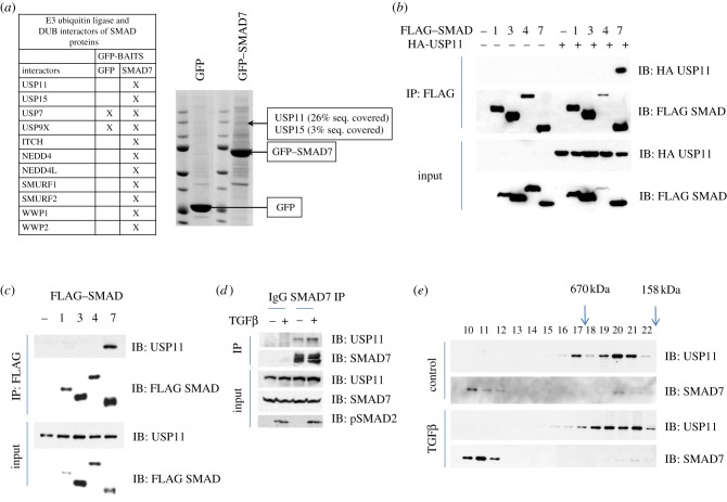
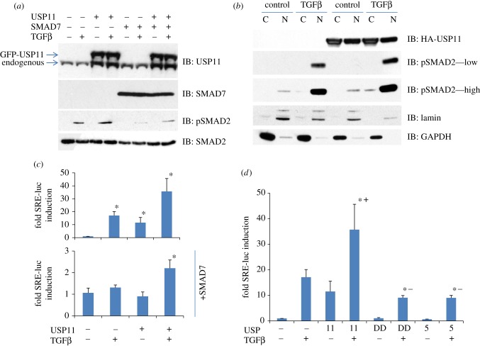
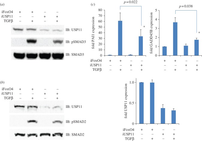
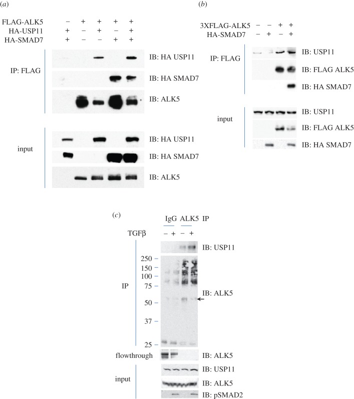
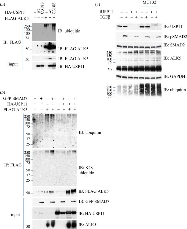
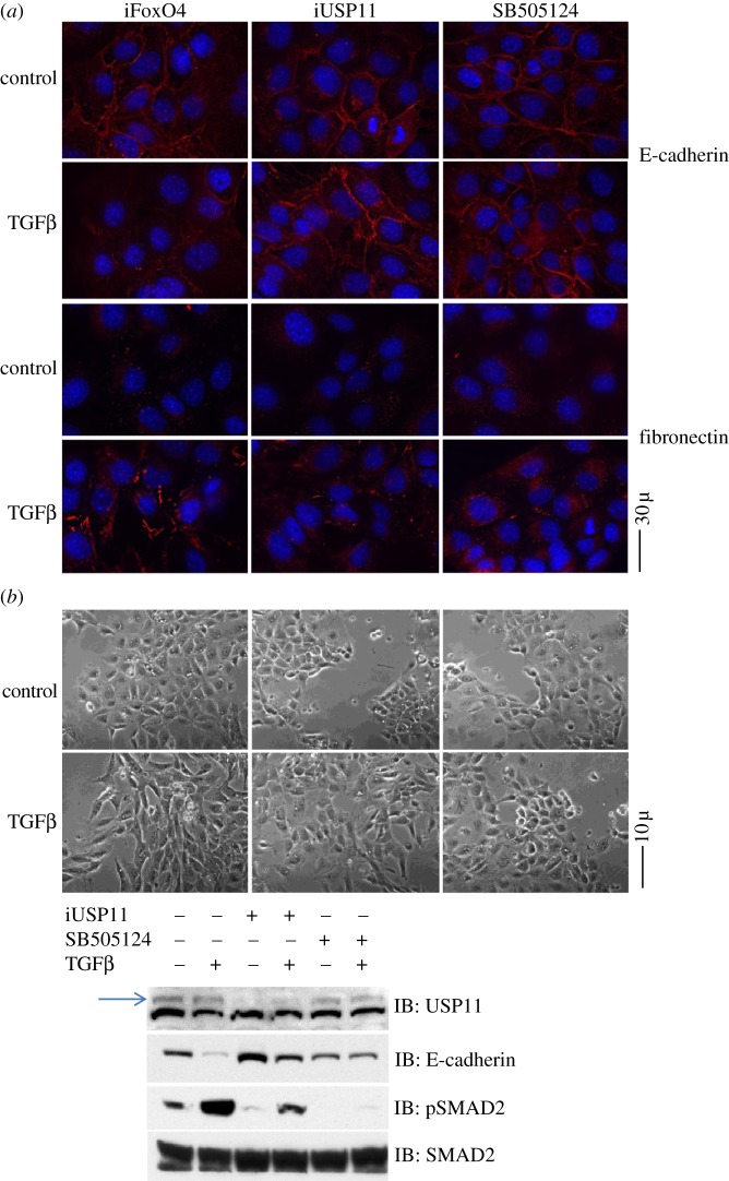
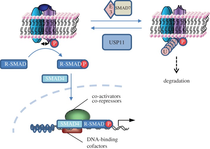
The TGFβ receptors signal through phosphorylation and nuclear translocation of SMAD2/3. SMAD7, a transcriptional target of TGFβ signals, negatively regulates the TGFβ pathway by recruiting E3 ubiquitin ligases and targeting TGFβ receptors for ubiquitin-mediated degradation. In this report, we identify a deubiquitylating enzyme USP11 as an interactor of SMAD7. USP11 enhances TGFβ signalling and can override the negative effects of SMAD7. USP11 interacts with and deubiquitylates the type I TGFβ receptor (ALK5), resulting in enhanced TGFβ-induced gene transcription. The deubiquitylase activity of USP11 is required to enhance TGFβ-induced gene transcription. RNAi-mediated depletion of USP11 results in inhibition of TGFβ-induced SMAD2/3 phosphorylation and TGFβ-mediated transcriptional responses. Central to TGFβ pathway signalling in early embryogenesis and carcinogenesis is TGFβ-induced epithelial to mesenchymal transition. USP11 depletion results in inhibition of TGFβ-induced epithelial to mesenchymal transition.

Keywords: ALK5; TGFβ; USP11; USP15; cancer; ubiquitin.


Figures

Figure 1.
Figure 1.
Identification and characterization of USP11 as an interactor of SMAD7. (a) Representative Coomassie-stained gels showing anti-GFP IPs from HEK293 extracts expressing GFP-alone or GFP-SMAD7. The interacting proteins were excised as 2 mm gel pieces, digested with trypsin and identified by mass spectrometry. The gel piece from which USP11 was identified is indicated. A summary table of various Smad-interacting E3 ubiquitin ligases and DUBs identified by mass spectrometry is included. The sequence coverage of USP11 and USP15 in GFP-SMAD7 IPs is indicated. (b) HEK293 cells were co-transfected transiently with HA-USP11 and FLAG–SMADs. FLAG IPs and lysate inputs were immunoblotted with FLAG and HA antibodies as indicated. (c) HEK293 cells were transiently transfected with FLAG–SMADs only. FLAG IPs and lysate inputs were immunoblotted with FLAG and endogenous USP11 antibodies. (d) Lysates from HEK293 cells treated with vehicle or TGFβ (50 pM 45 min) were immunoprecipitated using pre-immune IgG or a SMAD7 antibody covalently bound to Dynabeads (Invitrogen). IPs and lysate inputs were immunoblotted with endogenous USP11, SMAD7 and phospho-SMAD2 antibodies. (e) Extracts from HaCaT cells starved for 4 h and stimulated with or without 50 pM TGFβ for 1 h were separated by size-exclusion gel chromatography. The collected fractions were immunoblotted with anti-USP11 and anti-SMAD7 antibodies.
Figure 2.
Figure 2.
USP11 enhances TGFβ pathway signalling. (a) HEK293 cells stably expressing GFP or GFP-USP11 were transfected with HA empty vector or HA-SMAD7, starved for 4 h and stimulated with 50 pM TGFβ for 1 h prior to lysis. Extracts were resolved by SDS–PAGE and immunoblotted with antibodies against GFP-USP11, HA-SMAD7, endogenous phospho-SMAD2 and SMAD2. (b) HEK293 cells transiently transfected with or without HA-USP11 were starved for 4 h and stimulated with 50 pM TGFβ for 1 h prior to separation into cytoplasmic and nuclear fractions. The fractions were resolved by SDS–PAGE and immunoblotted with antibodies against HA-USP11, lamin, GAPDH, endogenous phospho-SMAD2 and SMAD2. All immunoblots are representative of at least three biological replicates. (c) TGFβ transcriptional reporter activity (using a SMAD responsive element (SRE) luciferase reporter assay) normalized to renilla-luciferase in HEK293 cells transiently transfected with SRE-luciferase, renilla-luciferase, HA-USP11, FLAG–SMAD7 and stimulated for 6 h with or without 50 pM TGFβ, as indicated. Results are average of five biological replicates. Asterisk denotes statistical significance over vector transfected and unstimulated cells. (d) TGFβ transcriptional reporter activity (using an SRE luciferase reporter assay) normalized to renilla-luciferase in HEK293 cells transiently transfected with SRE-luciferase, renilla-luciferase, HA-USP11, HA-C318S USP11 (DD), HA-USP5 and stimulated for 6 h with or without 50 pM TGFβ, as indicated. Results are average of three biological replicates. Asterisk denotes statistical significance over vector transfected and unstimulated cells. Plus symbols denote positive divergence, minus symbols denote negative divergence.
Figure 3.
Figure 3.
RNAi depletion of USP11 inhibits TGFβ pathway signalling. (a) HEK293 cells were transiently transfected with siRNA targeting FoxO4 as control or USP11, starved for 4 h and stimulated with 50 pM TGFβ for 1 h prior to lysis. Extracts were resolved by SDS–PAGE and immunoblotted with antibodies against endogenous USP11, phospho-SMAD3 and SMAD3. (b) As in A except that HEK293 cells were transiently transfected with esiRNA targeting FoxO4 as control or USP11 and immunoblots against phospho-SMAD2 and SMAD2 were performed. Immunoblots are representative of two biological replicates each, using two sets of RNAi. (c) HEK293 cells were transiently transfected with siRNA targeting FoxO4 as control or USP11, starved overnight and stimulated for 4 h with 50 pM TGFβ. The expression of TGFβ-target genes PAI1 and GADD45B as well as USP11 knockdown were assessed by semiquantitative RT-PCR. Results are average of three biological replicates. Asterisk denotes statistical significance.
Figure 4.
Figure 4.
USP11 interacts with ALK5. (a) HEK293 cells were transiently transfected with FLAG-ALK5, HA-USP11 and/or HA-SMAD7, as indicated. Extracts or FLAG IPs were resolved by SDS–PAGE and immunoblotted with antibodies against HA-USP11, HA-SMAD7 and ALK5. (b) HEK293 cells were transiently transfected with 3XFLAG-ALK5, and HA-SMAD7, as indicated. Extracts or FLAG IPs were resolved by SDS–PAGE and immunoblotted with antibodies against endogenous USP11, HA-SMAD7 and 3XFLAG-ALK5. All immunoblots are representative of at least three biological replicates. (c) Lysates from HEK293 cells treated with vehicle or TGFβ (50 pM 45 min) were immunoprecipitated using pre-immune IgG or an ALK5 antibody covalently bound to Dynabeads. IPs, flow-through extracts and lysate inputs were immunoblotted with endogenous USP11, ALK5 and phospho-SMAD2 antibodies. The arrowhead denotes the native molecular weight ALK5.
Figure 5.
Figure 5.
USP11 deubiquitylates ALK5. (a) HEK293 cells were trasfected with FLAG-ALK5 with or without a wt or catalytically inactive mutant (C318S) of HA-USP11. The FLAG-ALK5 IPs and extracts were resolved by SDS–PAGE and immunoblotted with antibodies against ubiquitin, FLAG-ALK5 and HA-USP11. (b) HEK293 cells were trasfected with FLAG-ALK5 with or without HA-USP11 or GFP-SMAD7, as indicated. The FLAG-ALK5 IPs and extracts were resolved by SDS–PAGE and immunoblotted with antibodies against ubiquitin, K48-linked polyubiquitin chain, FLAG-ALK5, GFP-SMAD7 and HA-USP11. (c) HEK293 cells were trasfected with FLAG-ALK5 with FoxO4 as control or USP11 siRNA, and treated with or without MG132 and/or TGFβ, as indicated. Extracts were resolved by SDS–PAGE and immunoblotted with antibodies against USP11, phospho-SMAD2, SMAD2, ALK5 and ubiquitin. All immunoblots are representative of at least three biological replicates.
Figure 6.
Figure 6.
USP11 knockdown inhibits epithelial to mesenchymal transition. NMuMG cells were transiently transfected with siRNA targeting mouse FoxO4 as control or USP11 before being treated with 75 pM TGFβ for 24 h in the presence or absence of 1 μM TGFβ inhibitor SB505124. (a) E-cadherin and fibronectin immunofluorescence after TGFβ treatment (b) light microscopy of cells after TGFβ treatment. Western blotting of extracts from cells pictured were resolved on SDS–PAGE gels and blotted for USP11 (the arrowhead denotes USP11; a non-specific band appeared below USP11 in mouse cell extracts that was not present in human cell extracts), phospho-SMAD2, SMAD2 and E-cadherin.
Figure 7.
Figure 7.
A schematic representation of the TGFβ pathway regulation by USP11. USP11 augments TGFβ signalling by deubiquitylating the type I TGFβ receptor, thereby counterbalancing the negative effect E3 ubiquitin ligases and SMAD7 have on the receptors.

References

    1. Massagué J. 1998. TGF-beta signal transduction. Annu. Rev. Biochem. 67, 753–79110.1146/annurev.biochem.67.1.753 (doi:10.1146/annurev.biochem.67.1.753) - DOI - DOI - PubMed
    1. Shi Y, Massagué J. 2003. Mechanisms of TGF-beta signaling from cell membrane to the nucleus. Cell 113, 685–70010.1016/S0092-8674(03)00432-X (doi:10.1016/S0092-8674(03)00432-X) - DOI - DOI - PubMed
    1. Ikushima H, Miyazono K. 2011. TGF-β signal transduction spreading to a wider field: a broad variety of mechanisms for context-dependent effects of TGF-β. Cell Tissue Res. 347, 37–4910.1007/s00441-011-1179-5 (doi:10.1007/s00441-011-1179-5) - DOI - DOI - PubMed
    1. Santibañez JF, Quintanilla M, Bernabeu C. 2011. TGF-β/TGF-β receptor system and its role in physiological and pathological conditions. Clin. Sci. (Lond.) 121, 233–25110.1042/CS20110086 (doi:10.1042/CS20110086) - DOI - DOI - PubMed
    1. Massagué J. 2008. TGFbeta in cancer. Cell 134, 215–23010.1016/j.cell.2008.07.001 (doi:10.1016/j.cell.2008.07.001) - DOI - DOI - PMC - PubMed

Publication types

MeSH terms

Substances

LinkOut - more resources