. 2012 Aug;169(2):156-63.
doi: 10.1111/j.1365-2249.2012.04607.x.

Macrophage migration inhibitory factor deficiency protects pancreatic islets from cytokine-induced apoptosis in vitro

Affiliations

Macrophage migration inhibitory factor deficiency protects pancreatic islets from cytokine-induced apoptosis in vitro

I Stojanovic et al. Clin Exp Immunol. 2012 Aug.

Abstract

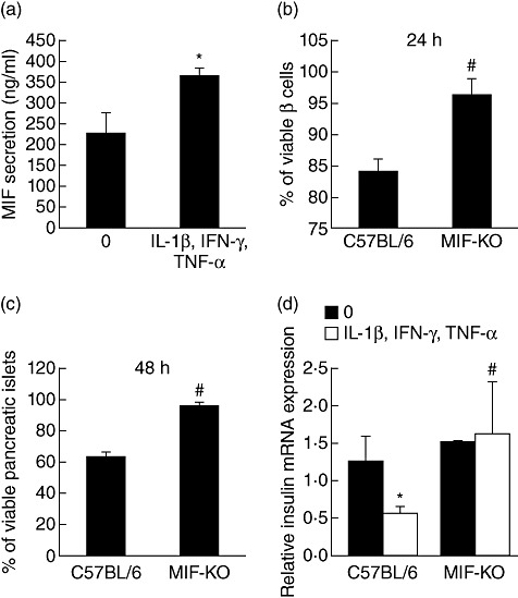
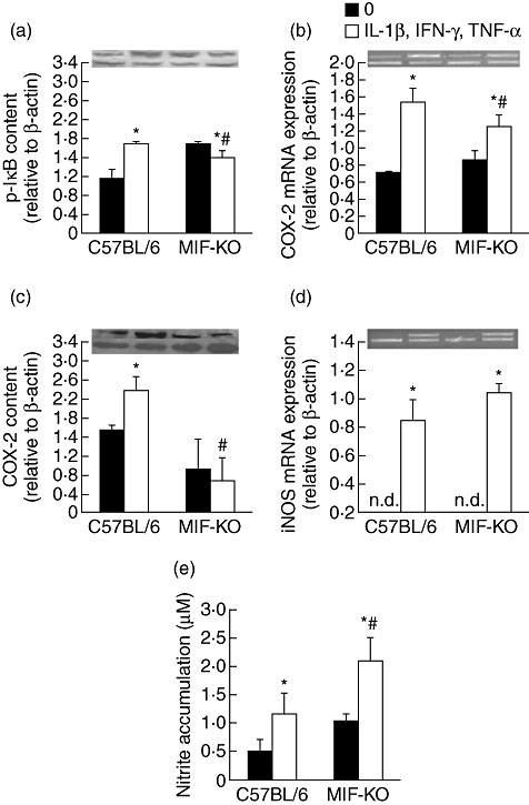
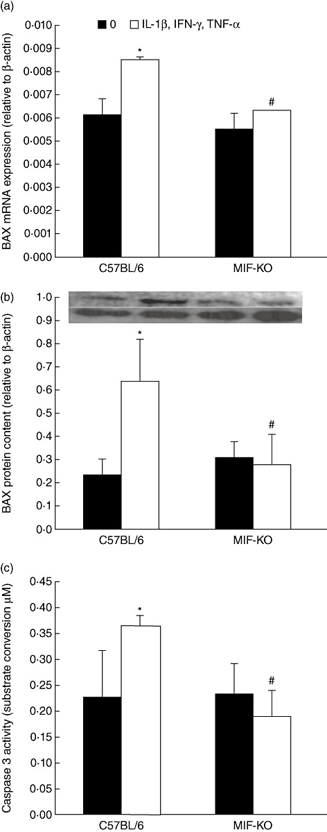
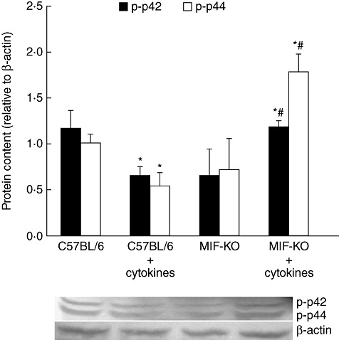
During pathogenesis of diabetes, pancreatic islets are exposed to high levels of cytokines and other inflammatory mediators that induce deterioration of insulin-producing beta cells. Macrophage migration inhibitory factor (MIF) plays a key role in the onset and development of several immunoinflammatory diseases and also controls apoptotic cell death. Because the occurrence of apoptosis plays a pathogenetic role in beta cell death during type 1 diabetes development and MIF is expressed in beta cells, we explored the influence of MIF deficiency on cytokine-induced apoptosis in pancreatic islets. The results indicated clearly that elevated MIF secretion preceded C57BL/6 pancreatic islets death induced by interferon (IFN)-γ + tumour necrosis factor (TNF)-α + interleukin (IL)-1β. Consequently, MIF-deficient [MIF-knock-out (KO)] pancreatic islets or islet cells showed significant resistance to cytokine-induced death than those isolated from C57BL/6 mice. Furthermore, upon exposure to cytokines pancreatic islets from MIF-KO mice maintained normal insulin expression and produced less cyclooxygenase-2 (COX-2) than those from wild-type C57BL6 mice. The final outcome of cytokine-induced islet apoptosis in islets from wild-type mice was the activation of mitochondrial membrane pore-forming protein Bcl-2-associated X protein and effector caspase 3. In contrast, these apoptotic mediators remained at normal levels in islets from MIF-KO mice suggesting that MIF absence prevented initiation of the mitochondrial apoptotic pathway. Additionally, the protection from apoptosis was also mediated by up-regulation of prosurvival kinase extracellular-regulated kinase 1/2 in MIF-KO islets. These data indicate that MIF is involved in the propagation of pancreatic islets apoptosis probably via nuclear factor-κB and mitochondria-related proteins.


Figures

Fig. 1
Fig. 1
Macrophage migration inhibitory factor (MIF) is a mediator of islet dysfunction and death. Pancreatic islets from C57BL/6 mice were incubated in groups of 20, in the presence or absence of cytokine mixture (interleukin-1β + interferon-γ + tumour necrosis factor-α, 10 ng/ml each) for 24 h and MIF concentration from cell-free supernatants was determined by enzyme-linked immunosorbent assay (a). Islet cells (1 × 10⁴/well) (b) or pancreatic islets (groups of 20) (c) isolated from C57BL/6 and MIF-knock-out (KO) mice were incubated in the presence or absence of cytokine mixture and their viability was assessed by MTT assay after 24 h or 48 h, respectively. Data are presented as % viable cells or islets compared to untreated cells or islets (100%). (d) Relative insulin mRNA expression was determined by real-time polymerase chain reaction in C57BL/6 and MIF-KO islets either left untreated or treated with cytokine mixture for 6 h. Results from three experiments were presented as mean ± standard deviation. *P < 0·05 cytokine-treated versus untreated (0). #P < 0·05 MIF-KO versus C57BL/6 islet cells or islets.
Fig. 2
Fig. 2
Macrophage migration inhibitory factor (MIF) absence confers resistance of islets through impairment of nuclear factor-κB and cyclooxygenase (COX)-2 activation. Pancreatic islets (in groups of 100) from C57BL/6 or MIF-knock-out (KO) mice were incubated in the presence or absence of cytokine mixture (interleukin-1β + interferon-γ + tumour necrosis factor-α, 10 ng/ml each) for determination of p-IκB protein content by Western blot after 24 h (a), COX-2 mRNA expression by polymerase chain reaction (PCR) after 6 h (b), COX-2 content by Western blot after 24 h (c), inducible nitric oxide synthase mRNA expression after 6 h (d) or nitric oxide production after 48 h (e); n.d. = not detectable. Data are presented as mean ± standard deviation from three separate experiments. *P < 0·05 cytokine-treated versus untreated (0); #P < 0·05 MIF-KO versus C57BL/6 islets. Photographs of PCR amplicons and Western blot are displayed above the adequate graphs; lower lane: β-actin, upper lane: gene or protein in question.
Fig. 3
Fig. 3
Macrophage migration inhibitory factor (MIF) absence protects islets through down-regulation of mitochondrial apoptotic pathway. Pancreatic islets from C57BL/6 or MIF-knock-out (KO) mice were incubated in the presence or absence of cytokine mixture (interleukin-1β + interferon-γ + tumour necrosis factor-α, 10 ng/ml each) for determination of BAX mRNA expression (in groups of 100) by real-time polymerase chain reaction after 6 h (a) or BAX protein content after 24 h (b), or caspase 3 activity (in groups of 10) after 24 h (c). Photograph of the blot is displayed above the adequate graph; lower lane: β-actin, upper lane: BAX protein. Data are presented as mean ± standard deviation from three experiments. *P < 0·05 cytokine-treated versus untreated (0); #P < 0·05 MIF-KO versus C57BL/6 islets.
Fig. 4
Fig. 4
Prosurvival p-extracellular-regulated kinase (ERK)1/2 is up-regulated in cytokine-stimulated macrophage migration inhibitory factor knock-out (MIF-KO) islets. Pancreatic islets (in groups of 100) from C57BL/6 or MIF-KO mice were incubated in the presence or absence of cytokine mixture (interleukin-1β + interferon-γ + tumour necrosis factor-α, 10 ng/ml each) for determination of phosphorylated ERK subunits (p-p42 and p-p44) after 24 h. Data are presented as mean ± standard deviation from two experiments. *P < 0·05 cytokine-treated versus untreated (0); #P < 0·05 MIF-KO versus C57BL/6 islets.

References

    1. Beyan H, Buckley LR, Yousaf N, Londei M, Leslie RD. A role for innate immunity in type 1 diabetes? Diabetes Metab Res Rev. 2003;19:89–100. - PubMed
    1. Kaminitz A, Stein J, Yaniv I, Askenasy N. The vicious cycle of apoptotic β-cell death in type 1 diabetes. Immunol Cell Biol. 2007;85:582–9. - PubMed
    1. Pirot P, Cardozo AK, Eizirik DL. Mediators and mechanisms of pancreatic beta-cell death in type 1 diabetes. Arq Bras Endocrinol Metabol. 2008;52:156–65. - PubMed
    1. Davi G, Falco A, Patrono C. Lipid peroxidation in diabetes mellitus. Antioxid Redox Signal. 2005;7:256–68. - PubMed
    1. Eizirik DL, Colli ML, Ortis F. The role of inflammation in insulitis and beta-cell loss in type 1 diabetes. Nat Rev Endocrinol. 2009;5:219–26. - PubMed

Publication types

MeSH terms