. 2012 Sep;122(6):1129-36.
doi: 10.1111/j.1471-4159.2012.07869.x. Epub 2012 Aug 3.

Urocortin 3 elevates cytosolic calcium in nucleus ambiguus neurons

Affiliations

Urocortin 3 elevates cytosolic calcium in nucleus ambiguus neurons

G Cristina Brailoiu et al. J Neurochem. 2012 Sep.

Abstract

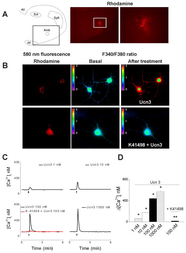
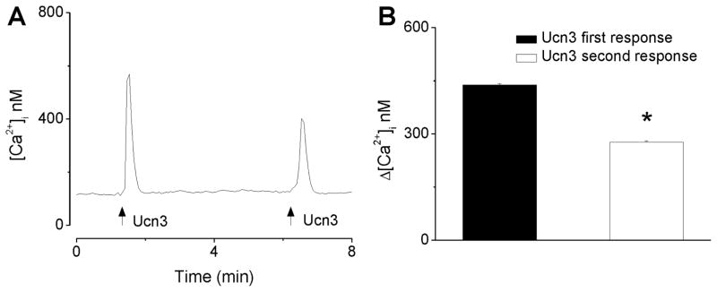
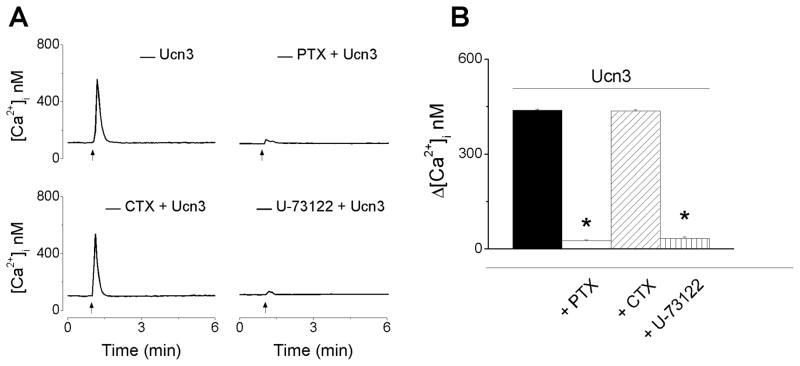
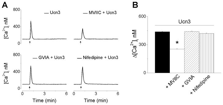
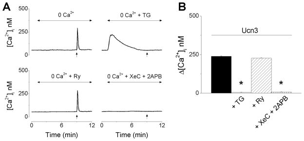
Urocortin 3 (also known as stresscopin) is an endogenous ligand for the corticotropin-releasing factor receptor 2 (CRF(2)). Despite predominant G(s) coupling of CRF(2), promiscuous coupling with other G proteins has been also associated with the activation of this receptor. As urocortin 3 has been involved in central cardiovascular regulation at hypothalamic and medullary sites, we examined its cellular effects on cardiac vagal neurons of nucleus ambiguus, a key area for the autonomic control of heart rate. Urocortin 3 (1 nM-1000 nM) induced a concentration-dependent increase in cytosolic Ca(2+) concentration that was blocked by the CRF(2) antagonist K41498. In the case of two consecutive treatments with urocortin 3, the second urocortin 3-induced Ca(2+) response was reduced, indicating receptor desensitization. The effect of urocortin 3 was abolished by pre-treatment with pertussis toxin and by inhibition of phospolipase C with U-73122. Urocortin 3 activated Ca(2+) influx via voltage-gated P/Q-type channels as well as Ca(2+) release from endoplasmic reticulum. Urocortin 3 promoted Ca(2+) release via inositol 1,4,5 trisphosphate receptors, but not ryanodine receptors. Our results indicate a novel Ca(2+) -mobilizing effect of urocortin 3 in vagal pre-ganglionic neurons of nucleus ambiguus, providing a cellular mechanism for a previously reported role for this peptide in parasympathetic cardiac regulation.


Conflict of interest statement

The authors have no conflict of interests.

Figures

Figure 1
Figure 1. Urocortin 3 increases cytosolic Ca2+ concentration in vagal preganglionic neurons via CRF2 activation
A, The left panel indicates the localization of nucleus ambiguus (Amb) in a medullary slice; the boxed area indicates the level shown in the middle panel. Abbreviations: 4V, 4th ventricle; py, pyramidal tract; Sol, nucleus of the solitary tract, and Sp5, spinal trigeminal nucleus. Lower magnification (middle panel) and higher magnification (right panel) of a medullary slice illustrating retrogradely labeled rhodamine-containing cardiac vagal neurons of nucleus ambiguus. B, Fura-2 AM fluorescence ratio (F340/F380 nm) of rhodamine-labeled neurons, before and after treatment with urocortin 3 (Ucn3, 100 nM) in the absence (top panels) and presence of CRF2 antagonist K41498 (10 μM). C, Representative examples of increases in cytosolic Ca2+ concentration, [Ca2+]i, in response to different concentrations of urocortin 3 (1–1000 nM) and urocortin 3 (100 nM) after pretreatment with K41498 (10 μM.); the arrows indicate the application of Ucn3. D, Comparison of mean amplitude ± SEM of [Ca2+]i increase produced by urocortin 3 (1–1000 nM) and urocortin 3 (100 nM) after pretreatment with K41498 (10 μM); urocortin 3 induced a concentration-dependent increase in [Ca2+]i; the effect was abolished by K41498. P < 0.05 compared to basal [Ca2+]i (*) or to urocortin 3 (100 nM)-induced increase in [Ca2+]i (**).
Figure 2
Figure 2. Urocortin 3-induced increase in [Ca2+]i is subject to tachyphylaxis
A, Representative example of [Ca2+]i increases produced by two consecutive applications of urocortin 3 (100 nM), 5 min apart, in vagal preganglionic neurons of nucleus ambiguus; the arrows indicate the application of urocortin 3 (Ucn3). B, Comparison of the mean amplitude ± SEM of [Ca2+]i increase produced by the first and second application of urocortin 3 (100 nM); * P < 0.05 compared to the first response.
Figure 3
Figure 3. Urocortin 3-induced increase in [Ca2+]i is inhibited by pertussis toxin and U-73122
A, Representative examples of urocortin 3 (100 nM)-induced increase in [Ca2+]i in the absence (top left trace) and presence of pertussis toxin (PTX) (100 nM, top right), a Gi/o inhibitor; cholera toxin (CTX) (100 nM, bottom left), a Gs inhibitor, and U-73122 (1 μM, bottom right), a PLC inhibitor. B, Comparison of mean amplitude ± SEM of [Ca2+]i increase produced by urocortin 3 (100 nM) in the absence or presence of PTX, CTX and U-73122 (* P < 0.05 compared to the response to urocortin 3 alone). Pertussis toxin and U-73122 abolished urocortin 3-induced increase in [Ca2+]i
Figure 4
Figure 4. Urocortin 3 promotes Ca2+ influx via voltage-gated P and Q channels
A, Representative examples of urocortin 3 (100 nM)-induced increase in [Ca2+]i in the absence (top left trace) and presence of ω-conotoxin MVIIC (MVIIC, 100 nM, top right), a P/Q-type inhibitor; ω-conotoxin GVIA (GIVA, 100 nM bottom left), a N-type Ca2+ channel inhibitor, or nifedipine (1 μM, bottom right), a L-type Ca2+ channel. B, Comparison of mean amplitude ± SEM of [Ca2+]i increase produced by urocortin 3 (100 nM) in the absence or presence of MVIIC, GVIA or nifedipine (* P < 0.05 compared to the response to urocortin 3 alone).
Figure 5
Figure 5. Urocortin 3 promotes Ca2+ release via IP3-dependent mechanisms
A, Representative examples of increases in [Ca2+]i produced by urocortin 3 (100 nM) in Ca2+-free saline alone before (top left) or after treatment with thapsigargin (TG, 1 μM, top right), ryanodine (Ry, 10 μM, bottom left) or xestospongin C (XeC, 1 μM) and 2- aminoethoxydiphenyl borate (2APB, 100 μM, bottom right); the arrows indicate the treatment with Ucn3. B, Comparisons of mean amplitude ± SEM of [Ca2+]i increase produced by urocortin 3 (100 nM) in Ca2+-free saline alone or in presence of TG, Ry or XeC + 2APB (* P < 0.05 compared to the response to urocortin 3 alone).

Similar articles

Cited by

References

    1. Bale TL, Vale WW. CRF and CRF receptors: role in stress responsivity and other behaviors. Annu Rev Pharmacol Toxicol. 2004;44:525–557. - PubMed
    1. Berridge MJ. Neuronal calcium signaling. Neuron. 1998;21:13–26. - PubMed
    1. Berridge MJ. The endoplasmic reticulum: a multifunctional signaling organelle. Cell Calcium. 2002;32:235–249. - PubMed
    1. Blank T, Nijholt I, Grammatopoulos DK, Randeva HS, Hillhouse EW, Spiess J. Corticotropin-releasing factor receptors couple to multiple G-proteins to activate diverse intracellular signaling pathways in mouse hippocampus: role in neuronal excitability and associative learning. J Neurosci. 2003;23:700–707. - PMC - PubMed
    1. Bouairi E, Kamendi H, Wang X, Gorini C, Mendelowitz D. Multiple types of GABAA receptors mediate inhibition in brain stem parasympathetic cardiac neurons in the nucleus ambiguus. J Neurophysiol. 2006;96:3266–3272. - PubMed

Publication types

MeSH terms