Review
. 2013 Jan;15(1):15-27.
doi: 10.1111/j.1463-1326.2012.01663.x. Epub 2012 Aug 1.

GLP-1 receptor activated insulin secretion from pancreatic β-cells: mechanism and glucose dependence

Affiliations
Free PMC article
Review

GLP-1 receptor activated insulin secretion from pancreatic β-cells: mechanism and glucose dependence

A R Meloni et al. Diabetes Obes Metab. 2013 Jan.
Free PMC article

Abstract

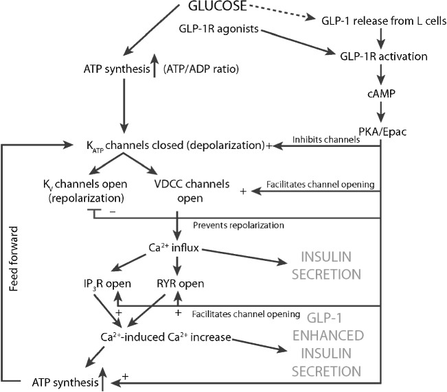
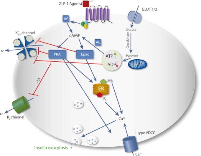
The major goal in the treatment of type 2 diabetes mellitus is to control the hyperglycaemia characteristic of the disease. However, treatment with common therapies such as insulin or insulinotrophic sulphonylureas (SU), while effective in reducing hyperglycaemia, may impose a greater risk of hypoglycaemia, as neither therapy is self-regulated by ambient blood glucose concentrations. Hypoglycaemia has been associated with adverse physical and psychological outcomes and may contribute to negative cardiovascular events; hence minimization of hypoglycaemia risk is clinically advantageous. Stimulation of insulin secretion from pancreatic β-cells by glucagon-like peptide 1 receptor (GLP-1R) agonists is known to be glucose-dependent. GLP-1R agonists potentiate glucose-stimulated insulin secretion and have little or no activity on insulin secretion in the absence of elevated blood glucose concentrations. This 'glucose-regulated' activity of GLP-1R agonists makes them useful and potentially safer therapeutics for overall glucose control compared to non-regulated therapies; hyperglycaemia can be reduced with minimal hypoglycaemia. While the inherent mechanism of action of GLP-1R agonists mediates their glucose dependence, studies in rats suggest that SUs may uncouple this dependence. This hypothesis is supported by clinical studies showing that the majority of events of hypoglycaemia in patients treated with GLP-1R agonists occur in patients treated with a concomitant SU. This review aims to discuss the current understanding of the mechanisms by which GLP-1R signalling promotes insulin secretion from pancreatic β-cells via a glucose-dependent process.


Figures

Figure 1
Figure 1
Insulin secretion. Basal timepoints from −30 to 0 min. Infusion of exenatide or placebo commenced at 0 min as indicated by arrow. From 0 to 120 min, plasma glucose was ∼5.0 mmol/l (euglycaemia). At 120–180 min, plasma glucose was ∼4.0 mmol/l (hypoglycaemia). At 180–240 min, plasma glucose was ∼3.2 mmol/l ending in nadir of ∼2.8 mmol/l (hypoglycaemia). Recovery phase from 270 to 360 min. ○, placebo treatment arm;formula image
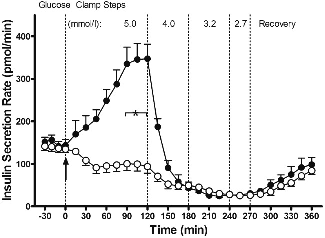
, exenatide treatment arm. Data are means ± s.e.; n = 11 per treatment arm. *p < 0.05, exenatide vs. placebo during steady state of a glycaemic interval. Reproduced with permission from Degn et al. [31].
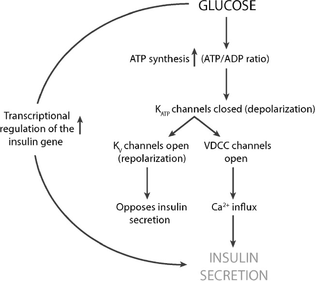
Figure 2
Figure 2
Signalling cascade of first-phase insulin secretion in the β-cell in response to glucose, without the contributions of GLP-1. Glucose metabolism triggers a cellular increase in the ATP/ADP ratio which inhibits K+ATP channels leading to depolarization. Depolarization triggers both the opening of the voltage-dependent K+ channels (Kv) to repolarize the β-cell and the activation of VDCCs. The resulting inward Ca2+ influx triggers exocytosis of insulin-containing granules leading to insulin secretion.
Figure 3
Figure 3
Phases of insulin secretion in response to a square wave of hyperglycaemia. Initial levels of basal insulin production are low. With the induction of hyperglycaemia, a large and rapid insulin secretion occurs that quickly peaks and then falls to levels above basal for an extended period of time.
Figure 4
Figure 4
Putative vesicle priming events that allow vesicles to become competent for exocytosis. Once primed, vesicles ‘move’ from the reserve pool to the readily releasable pool where the increase in Ca2+ influx through activated VDCCs promotes exocytosis. GLP-1 may promote Cl- ion pumping into a vesicle through Epac and a putative granule SUR protein that may regulate ClC-3 chloride channels. The influx of Cl- ions counters the charge promoted by the action of the H+-ATPase pump. The increase in H+ allows the decrease in intragranular pH necessary for exocytosis.
Figure 5
Figure 5
Signalling cascade of insulin secretion in the β-cells in response to glucose and GLP-1. Glucose triggers the cascade of events outlined in figure 1. GLP-1 potentiates the activity of glucose by inhibiting K+ATP channels, facilitating the opening of VDCCs, and inhibiting membrane repolarization via Kv channels. GLP-1 also sensitizes IP3 (IP3R) and ryanodine (RYR) receptors to the effects of Ca2+, facilitating Ca2+-induced Ca2+ release (CICR). CICR promotes enhanced insulin secretion and enhanced ATP production, the latter of which facilitates a feed-forward loop to promote further rounds of depolarization and insulin secretion.
Figure 6
Figure 6
A schematic overview of the main signalling cascades involved in insulin secretion. Glucose enters the β-cell through the GLUT1/2 transporters and is converted to ATP. GLP-1 secreted from L-cells in response to glucose and other nutrients, binds to the GLP-1 receptor, triggering an increase in cAMP and activation of Epac and PKA; cAMP is also produced from soluble adenylate cyclase adding to the overall concentration of cAMP. Glucose metabolism leads to an increase in ATP/ADP concentrations which act to inhibit KATP channels. The inability to pump K+ from the β-cell leads to membrane depolarization and opening of VDCC. GLP-1-activated PKA and Epac potentiate the effects of glucose by further inhibiting both the K+ATP and the Kv channels, preventing repolarization of the β-cell. PKA and Epac also sensitize multi-subunit calcium channels on the endoplasmic reticulum (ER) allowing the release of calcium from intracellular stores. This increase in free calcium concentration further aids in exocytosis of insulin.

Similar articles

Cited by

References

    1. DeFronzo RA. Banting lecture. From the triumvirate to the ominous octet: a new paradigm for the treatment of type 2 diabetes mellitus. Diabetes. 2009;58:773–795. - PMC - PubMed
    1. Inzucchi SE, Bergenstal RM, Buse JB, et al. Management of hyperglycemia in type 2 diabetes: a patient-centered approach: position statement of the American Diabetes Association (ADA) and the European Association for the Study of Diabetes (EASD) Diabetes Care. 2012;35:1577–1596. - PubMed
    1. Pettersson B, Rosenqvist U, Deleskog A, Journath G, Wändell P. Self-reported experience of hypoglycemia among adults with type 2 diabetes mellitus (Exhype) Diabetes Res Clin Pract. 2011;92:19–25. - PubMed
    1. Christensen TF, Randløv J, Kristensen LE, Eldrup E, Hejlesen OK, Struijk JJ. QT measurement and heart rate correction during hypoglycemia: is there a bias? Cardiol Res Pract. 2010;2010:961290. - PMC - PubMed
    1. Eckert B, Agardh CD. Hypoglycaemia leads to an increased QT interval in normal men. Clin Physiol. 1998;18:570–575. - PubMed

MeSH terms