. 2012 Jul 9:9:164.
doi: 10.1186/1742-2094-9-164.

Time-dependent effects of hypothermia on microglial activation and migration

Affiliations

Time-dependent effects of hypothermia on microglial activation and migration

Jung-Wan Seo et al. J Neuroinflammation. .

Abstract

Background: Therapeutic hypothermia is one of the neuroprotective strategies that improve neurological outcomes after brain damage in ischemic stroke and traumatic brain injury. Microglial cells become activated following brain injury and play an important role in neuroinflammation and subsequent brain damage. The aim of this study was to determine the time-dependent effects of hypothermia on microglial cell activation and migration, which are accompanied by neuroinflammation.

Methods: Microglial cells in culture were subjected to mild (33 °C) or moderate (29 °C) hypothermic conditions before, during, or after lipopolysaccharide (LPS) or hypoxic stimulation, and the production of nitric oxide (NO), proinflammatory cytokines, reactive oxygen species, and neurotoxicity was evaluated. Effects of hypothermia on microglial migration were also determined in in vitro as well as in vivo settings.

Results: Early-, co-, and delayed-hypothermic treatments inhibited microglial production of inflammatory mediators to varying degrees: early treatment was the most efficient, and delayed treatment showed time-dependent effects. Delayed hypothermia also suppressed the mRNA levels of proinflammatory cytokines and iNOS, and attenuated microglial neurotoxicity in microglia-neuron co-cultures. Furthermore, delayed hypothermia reduced microglial migration in the Boyden chamber assay and wound healing assay. In a stab injury model, delayed local hypothermia reduced migration of microglia toward the injury site in the rat brain.

Conclusion: Taken together, our results indicate that delayed hypothermia is sufficient to attenuate microglial activation and migration, and provide the basis of determining the optimal time window for therapeutic hypothermia. Delayed hypothermia may be neuroprotective by inhibiting microglia-mediated neuroinflammation, indicating the therapeutic potential of post-injury hypothermia for patients with brain damages exhibiting some of the inflammatory components.


Figures

Figure 1
Figure 1
Inhibition of microglial and astrocytic NO production by hypothermia. BV-2 microglial cells (A), primary microglia cultures (B), primary astrocyte cultures (C) (5 x 10⁴ cells per well in a 96-well plate) were incubated with 100 ng/ml of LPS under hypothermic (33 °C, 29 °C) or normothermic (37 °C) conditions for 0 to 72 hours. The amounts of nitrite in the culture media were measured with the Griess reagent (upper). Cell viability was examined by MTT assays and the results were expressed as the percentage of surviving cells over the controls (lower). Results are the mean ± SD (n = 3). *P < 0.05, **P < 0.01; significantly different from the LPS-treated group at the same time point under normothermic condition (37 °C). LPS, lipopolysaccharide; MTT, 3-(4, 5-dimethylthiazol-2-yl)-2, 5-diphenyltetrazolium bromide; n, number.
Figure 2
Figure 2
Time-dependent effects of hypothermia on NO production in LPS-stimulated microglial cells. Hypothermia was initiated at the different time points before or after LPS stimulation as shown in the experimental scheme (A). BV-2 microglial cells (5 x 10⁴ cells/well in a 96-well plate) were incubated with LPS (100 ng/ml) under hypothermic (33 °C, 29 °C) or normothermic (37 °C) conditions as indicated. The amount of nitrite in the culture media was measured with the Griess reagent at 24 hours after LPS stimulation (B). Cell viability was examined by MTT assays. The results represent the percentage of surviving cells compared to the control (C). Results are the mean ± SD (n = 3). *P < 0.05, **P < 0.01; significantly different from the LPS-treated group under normothermia (37 °C). LPS, lipopolysaccharide; MTT, 3-(4, 5-dimethylthiazol-2-yl)-2, 5-diphenyltetrazolium bromide; n, number.
Figure 3
Figure 3
Effects of delayed hypothermia on NO production in LPS-stimulated microglial cells. Hypothermia was initiated at the different time points with the same duration as indicated in the experimental scheme (A). BV-2 microglial cells (5 x 10⁴ cells per well in a 96-well plate) were treated with LPS (100 ng/ml) for 24 hours under normothermic (37 °C) or hypothermic (29 °C) conditions as indicated (conditions 1 to 7). The concentration of nitrite in the culture media was measured with the Griess reagent at the end of the incubation (B). Cell viability was examined by MTT assays and the results were expressed as the percentage of surviving cells over the controls (C). Results are the mean ± SD (n = 3). *P < 0.05, **P < 0.01; significantly different from LPS treatment under normothermia. LPS, lipopolysaccharide; MTT, 3-(4, 5-dimethylthiazol-2-yl)-2, 5-diphenyltetrazolium bromide; n, number.
Figure 4
Figure 4
Effect of hypothermia on pro-inflammatory gene expression in LPS-stimulated microglial cells. Primary microglial cells (5 x 10⁵ cells per well in a six-well plate) were treated with LPS (100 ng/ml) under hypothermia (29 °C) or normothermia (37 °C) as indicated (A). Total RNA was isolated at six hours after the LPS treatment. The levels of cytokine or chemokine mRNA were determined by real-time RT-PCR. The data were normalized to GAPDH (B). Results are mean ± SD (n = 3). *P <0.05, **P < 0.01; significantly different from the LPS-treated group under normothermia. LPS, lipopolysaccharide; n, number.
Figure 5
Figure 5
Effect of hypothermia on pro-inflammatory gene expression in LPS-stimulated astrocyte cultures. Primary astrocyte cultures (5 x 10⁵ cells per well in six-well plates) were treated with LPS (100 ng/ml) under hypothermia (29 °C) or normothermia (37 °C) as indicated (A). Total RNA was isolated at six hours after the LPS treatment. The levels of cytokine mRNA were determined by real-time RT-PCR. The data were normalized to GAPDH (B). Results are mean ± SD (n = 3). *P < 0.05, **P < 0.01; significantly different from the LPS-treated group under normothermia. LPS, lipopolysaccharide; n, number.
Figure 6
Figure 6
Effect of hypothermia on pro-inflammatory gene levels in LPS-stimulated microglial cells: conventional RT-PCR analysis. Primary microglial cells (5 x 10⁵ per well in six-well plates) were treated with LPS (100 ng/ml) under hypothermia (29 °C) or normothermia (37 °C) as indicated in the main text. Total RNA was isolated at six hours after the LPS treatment. The levels of cytokine and iNOS mRNA were determined by conventional RT-PCR (A). The data were normalized to GAPDH (B, C, and D). Results are mean ± SD (n = 3). *P < 0.05, **P <0.01; different from LPS-treated group under normothermia. LPS, lipopolysaccharide; n, number.
Figure 7
Figure 7
Effect of hypothermia on ROS production in LPS-stimulated microglial cells. BV-2 microglial cells (5 x 10⁴ cells per well in a 96-well plate) were treated with LPS (100 ng/ml) for six hours under normothermic (37 °C) or hypothermic (29 °C) conditions as indicated (A). Production of H2O2 was evaluated by Amplex Red assay (B). Results are the mean ± SD (n = 3). *P <0.05; significantly different from the LPS-treated group under normothermia. LPS, lipopolysaccharide; n, number; ROS, reactive oxygen species.
Figure 8
Figure 8
Effect of hypothermia on ROS production in LPS-stimulated microglial cells: DCFDA assay. BV-2 microglial cells (5 x 10⁴ cells per well in a 96-well plate) were treated with LPS (100 ng/ml) for six hours under normothermic (37 °C) or hypothermic (29 °C) conditions as indicated (A). Production of ROS was evaluated by using DCFDA staining (B). Results are mean ± SD (n = 3). *P < 0.05, **P < 0.01; significantly different from LPS treatment under normothermia. DCFDA, 2'-7'-dichlorodihydrofluorescein diacetate; LPS, lipopolysaccharide; n, number; ROS, reactive oxygen species.
Figure 9
Figure 9
Effect of hypothermia on iNOS gene expression in hypoxia-stimulated microglial cells. BV-2 microglial cells or primary microglial cells (5 x 10⁵ per well in six well plates) were exposed to LPS (100 ng/ml) or hypoxia under hypothermia (29 °C) or normothermia (37 °C) as described (A). Total RNA was isolated at six hours after LPS or hypoxic stimulation followed by reoxygenation. The levels of iNOS mRNA were determined by conventional RT-PCR. The data was normalized to GAPDH (B, C). Results are the mean ± SD (n = 3). *P < 0.05; significantly different from the hypoxia-treated group under normothermia. iNOS, inducible NO synthase; LPS, lipopolysaccharide; n, number.
Figure 10
Figure 10
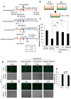
Protective effect of hypothermia against microglial neurotoxicity. Microglia/neuroblastoma co-culture scheme (A, B). BV-2 microglial cells were seeded in triplicate at a density of 1.5 x 10⁴ cells per well in a 96-well plate. B35 neuroblastoma cells (3.75 x 10⁴ cells per well) stably expressing EGFP were added onto the BV-2 microglial cells three hours prior to LPS (100 ng/ml) treatment. The co-culture of microglia and neuroblastomas was performed under normothermic or hypothermic conditions as indicated. At the end of the co-culturing, the EGFP-positive cells were counted under a fluorescence microscope to evaluate B35 neuroblastoma cell survival (C). **P < 0.01; different from the LPS-treated group under normothermia. LPS alone did not affect either B35 or BV-2 cell viability (E). Results are the mean ± SD (n = 3). Representative microscopic images are shown (D). Scale bar = 200 μm. EGFP, enhanced GFP; LPS, lipopolysaccharide; n, number; O/N, overnight.
Figure 11
Figure 11
Protective effect of hypothermia against microglial neurotoxicity: co-cultures of primary microglia and neuroblastoma cells. B35-EGFP neuroblastoma cells were added onto BV-2 microglial cells or primary microglia cultures three hours prior to LPS (100 ng/ml) treatment. The co-culture of microglia and neuroblastoma was performed under normothermia (37 °C) or hypothermia (33 °C, 29 °C) as indicated (A, B). At the end of the co-culture, the EGFP-positive cells were counted under fluorescence microscope to evaluate B35 neuroblastoma cell survival (C). Results are mean ± SD (n = 3). *P < 0.05, **P < 0.01; different from LPS-treated group under normothermia. LPS alone did not affect B35 cell viability (data not shown). Representative microscopic images are also shown (D). Scale bar = 200 μm. EGFP, enhanced GFP; LPS, lipopolysaccharide; n, number; O/N, overnight
Figure 12
Figure 12
Protective effect of hypothermia against microglial neurotoxicity in primary microglia and cortical neuron co-cultures. Co-cultures of primary microglia and cortical neurons were done using culture inserts as shown (A, B). Following LPS stimulation of the primary microglia/neuron co-cultures under normothermic or hypothermic condition, the co-cultures were further incubated for 48 hours. Afterwards, the culture inserts containing microglial cells were removed, and a MTT assay was performed to determine the viability of cortical neurons (C). Results are the mean ± SD (n = 3). **P < 0.01; significantly different from the LPS-treated group under normothermia. LPS, lipopolysaccharide; MTT, 3-(4, 5-dimethylthiazol-2-yl)-2, 5-diphenyltetrazolium bromide; n, number; O/N, overnight.
Figure 13
Figure 13
Protective effect of hypothermia against neurotoxicity of microglia-conditioned media. Microglia-conditioned media (MCM) were collected after stimulation of primary microglia cultures with LPS under normothermic or hypothermic condition as indicated (A, B). MCM were added to primary cortical neurons and the viability was assessed by a MTT assay 24 hours later (C). Results are mean ± SD (n = 3). *P < 0.05; different from LPS-treated group under normothermia. LPS, lipopolysaccharide; MTT, 3-(4, 5-dimethylthiazol-2-yl)-2, 5-diphenyltetrazolium bromide; n, number; O/N, overnight.
Figure 14
Figure 14
Neuroprotective effect of hypothermia against oxidative or nitrosative stress. B35-EGFP neuroblastoma cells were seeded in triplicate at a density of 5 x 10⁴ cells per well in 96-well plate, and then treated with H2O2 (0.5 mM) or SNP (0.1 mM) under normothermic (37 °C) or hypothermic (33 °C or 29 °C) conditions for 24 hours. After incubation B35-EGFP cells were counted (A, lower) or subjected to a MTT assay (B) for assessment of cell viability. Representative images of the fluorescence microscopy are shown (A, upper). Scale bar = 200 μm. Results are the mean ± SD (n = 3). *P < 0.05; significantly different from H2O2- or SNP-treated group under normothermia. EGFP, enhanced GFP; LPS, lipopolysaccharide; MTT, 3-(4, 5-dimethylthiazol-2-yl)-2, 5-diphenyltetrazolium bromide; n, number; SNP, sodium nitroprusside.
Figure 15
Figure 15
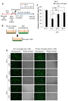
Effect of hypothermia on microglial cell migration: the Boyden chamber assay. Primary microglial cultures (A) or BV-2 microglial cells (1 x 10⁴ cells per well) (B) were seeded on the upper compartment of the Boyden chamber. After three hours (BV-2 microglial cells) or six hours (primary microglia) incubation under normothermic (37 °C) or hypothermic (33 C, 29 C) conditions, cells that migrated through the membrane were stained (A upper) and counted to evaluate the relative cell migration (A lower, B). A representative microscopic image for each condition is shown (magnification, x40 or x200) (A upper). Scale bar = 200 μm. Results are the mean ± SD (n = 3). **P < 0.01, *P < 0.05; significantly different from normothermia (37 C). n, number.
Figure 16
Figure 16
Effect of hypothermia on microglial cell migration: wound healing assay. Effect of hypothermia on microglial cell migration was investigated by the wound healing assay. When the cells reached 90 % confluence, a single wound was made in the center of the cell monolayer and cell debris was removed by washing with PBS. After 24 hours or 48 hours of incubation under normothermic or hypothermic condition as indicated (A), the wound closure areas were visualized under an inverted microscope and quantified (B). Results are the mean ± SD (n = 3). *P < 0.05, **P < 0.01; different from normothermia (37 °C) at the same time point. A representative microscopic image for each condition is shown (magnification, x100) (C). Scale bar = 200 μm. n, number; O/N, overnight.
Figure 17
Figure 17
Effect of hypothermia on microglial migrationin vivo. To assess the effect of therapeutic hypothermia on microglial migration in vivo, focal stab injury was created by placing a needle in the cortical area of the brain through a guide cannula. Local hypothermia (33 °C) using cold water circulation through a cooling coil was initiated either six hours before or after the needle injury, and maintained for 66 to 78 hours as indicated (A). Asterisks indicate the injury site. Horizontal sections of rat brain were subjected to microglial immunohistochemistry (Iba-1, green) (B). A representative microscopic image for each condition is shown (magnification, x40). Scale bar = 1 mm. Data acquisition and immunohistological intensity measurement of Iba-1 staining was performed with a NIH image J program (C). To count the Iba-1 positive cells, three concentric circles with constant interval were placed in the peri-region of the injury site (asterisk) in the sub-threshold image of the six independent sections. Three animals were used for each experimental group. The cells in the three circles were counted and statistically analyzed (D). Results are the mean ± SD. *P < 0.05, **P < 0.01; different from the same circle under normothermia (37 °C). #P < 0.05; different from the two conditions indicated.
Figure 18
Figure 18
Effects of local hypothermia on microglial proliferationin vivo. A focal needle injury and BrdU injection (i.p., 200 mg/kg) were done under normothermic or hypothermic condition as indicated (A). Iba-1 (red) or BrdU (green) immunochemistry was done to determine the effects of local hypothermia on microglial proliferation in vivo (B). Asterisks indicate the injury site. Arrows indicate Iba-1 and BrdU double positive cells in the merged images. A representative microscopic image for each condition is shown (magnification, x200 or x400). Scale bar = 200 μm. BrdU, 5-bromo-2’-deoxyuridine; i.p., intraperitoneally.
Figure 19
Figure 19
Inhibition of proinflammatory cytokine and iNOS expression by local hypothermia in the rat model of stab injury. After the focal stab injury in the cortex under normothermia or hypothermia as described in the main text (A), cortical tissues were prepared and subjected to RT-PCR analysis (B). Compared to normothermia, hypothermia diminished the injury-induced expression of TNF-α, IL-1β, and iNOS. iNOS, inducible NO synthase.

References

    1. Pedersen MO, Larsen A, Stoltenberg M, Penkowa M. Cell death in the injured brain: roles of metallothioneins. Prog Histochem Cytochem. 2009;44:1–27. doi: 10.1016/j.proghi.2008.10.002. - DOI - PubMed
    1. Lenzlinger PM, Morganti-Kossmann MC, Laurer HL, McIntosh TK. The duality of the inflammatory response to traumatic brain injury. Mol Neurobiol. 2001;24:169–181. doi: 10.1385/MN:24:1-3:169. - DOI - PubMed
    1. Baskaya MK, Rao AM, Dogan A, Donaldson D, Dempsey RJ. The biphasic opening of the blood–brain barrier in the cortex and hippocampus after traumatic brain injury in rats. Neurosci Lett. 1997;226:33–36. doi: 10.1016/S0304-3940(97)00239-5. - DOI - PubMed
    1. Ceulemans AG, Zgavc T, Kooijman R, Hachimi-Idrissi S, Sarre S, Michotte Y. The dual role of the neuroinflammatory response after ischemic stroke: modulatory effects of hypothermia. J Neuroinflammation. 2010;7:74. doi: 10.1186/1742-2094-7-74. - DOI - PMC - PubMed
    1. Simi A, Tsakiri N, Wang P, Rothwell NJ. Interleukin-1 and inflammatory neurodegeneration. Biochem Soc Trans. 2007;35:1122–1126. doi: 10.1042/BST0351122. - DOI - PubMed

Publication types