. 2012 Jul 9;2(1):37.
doi: 10.1186/2191-219X-2-37.

Synthesis and evaluation of 2-chloro N-[(S)-{(S)-1-[11 C]methylpiperidin-2-yl} (phenyl)methyl]3-trifluoromethyl-benzamide ([11 C]N-methyl-SSR504734) as a PET radioligand for glycine transporter 1

Affiliations

Synthesis and evaluation of 2-chloro N-[(S)-{(S)-1-[11 C]methylpiperidin-2-yl} (phenyl)methyl]3-trifluoromethyl-benzamide ([11 C]N-methyl-SSR504734) as a PET radioligand for glycine transporter 1

Takeshi Fuchigami et al. EJNMMI Res. .

Abstract

Background: Dysfunction of the glycine transporter 1 (GlyT1) has been suggested to be involved in psychiatric disorders such as schizophrenia. GlyT1 inhibitors have therefore been considered to have antipsychotic therapeutic potential. Positron emission tomography (PET) imaging probes for GlyT1 are, consequently, expected to be useful for investigating the mechanism of such disease conditions and for measuring occupancy of GlyT1 inhibitors in vivo. The aim of this study was to assess the potential of 2-chloro N-[(S)-{(S)-1-[11 C]methylpiperidin-2-yl} (phenyl)methyl] 3-trifluoromethyl-benzamide ([11 C]N-methyl-SSR504734) as a PET imaging agent for GlyT1.

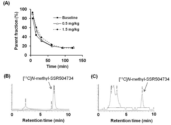
Methods: [11 C]N-methyl-SSR504734 was synthesized by N-[11 C]methylation of SSR504734 via [11 C]CH3OTf. In vitro brain distribution of [11 C]N-methyl-SSR504734 was tested in whole-hemisphere autoradiography (ARG) on human brain slices. Initial PET studies were performed using a cynomolgus monkey at baseline and after pretreatment with 0.1 to 1.5 mg/kg of SSR504734. Then, PET studies using rhesus monkeys were performed with arterial blood sampling at baseline and after pretreatment with 1.5 to 4.5 mg/kg SSR504734. Distribution volumes (VT) were calculated with a two-tissue compartment model, and GlyT1 occupancy by SSR504734 was estimated using a Lassen plot approach.

Results: [11 C]N-methyl-SSR504734 was successfully synthesized in moderate radiochemical yield and high specific radioactivity. In the ARG experiments, [11 C]N-methyl-SSR504734 showed specific binding in the white matter and pons. In the initial PET experiments in a cynomolgus monkey, [11 C]N-methyl-SSR504734 showed high brain uptake and consistent distribution with previously reported GlyT1 expression in vivo (thalamus, brainstem > cerebellum > cortical regions). However, the brain uptake increased after pretreatment with SSR504734. Further PET studies in rhesus monkeys showed a similar increase of brain uptake after pretreatment with SSR504734. However, the VT of [11 C]N-methyl-SSR504734 was found to decrease after pretreatment of SSR504734 in a dose-dependent manner. GlyT1 occupancy was calculated to be 45% and 73% at 1.5 and 4.5 mg/kg of SSR504734, respectively.

Conclusions: [11 C]N-methyl-SSR504734 is demonstrated to be a promising PET radioligand for GlyT1 in nonhuman primates. The present results warrant further PET studies in human subjects.


Figures

Figure 1
Figure 1
Chemical structure of GlyT1 inhibitors.
Figure 2
Figure 2
Chemical structure of PET radioligands for GlyT1.
Scheme 1
Scheme 1
Radiosynthesis of [11C] N-methyl-SSR504734.
Figure 3
Figure 3
In vitro postmortem autoradiography study of human brain. Autoradiogram from coronal (panels A, B, and C; 100-mm thickness) and horizontal whole-hemisphere (panels D, E, and F; 100-mm thickness) cryosections of the human brain incubated with [11 C]N-methyl-SSR504734 under baseline (panels A and D), blocked by non-radioactive N-methyl-SSR504734 (10 μM; panels B and E), and blocked by SSR504734 (10 μM; panels C and F) conditions. The quantified values of [11 C]N-methyl-SSR504734 (C) were expressed as (PSL-BG)/mm2 (panel G). TC, temporal cortex; PUT, putamen; CAU, caudate nucleus; CB, cerebellum; WM, white matter.
Figure 4
Figure 4
PET images of a cynomolgus monkey. PET images in the transaxial (left), the sagittal (middle), and the coronal (right) slices of the cynomolgus monkey, which were acquired from 9 to 123 min after intravenous injection with [11 C]N-methyl-SSR504734 under baseline (A), 0.5 mg/kg of pretreatment (B) and 1.5 mg/kg of pretreatment (C) conditions, and the corresponding MRI-T1 image (D).
Figure 5
Figure 5
Time-activity curves of brain radioactivity after intravenous injection of [11C] N -methyl-SSR504734 in a cynomolgus monkey. At baseline and pretreatment conditions (0.5 or 1.5 mg/kg of SSR504734). Whole brain uptake was expressed as percent injected dose (%ID) (A). Regional brain uptake was expressed as percent standard uptake value (%SUV) under baseline (B), pretreatment of SSR504734 (0.5 mg/kg) (C), and pretreatment of SSR504734 (1.5 mg/kg) conditions (D). Black square, thalamus; white circle, pons; black triangle, cerebellum; white diamond, putamen; asterisk, frontal cortex; cross, white matter; black circle, caudate; white square, anterior cingulate cortex; white triangle, temporal cortex.
Figure 6
Figure 6
Fraction of [11C] N -methyl-SSR504734 in the plasma during PET measurements. Percentage of unmetabolized [11 C]N-methyl-SSR504734 in the plasma of a cynomolgus monkey at baseline and pretreatment conditions (0.5 or 1.5 mg/kg of SSR504734) (A). HPLC analysis of plasma samples at 4 (C) and 90 min (D) after injection of [11 C]N-methyl-SSR504734 at baseline.
Figure 7
Figure 7
PET images of rhesus monkey 2. PET images in the transaxial (left), the sagittal (middle), and the coronal (right) slices of rhesus monkey 2 acquired from 9 to 123 min after intravenous injection with [11 C]N-methyl-SSR504734 at the baseline (A) and 4.5 mg/kg of pretreatment (B conditions, and the corresponding MRI-T1 image (C).
Figure 8
Figure 8
Time-activity curves of brain radioactivity after intravenous injection of [11C] N -methyl-SSR504734 in rhesus monkey 2. (A) %SUV in brain regions at baseline condition. (B) %SUV in brain regions at pretreatment conditions (4.5 mg/kg of SSR5047). Black square, thalamus; white circle, pons; black triangle, cerebellum; white diamond, putamen; asterisk, frontal cortex; cross, white matter; black circle, caudate; white square, anterior cingulate cortex; white triangle, temporal cortex. (C) Time-activity curve (%SUV) of metabolite-corrected radioactivity in the plasma under baseline condition and pretreatment (4.5 mg/kg of SSR504734) condition.
Figure 9
Figure 9
Lassen plot analysis applied to PET occupancy studies. Data were taken from [11 C]N-methyl-SSR504734 studies in the rhesus monkey brain at baseline and after administration of SSR504734 at doses of 1.5 mg/kg in rhesus monkey 1 (A) and 4.5 mg/kg in rhesus monkey 2 (B). Thalamus (white diamond) was not included for Lassen plot. (C) Relationship between GlyT1 occupancy (in percent) and dose of SSR504734. The relationship between GlyT1 occupancy (in percent) and dose of SSR504734 was expressed by the equation: Occupancy %=Omax×CpCp+Kd, where Omax is the assumed maximal occupancy (i.e., 100%), and Cp is the dose at which occupancy becomes 50%. Kd was estimated to be 1.7 mg/kg.

References

    1. Danysz W, Parsons CG. Glycine and N-methyl-D-aspartate receptors: physiological significance and possible therapeutic applications. Pharmacol Rev. 1998;50:597–564. - PubMed
    1. Legendre P. The glycinergic inhibitory synapse. Cell Mol Life Sci. 2001;58:760–793. doi: 10.1007/PL00000899. - DOI - PMC - PubMed
    1. Eulenburg V, Armsen W, Betz H, Gomeza J. Glycine transporters: essential regulators of neurotransmission. Trends Biochem Sci. 2005;30:325–333. doi: 10.1016/j.tibs.2005.04.004. - DOI - PubMed
    1. Zafra F, Aragon C, Olivares L, Danbolt NC, Gimenez C, Storm-Mathisen J. Glycine transporters are differentially expressed among CNS cells. J Neurosci. 1995;15:3952–3969. - PMC - PubMed
    1. Jursky F, Nelson N. Localization of glycine neurotransmitter transporter (GLYT2) reveals correlation with the distribution of glycine receptor. J Neurochem. 1995;64:1026–1033. - PubMed

LinkOut - more resources