. 2012 Jul 9:7:45.
doi: 10.1186/1750-1172-7-45.

Assessment of the structural and functional impact of in-frame mutations of the DMD gene, using the tools included in the eDystrophin online database

Affiliations

Assessment of the structural and functional impact of in-frame mutations of the DMD gene, using the tools included in the eDystrophin online database

Aurélie Nicolas et al. Orphanet J Rare Dis. .

Abstract

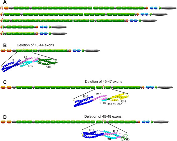
Background: Dystrophin is a large essential protein of skeletal and heart muscle. It is a filamentous scaffolding protein with numerous binding domains. Mutations in the DMD gene, which encodes dystrophin, mostly result in the deletion of one or several exons and cause Duchenne (DMD) and Becker (BMD) muscular dystrophies. The most common DMD mutations are frameshift mutations resulting in an absence of dystrophin from tissues. In-frame DMD mutations are less frequent and result in a protein with partial wild-type dystrophin function. The aim of this study was to highlight structural and functional modifications of dystrophin caused by in-frame mutations.

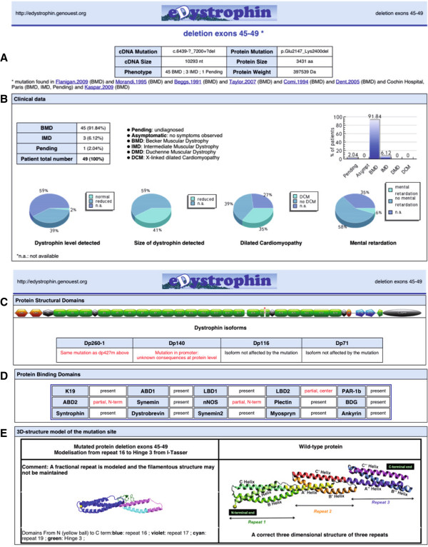
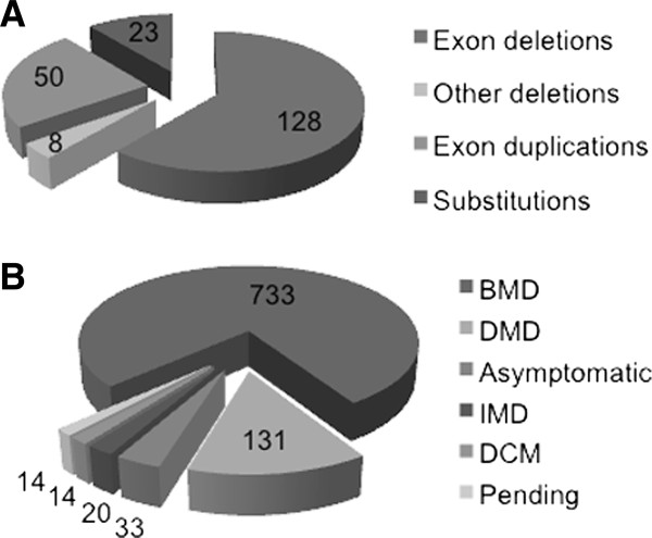
Methods and results: We developed a dedicated database for dystrophin, the eDystrophin database. It contains 209 different non frame-shifting mutations found in 945 patients from a French cohort and previous studies. Bioinformatics tools provide models of the three-dimensional structure of the protein at deletion sites, making it possible to determine whether the mutated protein retains the typical filamentous structure of dystrophin. An analysis of the structure of mutated dystrophin molecules showed that hybrid repeats were reconstituted at the deletion site in some cases. These hybrid repeats harbored the typical triple coiled-coil structure of native repeats, which may be correlated with better function in muscle cells.

Conclusion: This new database focuses on the dystrophin protein and its modification due to in-frame deletions in BMD patients. The observation of hybrid repeat reconstitution in some cases provides insight into phenotype-genotype correlations in dystrophin diseases and possible strategies for gene therapy. The eDystrophin database is freely available: http://edystrophin.genouest.org/.


Figures

Figure 1
Figure 1
The dystrophin molecule and its partners. (A) Schematic representation of the molecule with the structural domains CH1 and CH2, constituting actin-binding domain 1, hinges 1 to 4 (H1 to H4), spectrin-like repeats 1 to 24 (R1 to R24); the WW domain and EF hand–region constituting the Cys-rich domain; ZZ, the zinc finger domain; and the C-terminal domain (C term) and its partners. (B) Model of the three-dimensional structure of repeat 7 folded into a triple coiled-coil, consisting of three helices, A, B, and C, joined by two loops, AB and BC. (C) Model of the three-dimensional structure of tandem repeats R7-8 of the rod domain. Each repeat is composed of three alpha helices folded into a triple coiled-coil: R7 (helices A, B, and C, joined by loops AB and BC) and R8 (helices A’, B’ and helix C’, joined by loops A’B’ and B’C’). A long common helical linker is formed between the two repeats by R7 helix C and R8 helix A’.
Figure 2
Figure 2
Representation of the dystrophin proteins generated from genes with several types of deletions. (A) Illustration of the proteins produced from genes with deletions of exons corresponding to the 3’ terminus encoding actin-binding domain 1. The red spot indicates the deletion site. (B) Deletion of exons 35 to 44: representation of the entire molecule with a tag at the deletion site and a model of the three-dimensional structure at the deletion site, showing that a hybrid repeat is reconstituted from parts of repeats 2 and 17. (C) Deletion of exons 45 to 47: representation of the entire molecule with a tag at the deletion site and the homology model of three-dimensional structure at the deletion site, showing that the junction between the C-terminal part of repeat 17 and the N-terminal part of repeat 18 does not allow the reconstitution of a hybrid repeat. (D) Deletion of exons 45 to 48: representation of the entire molecule with a tag at the deletion site and the homology model of the three-dimensional structure at the deletion site, showing that a hybrid repeat is formed from parts of repeats 17 and 18.
Figure 3
Figure 3
Screenshot of “Search by mutation type/Deletions” tool. The user can select an exon and then a deletion involving this exon. On the right side of the page, the user can select a mutation of interest from a list, to obtain data about this mutation. The user can also save this list as a csv-format file.
Figure 4
Figure 4
Screenshot of data available for the deletion of exons 45 to 49. (A) A general view of the webpage after loading. Description of the mutation at the nucleotide and protein levels, cDNA and protein size, the molecular weight of the mutated protein, a link to cDNA and protein sequences and a list of references reporting patients carrying the mutation are available in table form. Detailed information about clinical data, structural and binding domains and models of three-dimensional structure can be obtained by clicking on the boxes below. (B) The “Clinical data” tab on the first line provides access to a table showing the distribution of phenotypes. In the second line, pie-charts showing the amount and size of dystrophin, as determined by western blotting, the presence of cardiomyopathy and mental retardation are given. (C) The “Structural domains” tab provides a schematic representation of the mutated dystrophin. The sequences of each protein domain are available. (D) The “Binding domains” tab indicates, in red, the changes to the binding domains caused by the mutations listed in the table. (E) The “3D-structure model of the mutation site tab shows the model of the three-dimensional structure of the domains around the mutation junctions (here R16, R17, R19 and H3). The secondary structure predicted by I-TASSER is displayed above the model. The PDB file and the Ramachandran plot are also available. The modeling tab is available only for the deletion of exons encoding part of the central rod domain. All information can be saved down in the form of PDF files.
Figure 5
Figure 5
Statistics for mutations recorded in the eDystrophin database. (A) Mutation types: the number of cases is shown for each of 209 different mutations. (B) Phenotype distribution: for each phenotype, the number of patients concerned, from a total of 945 patients, is shown.
Figure 6
Figure 6
Fusion of the exon borders and repeat alignment. (A) The 24 repeats are represented by the exons encoding them (not by their helices) as rectangles with the following color code: orange for even-numbered exons and light yellow for odd-numbered exons. The frameshifting exon borders are shown as blue triangles. The approximate position of the helices is indicated above the figure. (B) Focus on the deletion site for the deletion of exons 13 to 44, showing the reconstitution of a hybrid repeat by the joining of exons 12 and 45 (in green), maintaining the phasing of exon coding for a reconstituted B helix. (C) Focus on the deletion site for the deletion of exons 45 to 47, showing how the joining of exons 44 and 48 (in red) does not respect the phasing of the repeats and the presence of an extra sequence inconsistent with a repeat. (D) Focus on the deletion site for the deletion of exons 45 to 48, showing how the hybrid repeat can be reconstituted by the joining of exons 44 and 49 (in green), maintaining the phasing of the exons encoding a reconstituted B helix

Similar articles

Cited by

References

    1. Mendelian Inheritance in Man, [http://www.ncbi.nlm.gov/omim]
    1. Hoffman EP, Brown RH, Kunkel LM. Dystrophin: the protein product of the Duchenne muscular dystrophy locus. Cell. 1987;51:919–928. doi: 10.1016/0092-8674(87)90579-4. - DOI - PubMed
    1. Koenig M, Hoffman EP, Bertelson CJ, Monaco AP, Feener C, Kunkel LM. Complete cloning of the Duchenne muscular dystrophy (DMD) cDNA and preliminary genomic organization of the DMD gene in normal and affected individuals. Cell. 1987;50:509–517. doi: 10.1016/0092-8674(87)90504-6. - DOI - PubMed
    1. Roberts RG, Bobrow M. Dystrophins in vertebrates and invertebrates. Hum Mol Genet. 1998;7:589–595. doi: 10.1093/hmg/7.4.589. - DOI - PubMed
    1. Segalat L. Dystrophin and functionally related proteins in the nematode Caenorhabditis elegans. Neuromuscul Disord. 2002;12(Suppl 1):S105–S109. - PubMed

Publication types

LinkOut - more resources