Review
. 2013 Jun:69:37-44.
doi: 10.1016/j.neuropharm.2012.06.044. Epub 2012 Jul 6.

Does interictal synchronization influence ictogenesis?

Affiliations
Review

Does interictal synchronization influence ictogenesis?

Massimo Avoli et al. Neuropharmacology. 2013 Jun.

Abstract

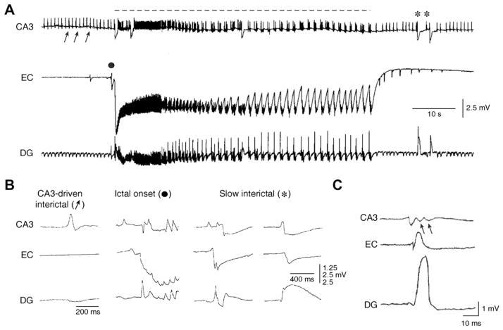
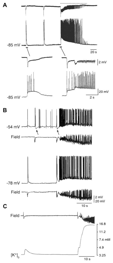
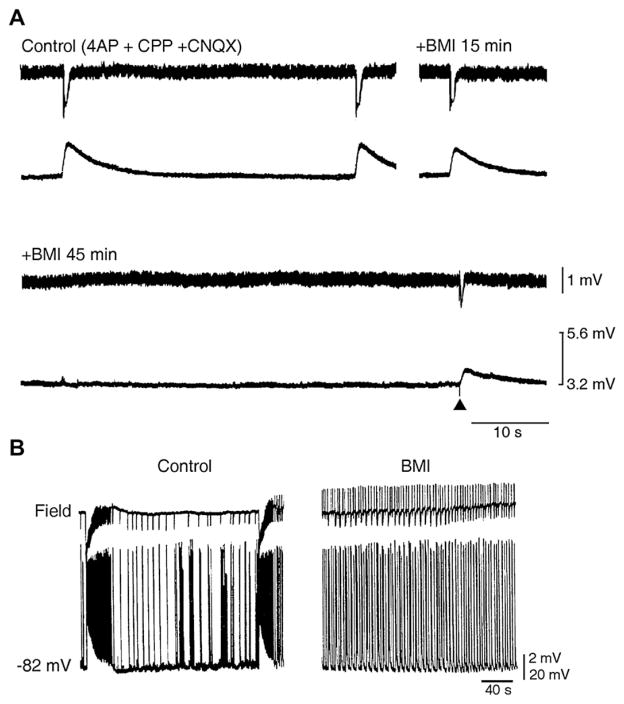
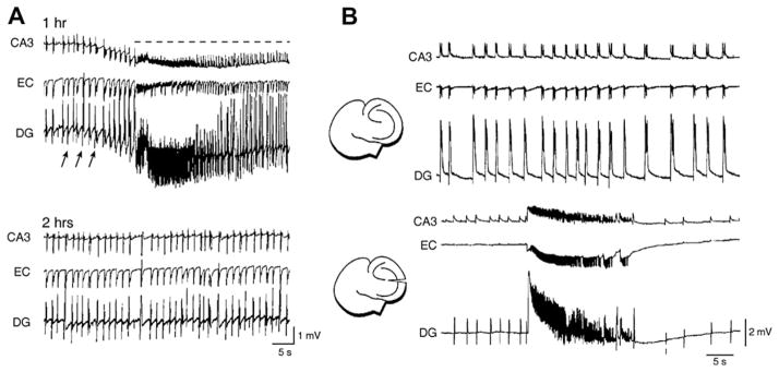
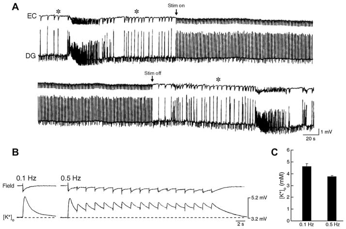
The EEG recorded from epileptic patients presents with interictal discharges that are not associated with detectable clinical symptoms but are valuable for diagnostic purposes. Experimental studies have shown that interictal discharges and ictal events (i.e., seizures) are characterized intracellularly by similar (but for duration) neuronal depolarizations leading to sustained action potential firing, thus indicating that they may share similar cellular and pharmacological mechanisms. It has also been proposed that interictal discharges may herald the onset of electrographic seizures, but other studies have demonstrated that interictal events interfere with the occurrence of ictal activity. The relationship between interictal and ictal activity thus remains ambiguous. Here we will review this issue in animal models of limbic seizures that are electrographically close to those seen in TLE patients. In particular we will: (i) focus on the electrophysiological and pharmacological characteristics of, at least, two types of interictal discharge; (ii) propose that they play opposite roles in leading to ictogenesis; and (iii) discuss the possibility that mimicking one of these two types of interictal activity by low frequency repetitive stimulation can control ictogenesis. Finally, we will also review evidence indicating that specific types of interictal discharge may play a role in epileptogenesis. This article is part of the Special Issue entitled 'New Targets and Approaches to the Treatment of Epilepsy'.


Figures

Fig. 1
Fig. 1
Epileptiform patterns induced by 4AP in rodent hippocampus-entorhinal cortex slices. (A) Field potential recordings performed simultaneously in the rat CA3 subfield, EC and dentate gyrus demonstrate the occurrence of three different types of activity; the first (discontinuous line) is recorded synchronously in all areas and consists of a sustained ictal-like epileptiform discharge; the second type (arrows) consists of continuous fast interictal-like events, and it is seen in the CA3 and dentate areas only; the third type (asterisk) is recorded in all areas and is characterized by a slow field potential. (B) Expanded traces of the field potential recordings illustrated in A show the modalities of onset and spread of CA3-drive interictal events, ictal discharge onset, and slow interictal discharge; note that the ictal discharge initiates in the EC while different sites of origin characterize the two examples of slow interictal events. (C) Expanded traces of a CA3-driven interictal discharge recorded from a mouse hippocampus-EC slice; note that this interictal discharge initiates in the CA3 region and propagates to the EC and DG; arrows point at the “late” components of the CA3-driven interictal discharge recorded in CA3, presumably representing the reentry of synchronous activity from the DG. In this and following figures abbreviations are: entorhinal cortex (EC); dentate gyrus (DG).
Fig. 2
Fig. 2
Relationship between slow interictal spikes and ictal discharges recorded from the EC during 4AP application. (A) Simultaneous extracellular (Field) and “sharp” intracellular (−85 mV) recordings obtained from the rat EC during 4AP application reveal slow interictal spikes and an ictal discharge. Note in the expanded traces that the ictal discharge appears to be initiated by an event similar to that associated to the interictal spike. (B) Field and intracellular characteristics of slow interictal events and ictal discharge onset in an EC neuron that was recorded with K-acetate-filled microelectrode during 4AP application both at resting membrane potential (−78 mV) and during intracellular injection of steady positive current (−54 mV). Note that this procedure causes a decrease of the ictal depolarization that is preceded by a hyperpolarizing event similar to what seen during the slow interictal event (arrows). (C) Simultaneous field and [K+]o recordings obtained from the rat EC show that the slow interictal spike is associated with a transient increases in [K+]o, while a sustained elevation is seen during the tonic phase of the ictal discharge; note that the ictal discharge onset is characterized by a “transient-like” increase in [K+]o that is larger than what observed during the isolated slow interictal discharges.
Fig. 3
Fig. 3
GABAA receptors contribute to the [K+]o increases associated with slow interictal discharges and to ictogenesis. (A) Simultaneous field and [K+]o recordings performed in the rat EC during application of 4AP and glutamatergic receptor antagonists (Control) and at different times after addition of the GABAA receptor antagonist bicuculline methiodide (BMI). Note that after 45 min of BMI application, no spontaneous events can be observed as well as that the response induced by an electrical stimulus delivered close to the recording site induces reduced field and [K+]o signals. (B) Application of BMI abolishes slow interictal and ictal discharges recorded from an EC slice with simultaneous extracellular (Field trace) and intracellular microelectrodes (−78 mV trace) while disclosing rhythmic short-lasting epileptiform events.
Fig. 4
Fig. 4
(A) Epileptiform activity recorded 1 and 2 h after initiation of continuous bath application of 4AP from a combined mouse hippocampus-EC slice. Note that ictal (dotted line) and fast-developing interictal (arrows) activity is recorded at 1 h; at 2 h of 4AP application, ictal discharges disappear. (B) Spontaneous epileptiform activity induced by super fusing a combined mouse hippocampus-EC slice with Mg2+-free medium before and after Schaffer collateral cut. Note that before the lesion (top panel), interictal discharges are recorded from all limbic structures, while after sectioning the Schaffer collaterals (bottom panel) interictal discharges disappear in the EC and ictal activity, which is recorded in the three areas, is disclosed.
Fig. 5
Fig. 5
(A) Effect induced by stimulation at 1 Hz delivered in the CA1 on the 4AP-induced epileptiform activity recorded after Schaffer collateral cut from a combined mouse hippocampus-EC slice. Note that ictal discharge is abolished during the stimulation period and re-appears on termination of the stimulation; note also the persistence of interictal discharges (asterisks), presumably of EC origin, before and after the ictal activity. (B) Simultaneous field and [K+]o recordings obtained from the rat EC during application of 4AP and glutamatergic receptor antagonists. Electrical stimuli were delivered in the EC (subicular side) at 0.1 and 0.5 Hz. Note that the peak elevations in [K+]o, were smaller during the latter protocol. (C) Histogram of the peak elevations in [K+]o induced by 0.1 and 0.5 Hz stimulating protocols during 6 trials in two experiments; note that the peak value is smaller during repetitive stimuli delivered at 0.5 Hz. Values are mean ± SEM.

References

    1. Avoli M, de Curtis M. GABAergic synchronization in the limbic system and its role in the generation of epileptiform activity. Prog Neurobiol. 2011;95:104–132. - PMC - PubMed
    1. Avoli M, Louvel J, Kurcewicz I, Pumain R, Barbarosie M. Extracellular free potassium and calcium during synchronous activity induced by 4-aminopyridine in the juvenile rat hippocampus. J Physiol. 1996;493:707–717. - PMC - PubMed
    1. Ayala GF, Dichter M, Gumnit RJ, Matsumoto H, Spencer WA. Genesis of epileptic interictal spikes. New knowledge of cortical feedback systems suggests a neurophysiological explanation of brief paroxysms. Brain Res. 1973;1973(52):1–17. - PubMed
    1. Barbarosie M, Avoli M. CA3-driven hippocampal-entorhinal loop controls rather than sustains in vitro limbic seizures. J Neurosci. 1997;17:9308–9314. - PMC - PubMed
    1. Barbarosie M, Louvel J, D’Antuono M, Kurcewicz I, Avoli M. Masking synchronous GABA-mediated potentials controls limbic seizures. Epilepsia. 2002;43:1469–1479. - PubMed

MeSH terms