. 2012 Oct;343(1):157-66.
doi: 10.1124/jpet.112.195206. Epub 2012 Jul 9.

Cannabinoid receptor activation correlates with the proapoptotic action of the β2-adrenergic agonist (R,R')-4-methoxy-1-naphthylfenoterol in HepG2 hepatocarcinoma cells

Affiliations

Cannabinoid receptor activation correlates with the proapoptotic action of the β2-adrenergic agonist (R,R')-4-methoxy-1-naphthylfenoterol in HepG2 hepatocarcinoma cells

Rajib K Paul et al. J Pharmacol Exp Ther. 2012 Oct.

Abstract

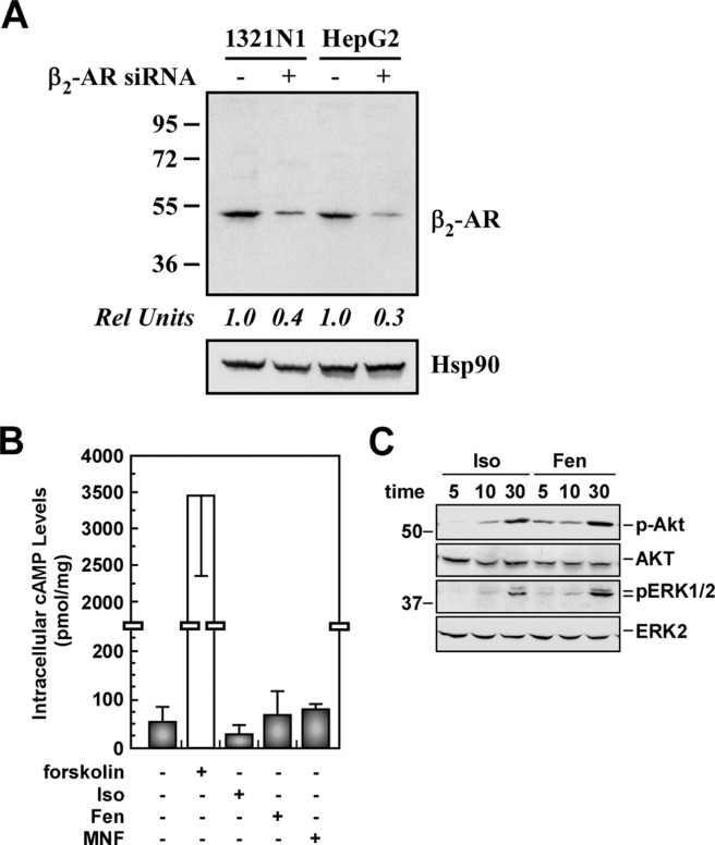
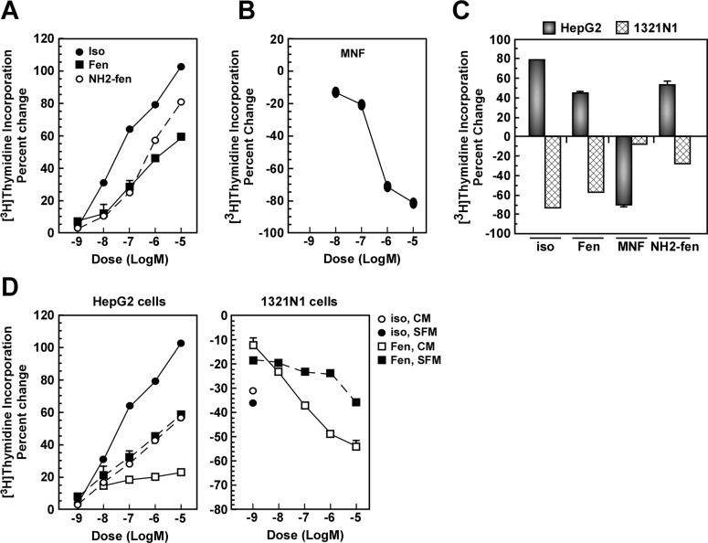
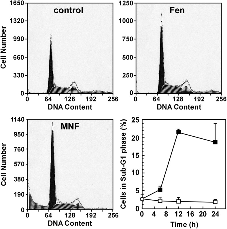
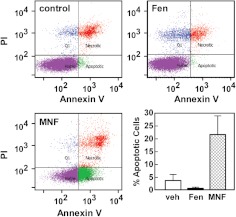
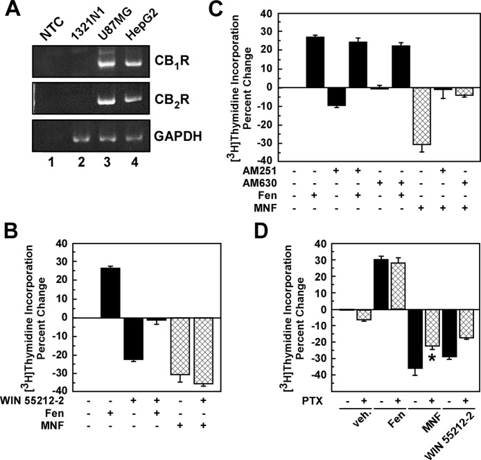
Inhibition of cell proliferation by fenoterol and fenoterol derivatives in 1321N1 astrocytoma cells is consistent with β(2)-adrenergic receptor (β(2)-AR) stimulation. However, the events that result in fenoterol-mediated control of cell proliferation in other cell types are not clear. Here, we compare the effect of the β(2)-AR agonists (R,R')-fenoterol (Fen) and (R,R')-4-methoxy-1-naphthylfenoterol (MNF) on signaling and cell proliferation in HepG2 hepatocarcinoma cells by using Western blotting and [(3)H]thymidine incorporation assays. Despite the expression of β(2)-AR, no cAMP accumulation was observed when cells were stimulated with isoproterenol or Fen, although the treatment elicited both mitogen-activated protein kinase and phosphatidylinositol 3-kinase/Akt activation. Unexpectedly, isoproterenol and Fen promoted HepG2 cell growth, but MNF reduced proliferation together with increased apoptosis. The mitogenic responses of Fen were attenuated by 3-(isopropylamino)-1-[(7-methyl-4-indanyl)oxy]butan-2-ol (ICI 118,551), a β(2)-AR antagonist, whereas those of MNF were unaffected. Because of the coexpression of β(2)-AR and cannabinoid receptors (CBRs) and their impact on HepG2 cell proliferation, these Gα(i)/Gα(o)-linked receptors may be implicated in MNF signaling. Cell treatment with (R)-(+)-[2,3-dihydro-5-methyl-3-(4-morpholinylmethyl)pyrrolo[1,2,3-de]-1,4-benzoxazin-6-yl]-1-napthalenylmethanone (WIN 55,212-2), a synthetic agonist of CB(1)R and CB(2)R, led to growth inhibition, whereas inverse agonists of these receptors blocked MNF mitogenic responses without affecting Fen signaling. MNF responses were sensitive to pertussis toxin. The β(2)-AR-deficient U87MG cells were refractory to Fen, but responsive to the antiproliferative actions of MNF and WIN 55,212-2. The data indicate that the presence of the naphthyl moiety in MNF results in functional coupling to the CBR pathway, providing one of the first examples of a dually acting β(2)-AR-CBR ligand.


Figures

Fig. 1.
Fig. 1.
Responses of HepG2 cells to β-agonist stimulation. A, HepG2 and 1321N1 cells were transfected with the negative control siRNA (−) or β2-AR siRNA (+) for 48 h. Cell lysates were immunoblotted with a specific anti-β2-AR antibody (ab40834), using Hsp90 as a loading control. B, increase in cAMP accumulation in HepG2 cells was observed with forskolin (10 μM), but not with 1 μM of either isoproterenol (Iso), Fen, or MNF. Data shown are from a single experiment conducted in quadruplicate. Error bars indicate mean ± S.D. C, serum-starved HepG2 cells were incubated in the presence of Iso (1 μM) or Fen (1 μM) for 5, 10, and 30 min. Cell lysates were immunoblotted with antibodies against phosphorylated Akt (p-Akt; Ser473) and total Akt, as well as phosphorylated ERK1/2 (p-ERK1/2) and total ERK2. The experiments shown in B and C were repeated twice with comparable results. The positions of molecular mass markers (in kilodaltons) are shown to the left of the immunoblots.
Fig. 2.
Fig. 2.
The effects of isoproterenol, Fen, and fenoterol derivatives on cell growth are cell type specific. A and B, serum-starved HepG2 cells were incubated with vehicle or the indicated concentrations of Iso, Fen, or (R,R′)-aminofenoterol (NH2-fen) (A) or MNF (B) for 24 h, and the levels of [3H]thymidine incorporation were measured; see Materials and Methods for experimental details. Representative concentration-response curves are shown. C, HepG2 cells in serum-depleted medium and 1321N1 cells in complete medium were treated with compounds at 1 μM for 24 h. D, HepG2 and 1321N1 cells were incubated without (SFM) or with serum (CM) in the presence of the indicated concentrations of Iso or Fen. Quantification of percentage change in [3H]thymidine incorporation versus control is expressed as means ± S.E. and represents results from two to six independent experiments, each performed in triplicate dishes. In most instances, error bars are smaller than the symbols.
Fig. 3.
Fig. 3.
β2-AR antagonist does not inhibit the antiproliferative action of MNF in HepG2 cells. A and B, serum-depleted HepG2 cells were incubated with the indicated concentrations of the β-AR antagonist ICI-118,551 (ICI) for 1 h followed by the addition of vehicle (A), Fen (B, left), or MNF (B, right) for 24 h, and levels of [3H]thymidine incorporation were measured. *, p < 0.05. Representative concentration-response curves for Fen and MNF are shown in B. C and D, quantification of percentage change in [3H]thymidine incorporation versus control is expressed as means ± S.E. and represents results from three independent experiments, each performed in triplicate dishes. C, Fen. D, MNF.
Fig. 4.
Fig. 4.
MNF increases the number of sub-G1 events in HepG2 cells. Serum-depleted HepG2 cells were harvested after 6-, 12-, and 24-h treatment with vehicle (top left), Fen (1 μM) (top right), or MNF (1 μM) (bottom left). Cells were fixed, stained and then analyzed for DNA content by using flow cytometry. Representative DNA content analysis in various phases of the cell cycle after 24-h treatment with vehicle, Fen, or MNF is shown. The number of sub-G1 events, which displays cells in late-stage apoptosis or already dead, in function of treatment duration was quantified and represents results from two independent experiments, each performed in duplicate dishes (bottom right). Data are expressed as means ± S.E. (n = 4).
Fig. 5.
Fig. 5.
MNF induces apoptosis in HepG2 cells. Serum-depleted HepG2 cells were treated with vehicle (top left), Fen (1 μM) (top right), or MNF (1 μM) (bottom left) for 24 h, stained with Annexin V and PI, and then analyzed by flow cytometry. Representative profiles are shown. The fraction of annexin V-positive HepG2 cells that were apoptotic was quantitated and represents results from two independent experiments, each performed in duplicate dishes (lower right). Data are expressed as means ± S.E. (n = 4).
Fig. 6.
Fig. 6.
Role of CBR activation in the antiproliferative action of MNF in HepG2 cells. A, total RNA was extracted from HepG2, 1321N1, and U87MG cells, and then analyzed semiquantitatively by PCR. A nontemplate control (NTC) has been included (lane 1). GAPDH, glyceraldehyde-3-phosphate dehydrogenase. B and C, serum-depleted HepG2 cells were incubated with the CBR agonist WIN 55,212-2 (Win; 1 μM) (B) or antagonists AM251 (1 μM) or AM630 (0.5 μM) (C) for 1 h followed by the addition of vehicle, Fen (0.5 μM), or MNF (0.25 μM) for 24 h. D, serum-depleted HepG2 cells were pretreated without or with pertussis toxin (PTX; 50 ng/ml) for 16 h followed by the addition of vehicle, Fen (0.5 μM), MNF (0.5 μM), or WIN 55,212-2 (0.5 μM) for 24 h. In B to D, the levels of [3H]thymidine incorporation were measured. Quantification of percentage change in [3H]thymidine incorporation versus control is expressed as means ± S.D. and represents results from three (B and C) or two (D) independent experiments, each performed in triplicate dishes. *, p < 0.05.

References

    1. Agarwal D, Glasel JA. (1999) Differential effects of opioid and adrenergic agonists on proliferation in a cultured cell line. Cell Prolif 32:215–229 - PMC - PubMed
    1. Aguado T, Carracedo A, Julien B, Velasco G, Milman G, Mechoulam R, Alvarez L, Guzmán M, Galve-Roperh I. (2007) Cannabinoids induce glioma stem-like cell differentiation and inhibit gliomagenesis. J Biol Chem 282:6854–6862 - PubMed
    1. Ahn S, Maudsley S, Luttrell LM, Lefkowitz RJ, Daaka Y. (1999) Src-mediated tyrosine phosphorylation of dynamin is required for β2-adrenergic receptor internalization and mitogen- activated protein kinase signaling. J Biol Chem 274:1185–1188 - PubMed
    1. Audet M, Bouvier M. (2008) Insights into signaling from the β2-adrenergic receptor structure. Nat Chem Biol 4:397–403 - PubMed
    1. Biswas KK, Sarker KP, Abeyama K, Kawahara K, Iino S, Otsubo Y, Saigo K, Izumi H, Hashiguchi T, Yamakuchi M, et al. (2003) Membrane cholesterol but not putative receptors mediates anandamide-induced hepatocyte apoptosis. Hepatology 38:1167–1177 - PubMed

Publication types

MeSH terms