. 2012 Aug 17;111(5):553-63.
doi: 10.1161/CIRCRESAHA.112.274548. Epub 2012 Jul 9.

S-Nitrosation of β-catenin and p120 catenin: a novel regulatory mechanism in endothelial hyperpermeability

Affiliations

S-Nitrosation of β-catenin and p120 catenin: a novel regulatory mechanism in endothelial hyperpermeability

Natalie Marín et al. Circ Res. .

Abstract

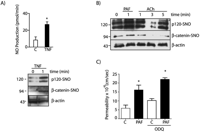
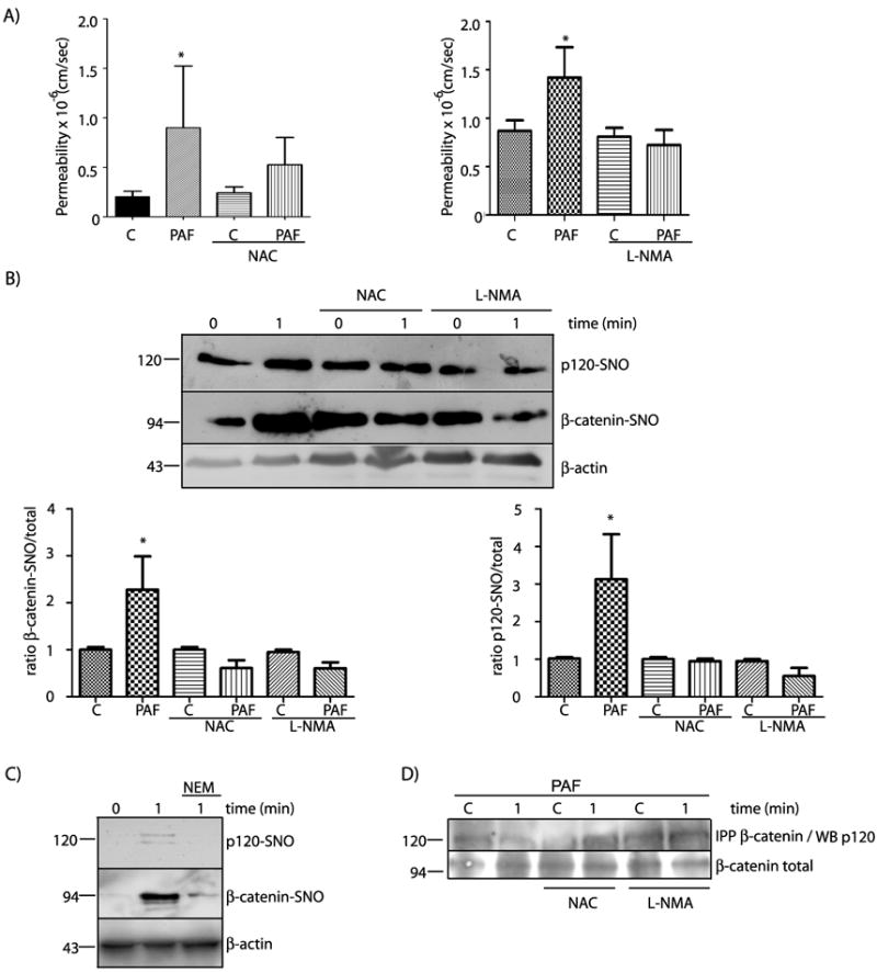
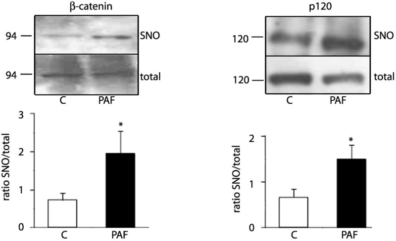
Rationale: Endothelial adherens junction proteins constitute an important element in the control of microvascular permeability. Platelet-activating factor (PAF) increases permeability to macromolecules via translocation of endothelial nitric oxide synthase (eNOS) to cytosol and stimulation of eNOS-derived nitric oxide signaling cascade. The mechanisms by which nitric oxide signaling regulates permeability at adherens junctions are still incompletely understood.

Objective: We explored the hypothesis that PAF stimulates hyperpermeability via S-nitrosation (SNO) of adherens junction proteins.

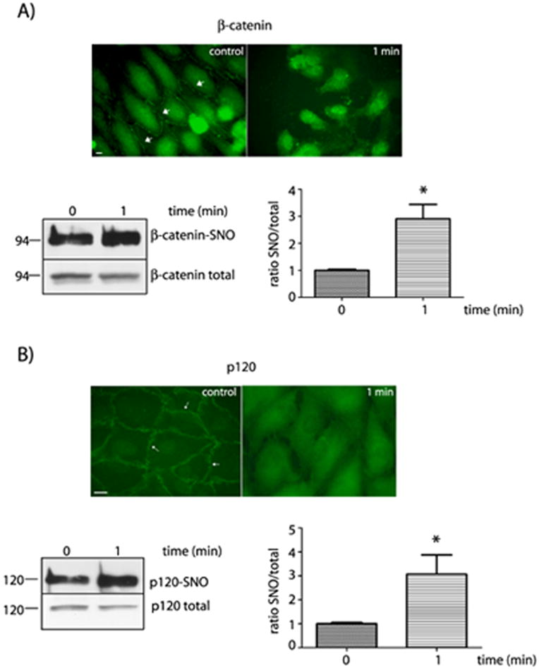
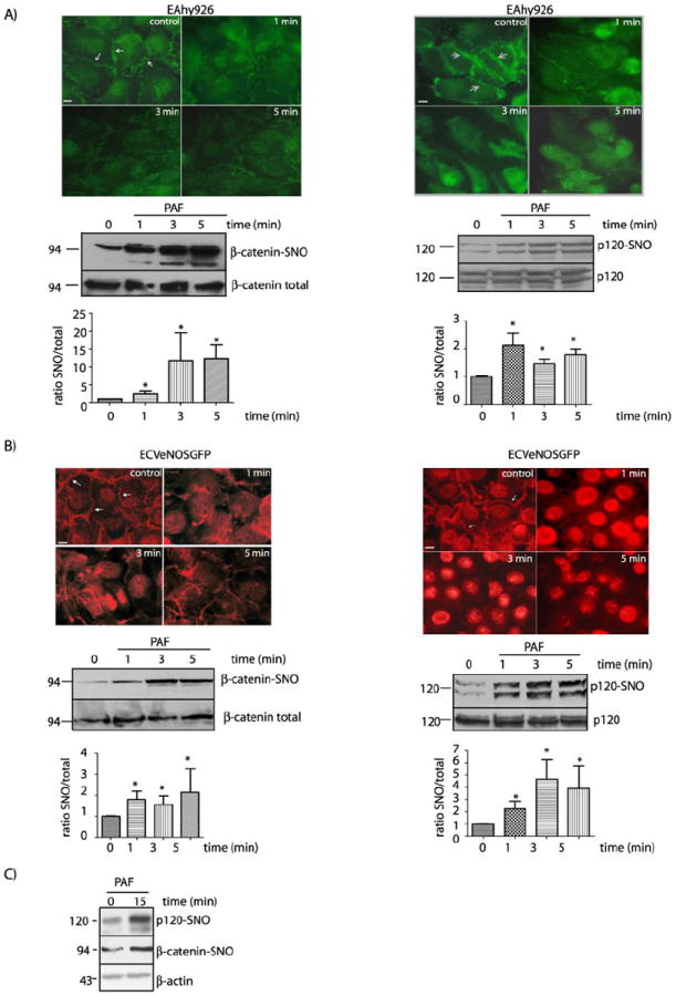
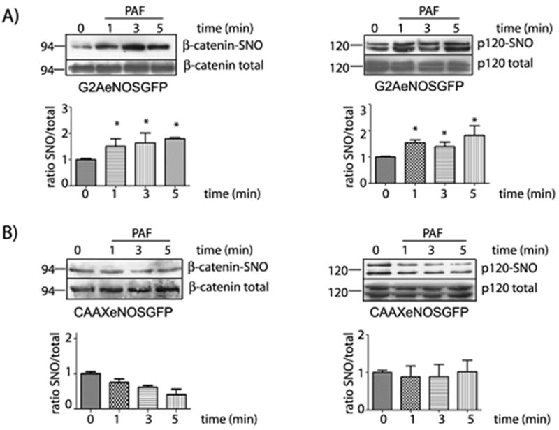
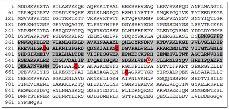
Methods and results: We measured PAF-stimulated SNO of β-catenin and p120-catenin (p120) in 3 cell lines: ECV-eNOSGFP, EAhy926 (derived from human umbilical vein), and postcapillary venular endothelial cells (derived from bovine heart endothelium) and in the mouse cremaster muscle in vivo. SNO correlated with diminished abundance of β-catenin and p120 at the adherens junction and with hyperpermeability. Tumor necrosis factor-α increased nitric oxide production and caused similar increase in SNO as PAF. To ascertain the importance of eNOS subcellular location in this process, we used ECV-304 cells transfected with cytosolic eNOS (GFPeNOSG2A) and plasma membrane eNOS (GFPeNOSCAAX). PAF induced SNO of β-catenin and p120 and significantly diminished association between these proteins in cells with cytosolic eNOS but not in cells wherein eNOS is anchored to the cell membrane. Inhibitors of nitric oxide production and of SNO blocked PAF-induced SNO and hyperpermeability, whereas inhibition of the cGMP pathway had no effect. Mass spectrometry analysis of purified p120 identified cysteine 579 as the main S-nitrosated residue in the region that putatively interacts with vascular endothelial-cadherin.

Conclusions: Our results demonstrate that agonist-induced SNO contributes to junctional membrane protein changes that enhance endothelial permeability.


Figures

Figure 1
Figure 1
Correlation between protein organization at the cell membrane and SNO of β-catenin and p120 in CVEC. A) Upper Panel: Indirect immunofluorescence staining for β-catenin in control conditions and after stimulation with 10-7 mol/L PAF. Arrows indicate the presence of β-catenin in the cell plasma membrane. Bar represents 10 μm. Lower Panel: SNO of β-catenin as measured by biotin switch. The quantification is shown as the ratio of the S-nitrosylated β-catenin to the total β-catenin on the right lower panel. * P < 0.05 compared to control; n = 3. B) Upper Panel: Indirect immunofluorescence staining for p120 in control conditions and after stimulation with 10-7 mol/L PAF. Left lower panel: SNO of p120 detected by biotin switch. The quantification is shown as the ratio of the S-nitrosylated p120 to the total p120 on the right lower panel. *P < 0.05 as compared with control; n = 3.
Figure 2
Figure 2
PAF induces internalization and SNO of β-catenin and p120 in EAhy926 cells and ECVeNOS-GFP. A) Experiments in EAhy926 cells. Upper Panel: Indirect immunofluorescence staining of EAhy926 cells for β-catenin and p120 after stimulation with 10-7 mol/L PAF. Middle Panel: SNO of β-catenin (left side) and p120 (right side) as detected by biotin switch. Lower panel: Bar graph showing the quantification of the SNO signal for β-catenin (left side) and p120 (right side). * P < 0.05 compared with control; n = 3. B) Experiments in ECVeNOS-GFP. Upper Panel: Indirect immunofluorescence staining of ECVeNOS-GFP for β-catenin (left side) and p120 (right side) in control and at different times after stimulation with 5×10⁻⁷ mol/L PAF. Middle Panel: SNO of β-catenin (left side) and p120 (right side) as detected by biotin switch. Lower Panel: Bar graph reporting the quantification of the SNO signal in Western blots for β-catenin (left side) and p120 (right side). * P < 0.05 as compared with control; n = 3. C) Prolonged application of PAF induces SNO of β-catenin and p120 in EAhy926 cells. PAF at 10-7 mol/L was applied for 15 minutes. Protein extracts were processed for biotin switch assay and probed with anti β-catenin and anti p120 antibodies. β-actin was used as a load control. Bars in microphotographs represent 10μm.
Figure 3
Figure 3
SNO is a widespread regulatory mechanism specific for permeability and independent of sGC and PKG. A) TNF-α induces NO production and SNO of β-catenin and p120 in EAhy926 cells. TNF-α at 50 ng/mL was applied for 1 minute. Protein extracts were processed for biotin switch assay and probed with anti β-catenin and anti p120 antibodies. β-actin was used as a load control. B) ACh at 10-5 moles/L does not induce SNO of β-catenin and p120 in EAhy926 cells. C) PAF at 10-7 moles/L increases permeability to FITC-dextran-70 across confluent EAhy926 monolayers. PAF-stimulated hyperpermeability is not affected by inhibition of sGC with ODQ. * P < 0.05 compared with control; n=3.
Figure 4
Figure 4
Subcellular location of eNOS determines SNO of β-catenin and p120. Protein extracts from ECV-GFPeNOS-G2A and ECV-GFPeNOS-CAAX cells treated with 5×10⁻⁷ mol/L PAF for different times were processed for biotin switch assay and probed with anti β-catenin and anti p120 antibodies. A) Upper Panel: PAF-induced SNO of β-catenin and p120 in ECV-GFPeNOS-G2A cells. Lower Panel: Bar graph showing the quantification of the Western blots of β-catenin (left side) and p120 (right side). * P < 0.05 compared to control; n = 3. B) Upper Panel: PAF-induced SNO of β-catenin and p120 in ECV-GFPeNOS-CAAX cells. Lower Panel: Bar graph showing the quantification of the Western blots of β-catenin (left side) and p120 (right side). * P < 0.05 compared to control; n = 3.
Figure 5
Figure 5
SNO disrupts the association between β-catenin and p120. Protein extracts from ECV-GFPeNOS-G2A and ECV-GFPeNOS-CAAX cells, treated with 5×10⁻⁷ mol/L PAF for different times, were immunoprecipitated (IPP) with anti β-catenin antibodies and probed by Western blot with anti-p120 antibodies. A) Experiments in ECV-GFPeNOS-G2A cells. A representative Western blot assessing the association between β-catenin and p120 in ECV-GFPeNOS-G2A cells treated with PAF for different times is shown on the upper section, with corresponding statistical analysis illustrated below. Time indicates the duration of PAF application. * P < 0.05 as compared with control; n = 3. B) Experiments in ECV-GFPeNOS-CAAX cells. The upper section shows a representative Western blot assessing whether or not PAF stimulates the disruption of the association between β-catenin and p120 in ECV-GFPeNOS-CAAX cells. The statistical evaluation is illustrated in the lower section. No statistical significance was observed.
Figure 6
Figure 6
PAF-induced hyperpermeability to macromolecules correlates strongly with SNO of β-catenin and p120 in EAhy926 cells. A) PAF-induced hyperpermeability in EAhy926 cells is blocked by inhibition of SNO with NAC (left side) and by inhibition of eNOS with L-NMA (right side). C= control. Data are expressed as mean permeability ± SEM. * P < 0.05 relative to control, n = 3. B) PAF-induced SNO of β-catenin and p120 is prevented by NAC, a competitive inhibitor of SNO, and by L-NMA, an inhibitor of eNOS. * P < 0.05 as compared with control; n =3. C) PAF-induced SNO of β-catenin and p120 is prevented by NEM, a blocker of SH groups. D) Protein extracts from EAhy926 cells treated with 10-7 moles/L PAF for 1 minute, in the presence of NAC or L-NMA, were immunoprecipitated for β-catenin and probed by Western blot with anti-p120 antibodies. The representative blot shows that NAC and L-NMA blocked PAF-induced disruption of the association between β-catenin and p120.
Figure 7
Figure 7
PAF induces S-nitrosation of β-Catenin and p120 in the in vivo mouse cremaster muscle. PAF, at 10-7 moles/L, was applied to one cremaster muscle for 3 minutes while buffer was applied to the other cremaster (control) in the same animal. Homogenized tissues were prepared for biotin switch assay and streptavidin pull down to determine β-catenin and p120 S-nitrosation. The top panels show representative Western blot. The lower panels display the ratio of SNO to total protein (input) for each junctional protein. * P < 0.05 as compared with control; n =6.
Figure 8
Figure 8
Mapping of S-nitrosation sites in p120. Protein sequence from human p120. Purified p120 was S-nitrosated with GSNO for 30 minutes, subjected to biotin switch assay followed by in-solution trypsin digest. The region that potentially interacts with VE-cadherin is shown in gray background. Cysteine 579 (white letter, red background) was 100% S-nitrosated according to mass spectrometry. Other cysteines (red background) were not S-nitrosated in 100% of the assays. Unlabeled cysteines were not S-nitrosated. (See Supplemental Data for experimental details).

Similar articles

Cited by

References

    1. Hatakeyama T, Pappas PJ, Hobson RW, 2nd, Boric MP, Sessa WC, Duran WN. Endothelial nitric oxide synthase regulates microvascular hyperpermeability in vivo. The Journal of physiology. 2006;574:275–281. - PMC - PubMed
    1. Sanchez FA, Kim DD, Duran RG, Meininger CJ, Duran WN. Internalization of eNOS via caveolae regulates PAF-induced inflammatory hyperpermeability to macromolecules. American journal of physiology. Heart and circulatory physiology. 2008;295:H1642–1648. - PMC - PubMed
    1. Ramirez MM, Quardt SM, Kim D, Oshiro H, Minnicozzi M, Duran WN. Platelet activating factor modulates microvascular permeability through nitric oxide synthesis. Microvascular research. 1995;50:223–234. - PubMed
    1. Yuan SY. New insights into eNOS signaling in microvascular permeability. American journal of physiology. Heart and circulatory physiology. 2006;291:H1029–1031. - PubMed
    1. Mayhan WG. Role of nitric oxide in modulating permeability of hamster cheek pouch in response to adenosine 5’-diphosphate and bradykinin. Inflammation. 1992;16:295–305. - PubMed

Publication types

MeSH terms