. 2012 Aug 17;111(5):564-74.
doi: 10.1161/CIRCRESAHA.112.274597. Epub 2012 Jul 9.

LRP1-dependent endocytic mechanism governs the signaling output of the bmp system in endothelial cells and in angiogenesis

Affiliations

LRP1-dependent endocytic mechanism governs the signaling output of the bmp system in endothelial cells and in angiogenesis

Xinchun Pi et al. Circ Res. .

Abstract

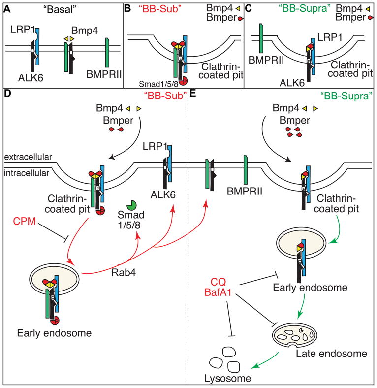
Rationale: Among the extracellular modulators of Bmp (bone morphogenetic protein) signaling, Bmper (Bmp endothelial cell precursor-derived regulator) both enhances and inhibits Bmp signaling. Recently we found that Bmper modulates Bmp4 activity via a concentration-dependent, endocytic trap-and-sink mechanism.

Objective: To investigate the molecular mechanisms required for endocytosis of the Bmper/Bmp4 and signaling complex and determine the mechanism of Bmper's differential effects on Bmp4 signaling.

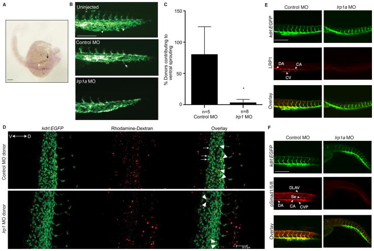
Methods and results: Using an array of biochemical and cell biology techniques, we report that LRP1 (LDL receptor-related protein 1), a member of the LDL receptor family, acts as an endocytic receptor for Bmper and a coreceptor of Bmp4 to mediate the endocytosis of the Bmper/Bmp4 signaling complex. Furthermore, we demonstrate that LRP1-dependent Bmper/Bmp4 endocytosis is essential for Bmp4 signaling, as evidenced by the phenotype of lrp1-deficient zebrafish, which have abnormal cardiovascular development and decreased Smad1/5/8 activity in key vasculogenic structures.

Conclusions: Together, these data reveal a novel role for LRP1 in the regulation of Bmp4 signaling by regulating receptor complex endocytosis. In addition, these data introduce LRP1 as a critical regulator of vascular development. These observations demonstrate Bmper's ability to fine-tune Bmp4 signaling at the single-cell level, unlike the spatial regulatory mechanisms applied by other Bmp modulators.


Figures

Figure 1
Figure 1. LRP1 associates with Bmper
(A) Lysates of Bmper-treated MECs were immunoprecipitated with an anti-Bmper antibody and stained with Coomassie blue to identify proteins. (B) Bmper-treated MECs (5 nmol/L for 1 minute) were stained with anti-Bmper and LRP1 antibodies and examined using confocal imaging. Scale bar: 10 μm. (C) Lysates of MECs were immunoprecipitated with anti-LRP1 antibody or mouse control IgG and analyzed by Western blotting with an biotinylated anti-Bmper antibody. (D) Non-denatured lysates of MECs were analyzed for ligand blotting. The membrane was incubated with recombinant Bmper and immunoblotted with anti-Bmper antibody (left panel). LRP1 and RAP1 interaction is included as a positive control. (E) Non-denatured lysates of HEK 293 cells transfected with mLRP1-4 were analyzed for ligand blotting with an anti-Bmper antibody (right top panel). Right bottom panel is included as a loading control. Left panel is a schematic representation of LRP1 and each mLRP1-4 construct.
Figure 2
Figure 2. LRP1 is required for Bmper endocytosis
(A) Time course of non-denatured cell lysates from Bmper-treated MECs (6 nmol/L) were analyzed by Western blotting. (B) Quantification of Bmper protein band intensity in A. *, compared to non-treated control sh-MECs, P<0.02, n=3; #, compared to LRP1-knockdown MECs treated similarly, P<0.05, n=3. (C) Non-denatured cell lysates from MECs treated with increasing doses of Bmper were analyzed by Western blotting. (D) Quantification of Bmper protein band intensity in C. *, compared to non-treated control sh-MECs, P<0.05; #, compared to LRP1-knockdown MECs treated similarly, P<0.02, n=3. (E) Bmper-treated MECs (15 minutes) were analyzed by confocal imaging for the co-localization of Bmper (green), LRP1 (red) and EEA1 (purple), indicated by the arrows. Scale bars: 5 μm.
Figure 3
Figure 3. The association of LRP1 and ALK6
(A) A schematic representation of wild type ALK6, its deletion mutants- ALK6-CTD and ALK6-PKD, and LRP1β constructs. (B) HEK 293 cells were transfected with CFP-ALK6 and YFP-mLRP2 and analyzed by FRET. The calibration bar indicates fluorescence signal level with white being highest intensity in normalized FRET channel (NFRET). Scale bar: 10 μm. (C) Lysates of HEK 293 cells transfected with plasmids encoding Flag-ALK6-WT and HA-mLRP1~4 were immunoprecipitated with an anti-Flag antibody and analyzed by Western blotting. * indicates precursor ‘ER’ and fully glycosylated ‘Golgi’ forms of mLRPs. (D) Lysates of HEK293 cells transfected with HA-ALK6-WT and Flag-LRP1β were immunoprecipitated with anti-HA or Flag antibody and analyzed by Western blotting. (E) Lysates of HEK 293 cells transfected with Flag-ALK6-CTD/PKD and HA-mLRP2 were immunoprecipitated with an anti-Flag antibody and analyzed by Western blotting.
Figure 4
Figure 4. LRP1-mediated endocytosis is required for the Bmper-dependent regulation of Bmp4 downstream signaling
(A) Endocytosis is required for both promoting- and inhibiting Bmp function of Bmper. MECs were pretreated with CPM or CQ and then treated with Bmp4 (0.6 nmol/L) and Bmper for 15 minutes (CPM) or 120 minutes (CQ) at 10 nmol/L (Bmper alone); 0.3 nmol/L (“BB-sub” = sub-stoichiometric Bmper); 10 nmol/L (“BB-supra” = Bmper suprastoichiometric). Cell lysates were analyzed by Western blotting. (B) Rab4 is required for Smad1/5/8 phosphorylation induced by Bmp4 and substoichiometric Bmper. Lysates of MECs transfected with control or Rab4 siRNA and treated with Bmp4 and Bmper (15 minutes) were analyzed by Western blotting. (C) LRP1 is required for Bmper/Bmp internalization and Bmper-dependent Bmp downstream signaling. Lysates of LRP1-knockdown or control MECs treated with Bmp4 and Bmper (30 minutes) were analyzed by Western blotting. (D) Lysates of MECs treated with Bmp4 and Bmper for 15 minutes were immunoprecipitated with an anti-BMPRII antibody and analyzed by Western blotting. (E) The association of LRP1 and ALK6 is regulated upon the different treatment of Bmp4 and Bmper. Lysates of MECs following Bmp4 and Bmper treatments for 15 minutes were immunoprecipitated with the mouse anti-LRP1β antibody and analyzed by Western blotting. (F) MECs were subjected to the in vitro Matrigel angiogenesis assay. *, compared to the same MECs at control condition, P<0.05. #, compared to the same MECs treated with Bmp4, P<0.01. n=3. Scale bar: 100 μm.
Figure 5
Figure 5. LRP1 is required for cardiovascular development in zebrafish
(A) RNA expression of lrp1a in a whole-mount zebrafish embryo, analyzed by in-situ hybridization using an lrp1a-specific antisense probe. The arrow represents the lateral dorsal aorta (LDA). (B) Loss of lrp1a results in a disrupted vascular phenotype. Images are lateral views of Tg(kdrl:EGFP)S843 zebrafish embryo tails at 24 hpf. The arrowheads represent the caudal vein plexus (CVP) with branches; the arrows represent filopodia located on the front edge of vessel plexus. (C) Quantitative analysis of the donor cells located at the tip cell region of cardinal vein plexus and contributing to ventral sprouting. *, compared to control MO-injected donor cells, P≤0.001. n is the number of recipient embryos. (D) Representative lateral views of wild-type recipient embryos of Tg(kdrl:EGFP)s843 fish tail at 34 hpf. Confocal imaging was performed. In the control MO-donor cells-transplanted embryo, three donor cells (white arrows) participated in active ventral sprouting. However, in the lrp1 MO-donor cells-transplanted embryo, donor cells remained in dorsal or axial vasculature (white arrowheads). (E, F) Confocal imaging analysis of whole mount Tg(kdrl:EGFP)s843 fish embryo tails at 48 hpf, with immunostaining for LRP1 (E) and phospho-Smad1/5/8 (F). Scale bars: 100 μm (A, B, E, F); 50 μm (D).
Figure 6
Figure 6. A schematic model shows how LRP1 is required for Bmp4/Bmper signaling
(A, left) In the absence of ligand, ALK6 and LRP1 are associated, blocking the assembly of an active BMPRII/ALK6 complex. In the presence of Bmp4 (A, middle), ALK6 dissociates from LRP1 and heterodimerizes with BMPRII. This Bmp4/BMPRII/ALK6 receptor complex is sequestered within endosomes where Bmp signaling occurs. (B) When the concentration of Bmper is substoichiometric, Bmper/LRP1 forms a transient holocomplex of Bmp/ALK6/BMPRII, which promotes Rab4-dependent endocytic fast recycling and enhances downstream Bmp signaling (D, red route). (C) When the concentration of Bmper is suprastoichiometric, the association of LRP1 with ALK6 increases, but that of ALK6 with BMPRII decreases. The LRP1-dependent endocytosis of a transient Bmper/Bmp/ALK6/LRP1 holocomplex leads to the degradation of the Bmper/Bmp signaling complex and termination of Bmp signaling activity (E, green route).

References

    1. Lowery JW, de Caestecker MP. BMP signaling in vascular development and disease. Cytokine Growth Factor Rev. 2010;21:287–298. - PMC - PubMed
    1. Umulis D, O’Connor MB, Blair SS. The extracellular regulation of bone morphogenetic protein signaling. Development. 2009;136:3715–3728. - PMC - PubMed
    1. Heinke J, Wehofsits L, Zhou Q, Zoeller C, Baar KM, Helbing T, Laib A, Augustin H, Bode C, Patterson C, Moser M. BMPER is an endothelial cell regulator and controls bone morphogenetic protein-4-dependent angiogenesis. Circ Res. 2008;103:804–812. - PubMed
    1. Moser M, Binder O, Wu Y, Aitsebaomo J, Ren R, Bode C, Bautch VL, Conlon FL, Patterson C. BMPER, a novel endothelial cell precursor-derived protein, antagonizes bone morphogenetic protein signaling and endothelial cell differentiation. Mol Cell Biol. 2003;23:5664–5679. - PMC - PubMed
    1. Moser M, Yu Q, Bode C, Xiong JW, Patterson C. BMPER is a conserved regulator of hematopoietic and vascular development in zebrafish. J Mol Cell Cardiol. 2007;43:243–253. - PMC - PubMed

Publication types

MeSH terms

Substances