. 2012 Sep;32(17):3594-607.
doi: 10.1128/MCB.00672-12. Epub 2012 Jul 9.

Multiple roles for the Ess1 prolyl isomerase in the RNA polymerase II transcription cycle

Affiliations

Multiple roles for the Ess1 prolyl isomerase in the RNA polymerase II transcription cycle

Zhuo Ma et al. Mol Cell Biol. 2012 Sep.

Abstract

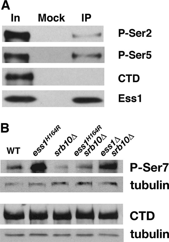
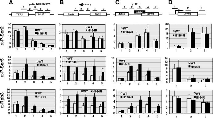
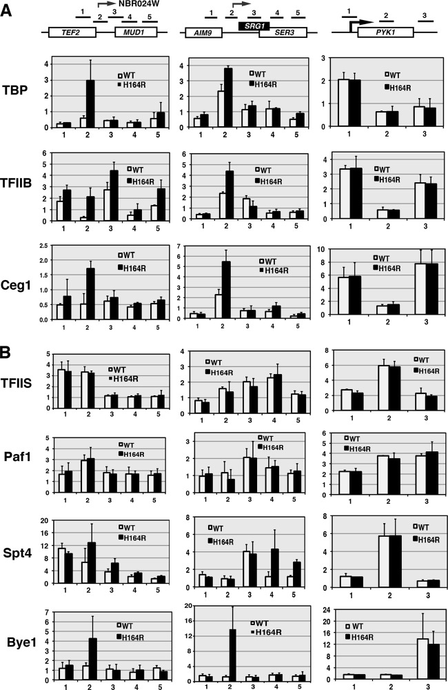
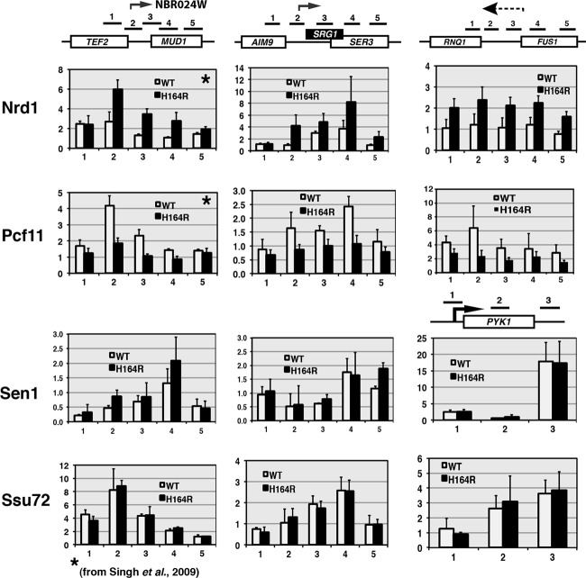
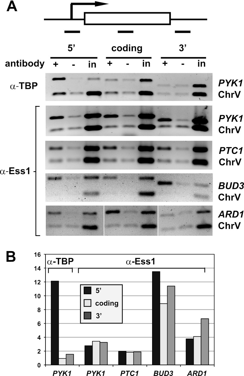
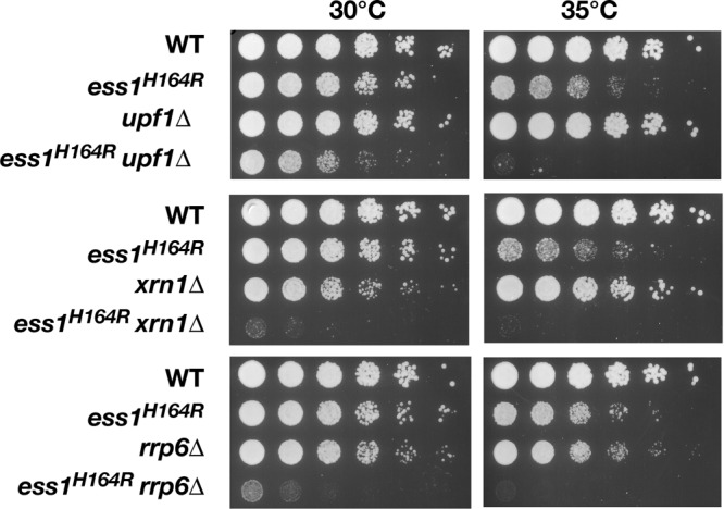
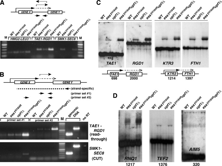
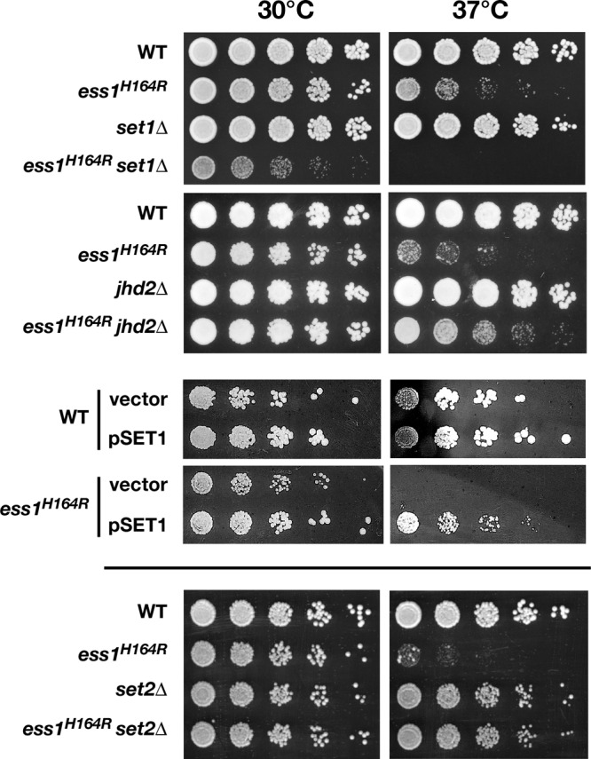
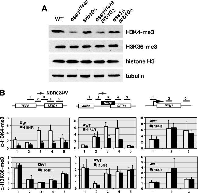
The Ess1 prolyl isomerase in Saccharomyces cerevisiae regulates RNA polymerase II (pol II) by isomerizing peptide bonds within the pol II carboxy-terminal domain (CTD) heptapeptide repeat (YSPTSPS). Ess1 preferentially targets the Ser5-Pro6 bond when Ser5 is phosphorylated. Conformational changes in the CTD induced by Ess1 control the recruitment of essential cofactors to the pol II complex and may facilitate the ordered transition between initiation, elongation, termination, and RNA processing. Here, we show that Ess1 associates with the phospho-Ser5 form of polymerase in vivo, is present along the entire length of coding genes, and is critical for regulating the phosphorylation of Ser7 within the CTD. In addition, Ess1 represses the initiation of cryptic unstable transcripts (CUTs) and is required for efficient termination of mRNA transcription. Analysis using strains lacking nonsense-mediated decay suggests that as many as half of all yeast genes depend on Ess1 for efficient termination. Finally, we show that Ess1 is required for trimethylation of histone H3 lysine 4 (H3K4). Thus, Ess1 has direct effects on RNA polymerase transcription by controlling cofactor binding via conformationally induced changes in the CTD and indirect effects by influencing chromatin modification.


Figures

Fig 1
Fig 1
Ess1 preferentially associates with pSer5 CTD in vivo and promotes dephosphorylation of pSer7 CTD. (A) Wild-type (WT) cells were subjected to immunoprecipitation (IP) with anti-Ess1 polyclonal antibodies, followed by Western analysis with monoclonal antibodies H5 (P-Ser2), H14 (P-Ser5), and 8WG16 (hypophosphorylated CTD). Sample inputs (In) are indicated. Note that the H14 antibody actually recognizes doubly phosphorylated pSer2/pSer5 (11). (B) Western analysis of protein (10 μg) from whole-cell extracts of the indicated strains grown at 30°C. The 4E12 monoclonal antibody was used to detect levels of pSer7 CTD, and the 8WG16 monoclonal antibody (hypophosphorylated CTD) as a control for overall levels of RNA pol II CTD. Antitubulin antibodies were used for detection of the loading control. Note that ESS1 is an essential gene but can be suppressed by deletion of SRB10 (62).
Fig 2
Fig 2
Ess1 mutants show increased recruitment to ncRNA loci of the initiation form (pSer5) of RNA pol II. Wild-type and ess1H164R mutant cells were grown to mid-logarithmic phase at 30°C. Chromatin immunoprecipitation was used to monitor recruitment of total RNA pol II complex (α-Rpb3) and the pSer2 or pSer5 forms of Rpb1. (A to C) Results of ChIP across three intergenic regions containing known CUTs (NBR024W and SRG1) or a suspected CUT (between RNQ1 and FUS1). Increased recruitment of the pSer5 but not the pSer2 form of pol II is detected in all three. (D) Results of control ChIP across the PYK1-coding gene locus. No increase in recruitment of either form of pol II is detected. Fold changes are relative to the results for a chromosome V control. Numbered horizontal bars represent approximate locations of qRT-PCR products (also in Fig. 3, 4, and 9). Error bars show standard deviations of three biological replicates.
Fig 3
Fig 3
Ess1 mutants show increased recruitment of the initiation factors but not general elongation factors to ncRNA loci. (A) Results of ChIP to detect recruitment of Ceg1 (capping enzyme), and initiation factors TBP and TFIIB to ncRNA loci in ess1H164R mutant cells. Increases are detected across intergenic regions of TEF2-MUD1 and SRG1 but not on the coding gene PYK1. (B) ChIP to monitor recruitment of elongation factors. No significant changes are detected in ess1H164R cells for TFIIS and Paf1. Spt4 shows a modest increase on SRG1. A larger increase in recruitment of the ess1-specific suppressor Bye1 (bypass of Ess1) is detected on ncRNA loci in ess1H164R cells.
Fig 4
Fig 4
Recruitment of some (Nrd1 and Pcf11) but not other termination factors (Sen1 and Ssu72) is altered in ess1 mutants. ChIP analysis of four termination factors was performed. No significant effects were detected on recruitment of termination factors (Sen1 and Ssu72) to the intergenic region of TEF2-MUD1 or to the SRG1 ncRNA gene or the PYK1 control gene. An increase in Nrd1 and a decrease in Pcf11 are observed at the 3′ regions of TEF2, SRG1, and RNQ1, similar to previous results for snoRNA loci (51). The inability to bind or release these factors in ess1 mutants may contribute to defective termination of these genes. Data in panels indicated by stars are reprinted from Molecular Cell (51) with permission of the publisher and are reproduced here for completeness.
Fig 5
Fig 5
Ess1 is present along transcribed protein-coding genes of all sizes. (A) Results of ChIP with antibodies to Ess1 to monitor the presence of Ess1 at 5′, coding, and 3′ regions of four protein-coding genes. No-antibody and input chromatin (in) controls are included. Shown are representative ethidium bromide-stained gels (image inverted) of PCR products for PYK1, PTC1, BUD3, and ARD1 genes, as well as an untranscribed region of chromosome V (ChrV). Results of a ChIP for TBP are also included. (B) Quantitation of the results in panel A, with ChIP signals normalized to input and ChrV signals [(IP/input)/(IP/inputChrV)]. Similar results were obtained when ChIP signals were normalized to those of no-antibody controls (not shown).
Fig 6
Fig 6
Ess1 mutants are synthetic lethal with NMD and RNA decay mutants. Serial dilutions (1:5, starting with cells at an OD600 of 0.5) of the wild type or the indicated mutant cells were grown at 30°C or 35°C on YEPD for 3 days. At both permissive (30°C) and semipermissive (35°C) temperatures, the growth defect of the ess1H164R mutant is strongly enhanced by upf1 (NMD pathway), xrn1 (cytoplasmic nuclease), and rrp6 (nuclear exonuclease) mutations. At 35°C, growth of the double mutants was negligible. Single upf1 mutants have no discernible growth defects, while xrn1 and rrp6 mutants have only minor growth defects at 30°C or 35°C, respectively.
Fig 7
Fig 7
Strong mRNA termination defects were detected in ess1 mutants using cells that lack nonsense-mediated decay. (A) Intergenic transcription is detected in ess1 mutants. Quantitative reverse transcription–RT-PCR was used to detect intergenic transcription. Results are shown for three representative loci, HMG2-LEU3, TAE1-RGD1, and SMK1-SEC8. Total RNA from the indicated strains was reverse transcribed using random hexamer primers, and the cDNA products were amplified by PCR for 30, 30, and 34 cycles, respectively, and visualized on an agarose gel. Intergenic primers were used for PCR as indicated in the schematic. (B) Ess1 mutants fail to terminate at mRNA genes, and the readthrough transcripts are degraded by NMD. In contrast to the experiment whose results are shown in panel A, gene-specific primers were used for first-strand cDNA synthesis (see schematic) to allow strand-specific detection of intergenic transcripts (primer set 1), as well as the longer fusion transcripts (primer set 2). The presence of long products at the TAE1-RGD1 locus in ess1H164R mutant cells indicates mRNA readthrough. Thirty cycles were used for PCR. Genomic DNA controls show the efficacy of the primers. Mixtures for control reactions without reverse transcription (no RT) contained 4× as much input template as the experimental samples. (C) Northern analysis of ess1 and ess1 upf1 double mutants and controls showing aberrantly long (readthrough) transcripts. Fifteen micrograms of total RNA was used. The 32P-labeled probes (specific activity, >5 × 10⁸/μg DNA) were generated from the open reading frames of the genes indicated. The numbers refer to the sizes of the open reading frames in base pairs. Both the upstream and downstream gene probes at each locus were used. (D) Additional loci are analyzed as described for panel C, except that only the upstream gene probe for each locus was used. Ten micrograms of total RNA was used for these samples.
Fig 8
Fig 8
Genetic interactions between ESS1 and SET1/JHD2 suggest that Ess1 is required for histone H3K4 methylation. Serial dilutions (1:5) starting at a 1:5 dilution (except for set1 mutant cells shown in the top panels, which started at 1:1) of wild-type or mutant cells at an OD600 of 0.5 were grown at 30°C or 37°C on YEPD for 2 to 3 days or, for the experiments using a plasmid overexpressing SET1 (pSET1), on CSM minus uracil (49) for 4 days. Deletion of the gene encoding the Set1 histone methyltransferase is synthetic lethal with ess1H164R, whereas deletion of JHD2, the gene encoding the H3K4 demethylase, rescues ess1H164R cells. The plasmid that overexpresses SET1 (pMP803-ADH/SET1) suppresses the growth defects of ess1H164R cells at permissive (30°C) and nonpermissive (37°C) temperatures, but the control vector (pRS416) does not. A set2Δ mutant rescues growth of ess1H164 cells at 37°C.
Fig 9
Fig 9
Levels of H3K4me3 are reduced in ess1 mutants. (A) Western analysis using protein from whole-cell extracts probed with the indicated antibodies (see Materials and Methods for details). Cells were grown at 30°C. Bulk levels of H3K4me3 are reduced in ess1 mutants. Levels of H3K36 methylation were unchanged, as were overall levels of histone H3. Tubulin was used as a loading control to assess protein integrity and concentration. Equal amounts of protein (10 μg) were loaded in all wells. Blots were generated individually and not reprobed. (B) ChIP analysis to monitor levels of H3K4me3 in ess1 mutants along individual gene loci TEF2-MUD1 and SRG1-SER3. Levels of H3K4me3 but not H3K36me3 are reduced across ncRNA loci. No effect was observed at PYK1. Fold changes are relative to the results for H3 after normalization to a chromosome V control (H3K36me3) or a tRNA gene control (H3K4me3). Error bars show standard deviations of three biological replicates.

Similar articles

Cited by

References

    1. Amrani N, et al. 2006. Aberrant termination triggers nonsense-mediated mRNA decay. Biochem. Soc. Trans. 34: 39–42 - PubMed
    1. Amrani N, et al. 1997. PCF11 encodes a third protein component of yeast cleavage and polyadenylation factor I. Mol. Cell. Biol. 17: 1102–1109 - PMC - PubMed
    1. Andrecka J, et al. 2008. Single-molecule tracking of mRNA exiting from RNA polymerase II. Proc. Natl. Acad. Sci. U. S. A. 105: 135–140 - PMC - PubMed
    1. Arevalo-Rodriguez M, Cardenas ME, Wu X, Hanes SD, Heitman J. 2000. Cyclophilin A and Ess1 interact with and regulate silencing by the Sin3-Rpd3 histone deacetylase. EMBO J. 19: 3739–3749 - PMC - PubMed
    1. Arevalo-Rodriguez M, Wu X, Hanes SD, Heitman J. 2004. Prolyl isomerases in yeast. Front. Biosci. 9: 2420–2446 - PubMed

Publication types

MeSH terms