. 2012 Sep;32(18):3707-17.
doi: 10.1128/MCB.00223-12. Epub 2012 Jul 9.

High-temperature requirement protein A4 (HtrA4) suppresses the fusogenic activity of syncytin-1 and promotes trophoblast invasion

Affiliations

High-temperature requirement protein A4 (HtrA4) suppresses the fusogenic activity of syncytin-1 and promotes trophoblast invasion

Liang-Jie Wang et al. Mol Cell Biol. 2012 Sep.

Abstract

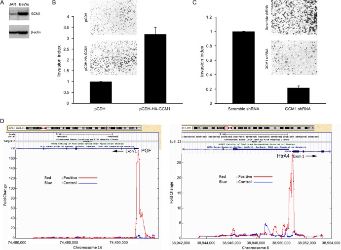
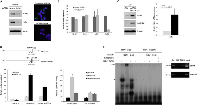
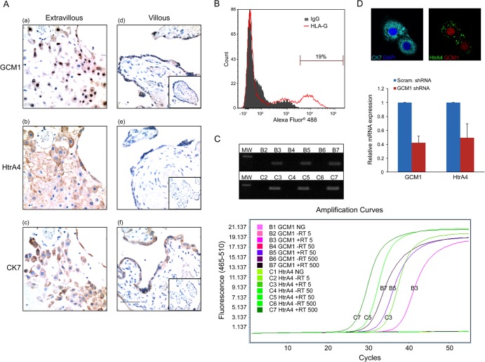
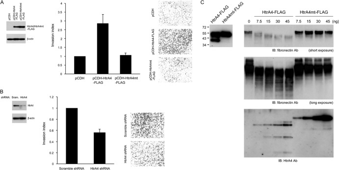
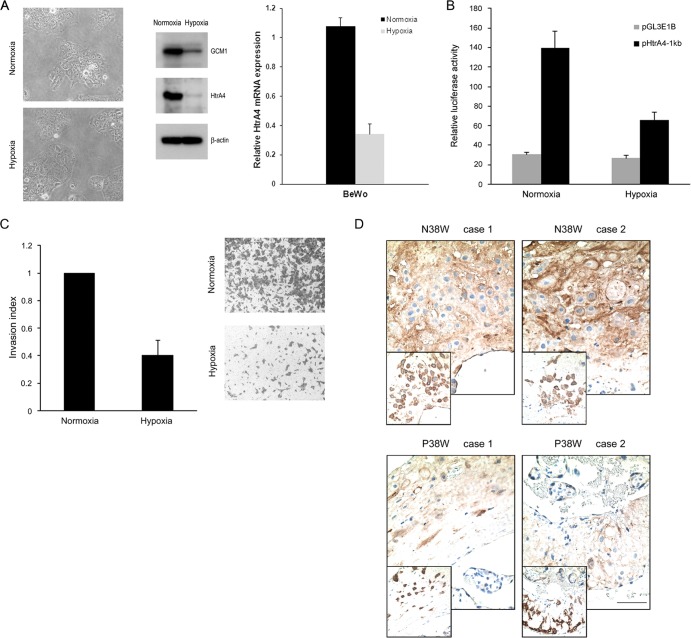
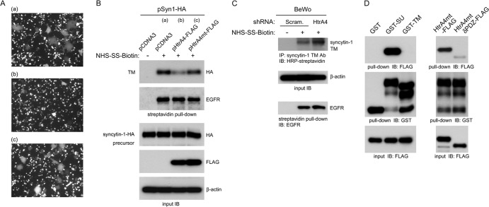
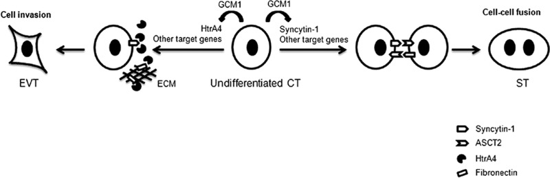
Cell-cell fusion and cell invasion are essential for placental development. Human cytotrophoblasts in the chorionic villi may undergo cell-cell fusion to form syncytiotrophoblasts to facilitate nutrient-gas exchange or differentiate into extravillous trophoblasts (EVTs) to facilitate maternal-fetal circulation. The placental transcription factor glial cells missing 1 (GCM1) regulates syncytin-1 and -2 expression to mediate trophoblast fusion. Interestingly, GCM1 and syncytin-1 are also expressed in EVTs with unknown physiological functions. In this study, we performed chromatin immunoprecipitation-on-chip (ChIP-chip) analysis and identified the gene for high-temperature requirement protein A4 (HtrA4) as a GCM1 target gene, which encodes a serine protease facilitating cleavage of fibronectin and invasion of placental cells. Importantly, HtrA4 is immunolocalized in EVTs at the maternal-fetal interface, and its expression is decreased by hypoxia and in preeclampsia, a pregnancy complication associated with placental hypoxia and shallow trophoblast invasion. We further demonstrate that HtrA4 interacts with syncytin-1 and suppresses cell-cell fusion. Therefore, HtrA4 may be crucial for EVT differentiation by playing a dual role in prevention of cell-cell fusion of EVTs and promotion of their invasion into the uterus. Our study reveals a novel function of GCM1 and HtrA4 in regulation of trophoblast invasion and that abnormal HrtA4 expression may contribute to shallow trophoblast invasion in preeclampsia.


Figures

Fig 1
Fig 1
GCM1 regulates placental cell invasion. (A) Expression of GCM1 in JAR and BeWo cells. JAR and BeWo cells were subjected to immunoblotting with GCM1 and β-actin Abs. (B) Stimulation of JAR cell invasion by GCM1. Mock (pCDH) and HA-GCM1-expressing (pCDH-HA-GCM1) JAR cells were plated in Matrigel-coated chambers for invasion analysis. Representative pictures of cells that migrated to and invaded the lower surface of filters are provided. Means and standard deviations (SD) obtained from three independent experiments are presented. (C) GCM1 knockdown suppresses BeWo cell invasion. BeWo cells stably expressing scrambled or GCM1 shRNA were subjected to invasion analysis as described for panel A. (D) Identification of GCM1 target genes. BeWo31 cells were treated with 50 μM forskolin for 24 h, followed by ChIP-chip analysis as described in Materials and Methods. HtrA4 was identified as a candidate GCM1 target gene with strong hybridization signals (red versus blue) in the region upstream of exon1 of the HtrA4 gene (right). PGF, encoded by a known GCM1 target gene, was also identified in the analysis (left).
Fig 2
Fig 2
Regulation of HtrA4 expression by GCM1. (A) GCM1 knockdown decreases HtrA4 expression. BeWo cells stably expressing scrambled or GCM1 shRNA were subjected to immunoblotting with GCM1, HtrA4, and β-actin Abs, respectively. In a separate experiment, cells were immunostained with HtrA4 Ab (green) and nuclei were stained by DAPI (blue), followed by confocal microscopy analysis. Note that HtrA4 signals in the cytoplasm were decreased in the GCM1 knockdown BeWo cells. (B) Expression of HtrA4, but not other HtrA family members, is decreased by GCM1 knockdown. BeWo cells stably expressing scrambled or GCM1 shRNA were harvested for quantitative PCR analysis of the transcript levels of HtrA family members. Means and SD obtained from three independent experiments are presented. (C) Overexpression of GCM1 stimulates HtrA4 expression. Mock and HA-GCM1-expressing JAR cells were harvested for immunoblotting and quantitative PCR analysis of HtrA4 protein and transcript levels, respectively. (D) Regulation of HtrA4 promoter activity by GCM1. Schematic representation of HtrA4 promoter region with the wild-type and mutant GCM1-binding site (GBS) is provided (top). 293T cells were transfected with pHtrA4-1kb or pHtrA4-1kbGBSmt with or without pHA-GCM1 expression plasmid (left). BeWo cells expressing scrambled or GCM1 shRNA were transfected with pHtrA4-1kb or pHtrA4-1kbGBSmt (right). At 48 h posttransfection, cells were harvested for luciferase assays. Means and SD obtained from three independent experiments are presented. (E) Interaction of GCM1 and the GBS in HtrA4 promoter. Recombinant GCM1-FLAG was incubated with radiolabeled HtrA4-GBS or HtrA4-GBSmt probes in the presence of GCM1 or syncytin-2 (Syn2) Ab in EMSA. The asterisk and arrow indicate the GCM1-FLAG-DNA complex and its supershifted complex, respectively. Association of GCM1 and HtrA4-GBS in BeWo cells was analyzed by ChIP using normal rabbit serum (NS) or GCM1 Ab for immunoprecipitation and designated primer pairs for PCR.
Fig 3
Fig 3
Coexpression of GCM1 and HtrA4 in the interstitial EVTs of human placenta. (A) Immunostaining of GCM1 and HtrA4 in term placenta. Term placental tissues sections were subjected to immunohistochemistry using GCM1 (a and d), HtrA4 (b and e), and CK7 (c and f) Abs and further counterstained with hematoxylin. Note that expression of GCM1 corresponded with that of HtrA4 in the CK7-positive EVTs in the consecutive sections of basal plate (a to c). The insets in panels d to f show sections immunostained with normal rabbit, guinea pig, and mouse serum, respectively. Bar, 100 μm. (B) Purification of EVTs. Primary trophoblast cells prepared from term placenta were subjected to flow cytometry analysis using HLA-G Ab as described in Materials and Methods. (C) Expression of HtrA4 and GCM1 in EVTs. HLA-G-positive EVTs were subjected to single cell RT-PCR as described in Materials and Methods. Amplification curves of real-time PCRs of increasing input cell numbers are presented. Of note, neither GCM1 nor HtrA4 transcript was detected in the samples without reverse transcriptase (RT) or in the mock reaction mixtures without input sample (NG). The PCR products were analyzed by agarose gel electrophoresis (top). MW, molecular weight marker. (D) Colocalization of GCM1 and HtrA4 in EVTs. HLA-G-positive EVTs were immunostained with GCM1, HtrA4, and CK7 Abs, followed by confocal microscopy analysis. In a separate experiment, cells were transduced with lentivirus harboring scrambled or GCM1 shRNA and subsequently harvested for quantitative PCR analysis of the HtrA4 and GCM1 transcripts. Means and SD from three independent experiments are presented.
Fig 4
Fig 4
HtrA4 regulates placental cell invasion. (A) Overexpression of HtrA4 stimulates placental cell invasion. Mock and HtrA4-HA-expressing JAR cells were plated in Matrigel-coated chambers for invasion analysis. Expression of HtrA4-HA was analyzed by immunoblotting with HA MAb (left). (B) HtrA4 knockdown suppresses placental cell invasion. BeWo cells stably expressing scrambled or HtrA4 shRNA were subjected to invasion analysis as described for panel A. Knockdown of HtrA4 expression in BeWo cells was analyzed by immunoblotting with HtrA4 Ab (left). (C) Proteolytic cleavage of fibronectin by HtrA4. Purified HtrA4-FLAG and HtrA4mt-FLAG proteins from culture medium of 293T cells transfected with pHtrA4-FLAG and pHtrA4mt-FLAG, respectively, were analyzed by immunoblotting with FLAG MAb (left). Fibronectin was incubated with increasing amounts of HtrA4-FLAG or HtrA4mt-FLAG at 37°C for 16 h. The reaction mixture was then subjected to immunoblotting with HtrA4 or fibronectin Ab. Of note, partial cleavage of HtrA4-FLAG, but not HtrA4mt-FLAG, in the reaction was detected (bottom).
Fig 5
Fig 5
HtrA4 expression under hypoxia and in preeclampsia. (A) Downregulation of HtrA4 expression by hypoxia. BeWo cells were incubated under normoxic or hypoxic conditions. After 72 h of incubation, cells were analyzed by immunoblotting with GCM1, HtrA4, and β-actin Abs, respectively. Phase-contrast images for the morphology of normoxic and hypoxic cells were provided (left). In a separate experiment, cells were subjected to quantitative PCR analysis of the HtrA4 transcript level. (B) Suppression of HtrA4 promoter activity by hypoxia. BeWo cells were transfected with pHtrA4-1kb and incubated under normoxic and hypoxic conditions and subsequently harvested for luciferase assays. (C) Suppression of placental cell invasion by hypoxia. BeWo cells plated in Matrigel-coated chambers were incubated under normoxic or hypoxic condition for invasion analysis. Means and SD from three independent experiments are presented in panels A to C. (D) Decreased HtrA4 expression in preeclamptic placentas. Sections of two normal (N) and two preeclamptic (P) placentas at 38 weeks of gestation were immunostained with HtrA4 Ab. Note that the insets show consecutive sections immunostained with CK7 Ab. Bar, 100 μm.
Fig 6
Fig 6
Regulation of syncytin-1-mediated cell-cell fusion by HtrA4. (A) HtrA4 suppresses cell-cell fusion mediated by syncytin-1. 293T cells coexpressing empty vector and EGFP (a), HtrA4-FLAG and EGFP (b), or HtrA4mt-FLAG and EGFP (c) were cocultured with 293T cells expressing syncytin-1–HA for 24 h. Cell-cell fusion was examined by fluorescence microscopy. (B) HtrA4 decreases the protein level of surface syncytin-1. A separate set of the cocultured 293T cells described for panel A were subjected to biotinylation, followed by streptavidin pulldown and immunoblotting with HA or EGFR MAb. As a loading control, whole-cell lysates were subjected to immunoblotting with HA, FLAG, and β-actin MAbs, respectively. (C) HtrA4 regulates syncytin-1 expression in placental cells. BeWo cells stably expressing scrambled or HtrA4 shRNA were subjected to biotinylation, followed by immunoprecipitation with a syncytin-1 TM Ab and then immunoblotting with HRP-conjugated streptavidin. Note that the surface EGFR protein level was not affected by HtrA4. (D) Characterization of interaction between HtrA4 and syncytin-1. Purified HtrA4mt-FLAG was incubated with agarose matrix preloaded with GST, GST-SU, or GST-TM, followed by immunoblotting with FLAG MAb. On the other hand, HtrA4mt-FLAG or HtrA4mtδPDZ-FLAG was subjected to pulldown analysis with GST-SU demonstrating that the PDZ domain of HtrA4 is critical for recognition of the SU domain of syncytin-1.
Fig 7
Fig 7
Model of regulation of EVT differentiation by GCM1. The mononuclear cytotrophoblasts (CT) in chorionic villi may proliferate and differentiate into a multinucleated syncytiotrophoblast (ST) layer or invasive extravillous trophoblasts (EVT). GCM1 can regulate ST differentiation through transactivation of syncytin-1, which interacts with its cognate receptor, the sodium-dependent neutral amino acid transporter type 2 (ASCT2), to facilitate cell-cell fusion. In addition, GCM1 can participate in EVT differentiation through transactivation of HtrA4, which may cleave fibronectin in the extracellular matrix (ECM) and facilitate cell migration and invasion. Importantly, HtrA4 may also cleave syncytin-1 to prevent cell-cell fusion of EVTs.

References

    1. Ajayi F, et al. 2008. Elevated expression of serine protease HtrA1 in preeclampsia and its role in trophoblast cell migration and invasion. Am. J. Obstet. Gynecol. 199:557.e1–557.e10 - PubMed
    1. Anson-Cartwright L, et al. 2000. The glial cells missing-1 protein is essential for branching morphogenesis in the chorioallantoic placenta. Nat. Genet. 25:311–314 - PubMed
    1. Baczyk D, et al. 2004. Complex patterns of GCM1 mRNA and protein in villous and extravillous trophoblast cells of the human placenta. Placenta 25:553–559 - PubMed
    1. Blaise S, de Parseval N, Benit L, Heidmann T. 2003. Genomewide screening for fusogenic human endogenous retrovirus envelopes identifies syncytin 2, a gene conserved on primate evolution. Proc. Natl. Acad. Sci. U. S. A. 100:13013–13018 - PMC - PubMed
    1. Blond JL, et al. 2000. An envelope glycoprotein of the human endogenous retrovirus HERV-W is expressed in the human placenta and fuses cells expressing the type D mammalian retrovirus receptor. J. Virol. 74:3321–3329 - PMC - PubMed

Publication types

MeSH terms