

Case Reports
doi: 10.1136/bcr.12.2009.2545.
A happy ending in hepatomegaly
Affiliations
- PMID: 22778246
- PMCID: PMC3027810
- DOI: 10.1136/bcr.12.2009.2545
Item in Clipboard
Case Reports
A happy ending in hepatomegaly
BMJ Case Rep.
.
No abstract available
Conflict of interest statement
Figures
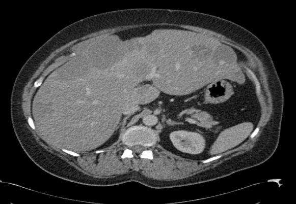
Portal phase CT post contrast showing multiple liver lesions with peripheral enhancement. The largest measured 10×8 cm.
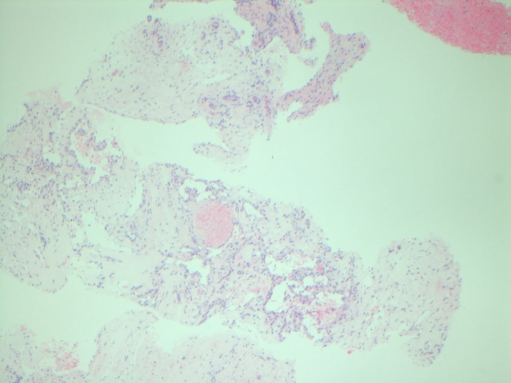
Low power image of the liver biopsy showing dilated thin-walled blood vessels and bile duct-like structures within fibrous stroma.
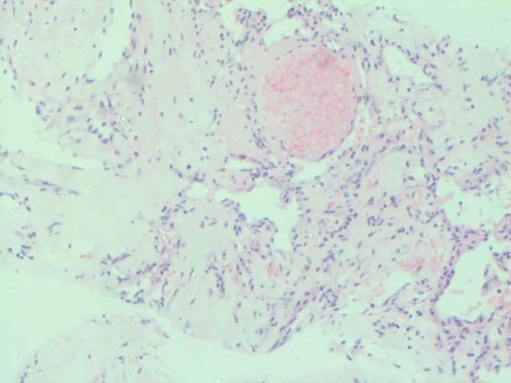
High power image of the liver biopsy showing dilated thin-walled blood vessels and bile duct-like structures within fibrous stroma. Cores of fibrous tissue can be seen containing ducts with bland cyto-architectural morphology resembling bile ducts within expanded portal tracts.
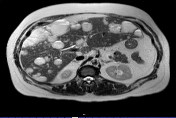
Coronal T2-weighted MRI showing multiple high signal liver lesions. The signal remained high on long echo time (TE) weighted images. Septae and fluid levels were noted but no significant fat content was seen.
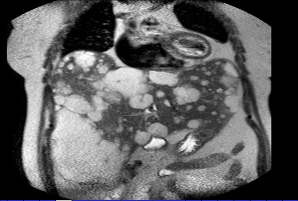
Axial T2-weighted MRI showing multiple high signal liver lesions.
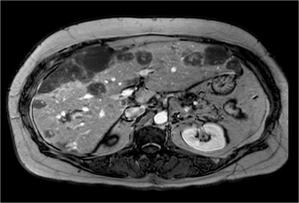
Axial T1-weighted MRI post-contrast (arterial phase) showing enhancement of the lesions. During the dynamic enhancement series several of the lesions demonstrate centripetal enhancement, which increased on delayed phase imaging.
Similar articles
-
Obscure hepatomegaly in clinical practice--a clinicopathological study.J Pak Med Assoc. 1980 Apr;30(4):84-7. J Pak Med Assoc. 1980. PMID: 6771429 No abstract available.
-
[Hepatomegaly in a 62-year-old woman].Rev Med Interne. 2012 Dec;33(12):713-7. doi: 10.1016/j.revmed.2012.07.003. Epub 2012 Sep 10. Rev Med Interne. 2012. PMID: 22974483 French. No abstract available.
-
Position of the duodenal bulb and liver size.Am J Roentgenol Radium Ther Nucl Med. 1973 Sep;119(1):78-84. doi: 10.2214/ajr.119.1.78. Am J Roentgenol Radium Ther Nucl Med. 1973. PMID: 4744732 No abstract available.
-
Hepatomegaly.Med Clin North Am. 1975 Jan;59(1):145-67. doi: 10.1016/s0025-7125(16)32059-4. Med Clin North Am. 1975. PMID: 162803 Review. No abstract available.
-
Accessory liver lobes: anatomical description and clinical implications.J Visc Surg. 2014 Dec;151(6):451-5. doi: 10.1016/j.jviscsurg.2014.09.013. Epub 2014 Oct 28. J Visc Surg. 2014. PMID: 25448768 Review.
References
-
- De Franco A, Monteforte MG, Maresca G, et al. [Integrated diagnosis of liver angioma: comparison of Doppler colour ultrasonography, computerized tomography, and magnetic resonance]. Radiol Med 1997;93:87–94 - PubMed
Publication types
MeSH terms
LinkOut - more resources
Full Text Sources