. 2012 Aug 17;287(34):28503-7.
doi: 10.1074/jbc.C112.387514. Epub 2012 Jul 9.

The autophagy-related protein kinase Atg1 interacts with the ubiquitin-like protein Atg8 via the Atg8 family interacting motif to facilitate autophagosome formation

Affiliations

The autophagy-related protein kinase Atg1 interacts with the ubiquitin-like protein Atg8 via the Atg8 family interacting motif to facilitate autophagosome formation

Hitoshi Nakatogawa et al. J Biol Chem. .

Abstract

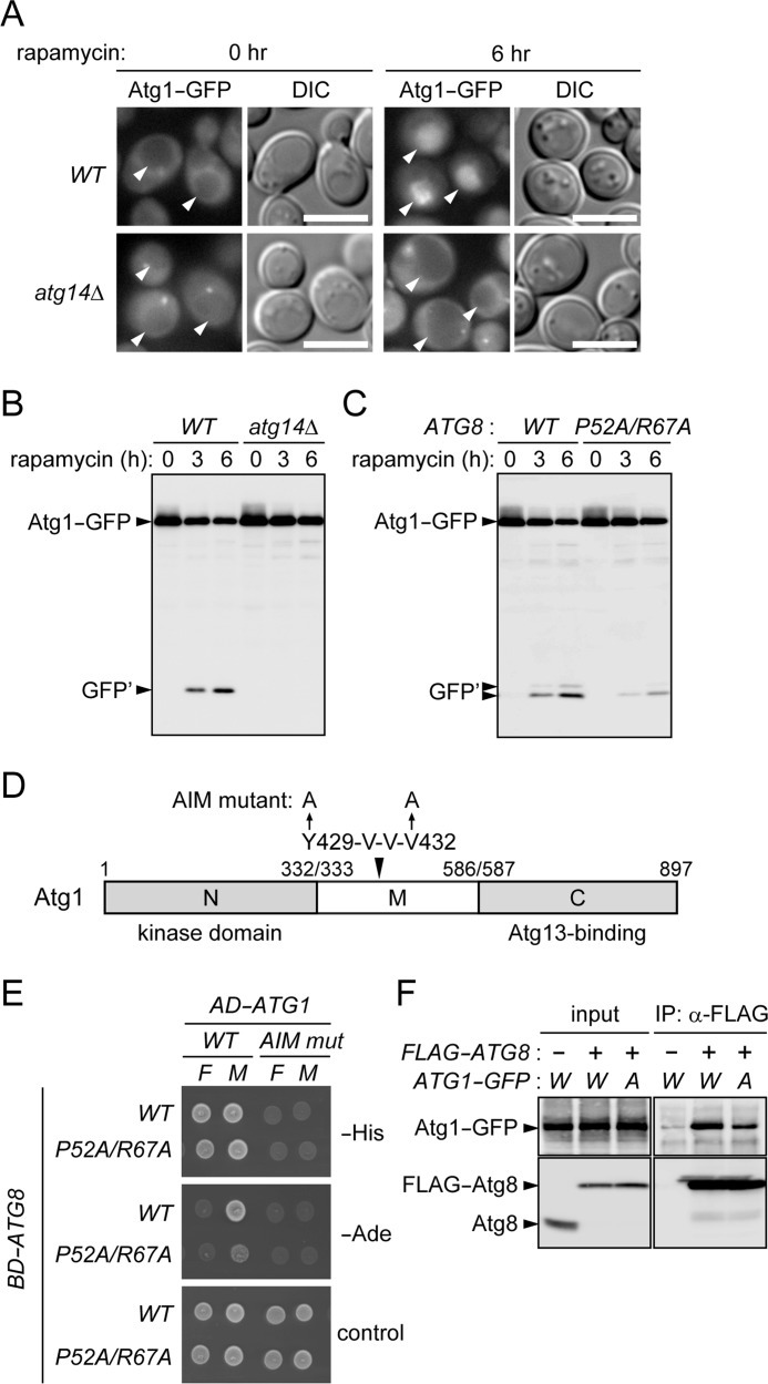
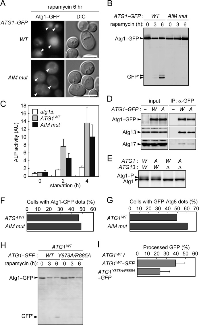
In autophagy, a cup-shaped membrane called the isolation membrane is formed, expanded, and sealed to complete a double membrane-bound vesicle called the autophagosome that encapsulates cellular constituents to be transported to and degraded in the lysosome/vacuole. The formation of the autophagosome requires autophagy-related (Atg) proteins. Atg8 is a ubiquitin-like protein that localizes to the isolation membrane; a subpopulation of this protein remains inside the autophagosome and is transported to the lysosome/vacuole. In the budding yeast Saccharomyces cerevisiae, Atg1 is a serine/threonine kinase that functions in the initial step of autophagosome formation and is also efficiently transported to the vacuole via autophagy. Here, we explore the mechanism and significance of this autophagic transport of Atg1. In selective types of autophagy, receptor proteins recognize degradation targets and also interact with Atg8, via the Atg8 family interacting motif (AIM), to link the targets to the isolation membrane. We find that Atg1 contains an AIM and directly interacts with Atg8. Mutations in the AIM disrupt this interaction and abolish vacuolar transport of Atg1. These results suggest that Atg1 associates with the isolation membrane by binding to Atg8, resulting in its incorporation into the autophagosome. We also show that mutations in the Atg1 AIM cause a significant defect in autophagy, without affecting the functions of Atg1 implicated in triggering autophagosome formation. We propose that in addition to its essential function in the initial stage, Atg1 also associates with the isolation membrane to promote its maturation into the autophagosome.


Figures

FIGURE 1.
FIGURE 1.
Atg1 interacts with Atg8 via the AIM. A, wild-type (YNH512) and atg14Δ (YNH731) cells expressing Atg1-GFP were grown to mid-log phase, treated with rapamycin for 6 h, and observed under a fluorescence microscope. Arrowheads indicate vacuoles. Images obtained using differential interference contrast (DIC) optics are also shown, with scale bars representing 5 μm. B, the same strains as in A were treated with rapamycin for the indicated time periods and subjected to immunoblotting analysis using monoclonal antibodies against GFP. GFP′ represents GFP-containing fragments generated by proteolysis of Atg1-GFP in the vacuole. C, ATG8WT (YNH695) and ATG8P52A/R67A (YNH696) cells expressing Atg1-GFP were analyzed as described in B. D, schematic diagram of Atg1. Atg1 consists of three regions: the N-terminal kinase domain (N), the middle region (M), and the C-terminal Atg13-binding region (C). The AIM mutant (AIM mut) of Atg1 contains two alanine substitutions at Tyr429 and Val432. E, the indicator strain AH109 was transformed with plasmids expressing a transcription activation domain (AD) fused with full-length Atg1 (F) or the middle region of Atg1 (M), with or without mutations at the AIM (AIM mut), in combination with plasmids expressing a DNA-binding domain (BD) fused with either Atg8WT or the Atg8P52A/R67A mutant. These strains were grown on SC-LTH (−His), SC-LTA (−Ade), and SC-LT (control) agar plates. F, ATG8 (YNH740) and 3×FLAG-ATG8 (YNH741) cells expressing Atg1WT-GFP or Atg1AIM mut-GFP from single-copy plasmids were treated with rapamycin for 2 h and disrupted to prepare cell lysates (input) for immunoprecipitation (IP) using a monoclonal antibody against the FLAG sequence. The immunoprecipitates were analyzed by immunoblotting with antibodies against GFP and Atg8.
FIGURE 2.
FIGURE 2.
Characterization of an AIM mutant of Atg1. A and B, atg1Δ cells (YNH204) expressing Atg1WT-GFP or Atg1AIM mut-GFP from single-copy plasmids were treated with rapamycin and analyzed by fluorescence microscopy (A) or immunoblotting with anti-GFP antibodies (B). Scale bars represent 5 μm. GFP′ represents GFP-containing fragments generated by proteolysis of Atg1-GFP in the vacuole. AIM mut, AIM mutant. C, ATG1WT (YNH738), ATG1AIM mut (YNH739), and atg1Δ (YNH621) cells expressing a mutant form of ALP were incubated in nitrogen-deprived medium and subjected to the ALP assay. The same experiments were repeated three times; data are represented as means with standard deviations. AU, arbitrary units. D, YNH740 cells that carried either plasmids expressing Atg1WT-GFP (W) or Atg1AIM mut-GFP (A) or empty vector (−) were treated with rapamycin for 1 h and subjected to coimmunoprecipitation (IP) analysis using anti-GFP antibodies. The immunoprecipitates were analyzed by immunoblotting using antibodies against Atg1, Atg13, and Atg17. E, ATG1WT (YNH736), ATG1AIM mut (YNH737), ATG1WT atg13Δ (YNH746), and ATG1AIM mut atg13Δ (YNH747) cells were treated with rapamycin for 1 h and examined by immunoblotting using anti-Atg1 antibodies. F, ATG1WT-GFP (YNH742) and ATG1AIM mut-GFP (YNH743) cells treated with rapamycin for 1 h were observed under a fluorescence microscope (the numbers of cells examined were 272 and 221, respectively); the percentages of cells with GFP dots are shown. G, dot formation of GFP-Atg8 in ATG1WT (YNH744) and ATG1AIM mut (YNH745) cells was examined as described in F (the numbers of cells examined were 307 and 246, respectively). H, the vacuolar transport of Atg1WT-GFP and Atg1Y878A/R885A-GFP expressed from single-copy plasmids in wild-type cells (BY4741) was examined by immunoblotting analysis as described in legend for Fig. 1. I, the same experiments as in H were repeated three times, and the intensities of the bands of Atg1-GFP and GFP′ at the time point of 6 h were measured to estimate the efficiency of the production of processed GFP fragments (the intensities of GFP′ were divided by the sum of those of Atg1-GFP and GFP′); results are represented as means with standard deviations.

Similar articles

Cited by

References

    1. Nakatogawa H., Suzuki K., Kamada Y., Ohsumi Y. (2009) Dynamics and diversity in autophagy mechanisms: lessons from yeast. Nat. Rev. Mol. Cell Biol. 10, 458–467 - PubMed
    1. Weidberg H., Shvets E., Elazar Z. (2011) Biogenesis and cargo selectivity of autophagosomes. Annu. Rev. Biochem. 80, 125–156 - PubMed
    1. Johansen T., Lamark T. (2011) Selective autophagy mediated by autophagic adapter proteins. Autophagy 7, 279–296 - PMC - PubMed
    1. Ichimura Y., Kirisako T., Takao T., Satomi Y., Shimonishi Y., Ishihara N., Mizushima N., Tanida I., Kominami E., Ohsumi M., Noda T., Ohsumi Y. (2000) A ubiquitin-like system mediates protein lipidation. Nature 408, 488–492 - PubMed
    1. Kirisako T., Baba M., Ishihara N., Miyazawa K., Ohsumi M., Yoshimori T., Noda T., Ohsumi Y. (1999) Formation process of autophagosome is traced with Apg8/Aut7p in yeast. J. Cell Biol. 147, 435–446 - PMC - PubMed

Publication types

MeSH terms

Substances