. 2012 Aug 24;287(35):29429-41.
doi: 10.1074/jbc.M112.366807. Epub 2012 Jul 9.

The ubiquitin-specific protease 12 (USP12) is a negative regulator of notch signaling acting on notch receptor trafficking toward degradation

Affiliations

The ubiquitin-specific protease 12 (USP12) is a negative regulator of notch signaling acting on notch receptor trafficking toward degradation

Julien Moretti et al. J Biol Chem. .

Abstract

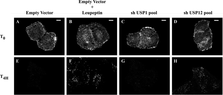
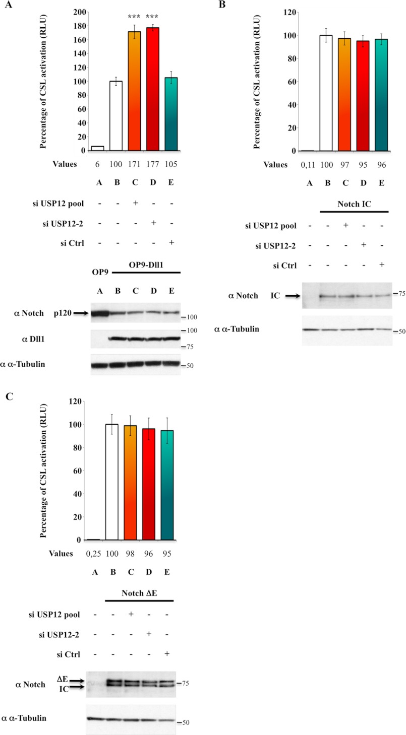
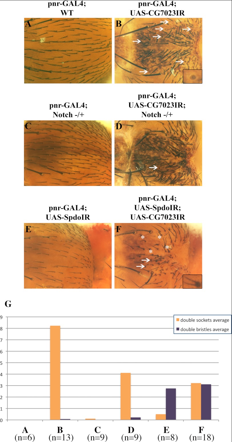
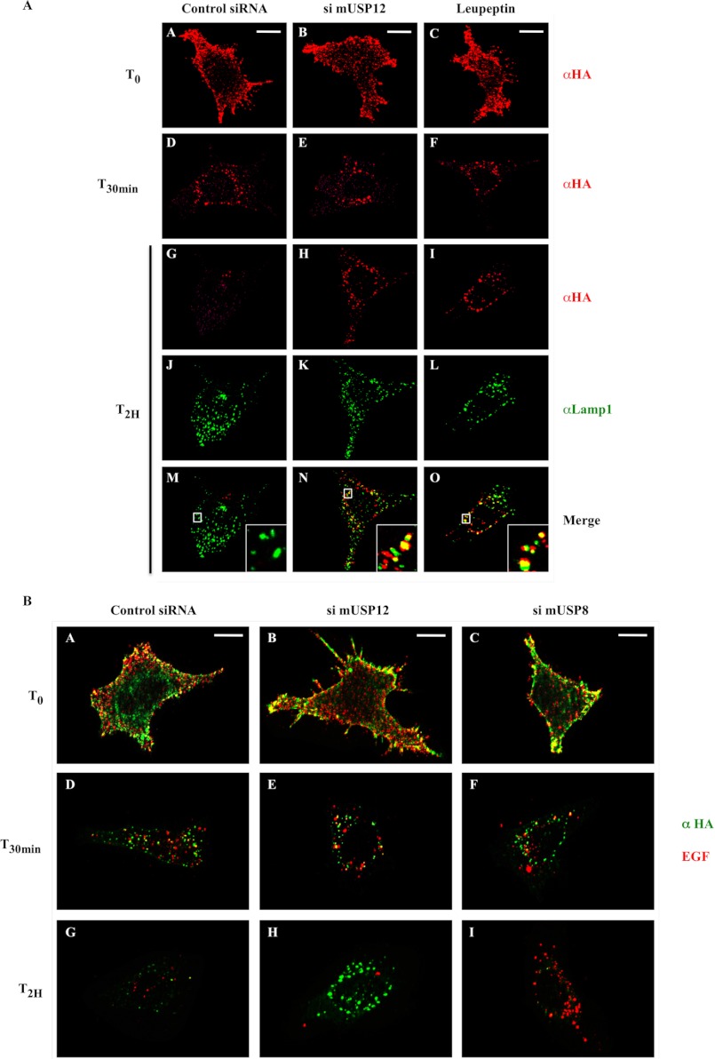
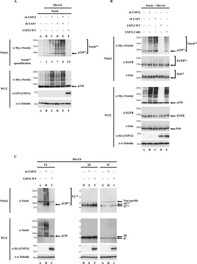
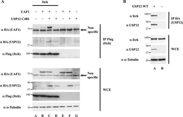
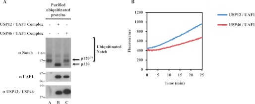
Notch signaling is critical for development and adult tissue physiology, controlling cell fate in a context-dependent manner. Upon ligand binding, the transmembrane Notch receptor undergoes two ordered proteolytic cleavages releasing Notch intracellular domain, which regulates the transcription of Notch target genes. The strength of Notch signaling is of crucial importance and depends notably on the quantity of Notch receptor at the cell surface. Using an shRNA library screen monitoring Notch trafficking and degradation in the absence of ligand, we identified mammalian USP12 and its Drosophila melanogaster homolog as novel negative regulators of Notch signaling. USP12 silencing specifically interrupts Notch trafficking to the lysosomes and, as a consequence, leads to an increased amount of receptor at the cell surface and to a higher Notch activity. At the biochemical level, USP12 with its activator UAF1 deubiquitinate the nonactivated form of Notch in cell culture and in vitro. These results characterize a new level of conserved regulation of Notch signaling by the ubiquitin system.


Figures

FIGURE 1.
FIGURE 1.
Identification of USP12. U2OS-FL cells stably expressing HA-tagged Notch were transfected with empty vector (A, B, E, F) or each one of the 91 pools of shRNAs targeting the human DUBs (USP1 in C and G as a representative example; USP12 in D and H). Living cells were labeled with an anti-HA antibody coupled to Alexa Fluor 594 (shown in white), washed, fixed directly (A–D, T0), or incubated during 4 h at 37 °C before fixation and immunofluorescence analysis (E–H, T4H). When indicated (B and F), cells were treated with 100 μm leupeptin 1 h before adding the antibody and until fixation. Scale bar: 10 μm.
FIGURE 2.
FIGURE 2.
USP12 is a negative regulator of Notch signaling in mammals. A, USP12 silencing increases Notch reporter activity in Notch-expressing cells cocultured with Dll1-expressing cells. U2OS-FL cells were first transfected with siRNA control (si Ctrl) or targeting USP12 (siUSP12 pool or siUSP12-2, see supplemental Figs. S1A and S2A) and then with both CSL-firefly luciferase (Notch reporter) and TK-Renilla luciferase (internal control). The CSL-reporter activation corresponds to the ratio between firefly and Renilla luciferase activities. It was defined as 100% in the presence of Dll1-expressing cells (lane B), corresponding to a 17-fold increased as compared with coculture with control OP9 cells (lane A). B and C, USP12 silencing does not affect the activity of Notch-IC and Notch-ΔE. U2OS cells were first transfected with the same doses of siRNA control or targeting USP12 as above and then with Notch and control reporters together with Notch-IC (B) or Notch-ΔE (C). The relative luciferase activity in the absence of any siRNA (lane B) was defined as 100%. In all panels, the CSL-activation values are indicated for each sample under the graph. Error bars represent the S.E. of triplicate experiments, and the Western blot analysis of a representative experiment is shown using α-tubulin as a loading control. In panel A, p values calculated with Student's t test are indicated (***, p < 0.0001 as compared with siRNA control (lane E)). In panel C, Notch-IC originates from the constitutive γ-secretase cleavage of ΔE. The molecular masses (kDa) are also indicated on the right.
FIGURE 3.
FIGURE 3.
CG7023 is a negative regulator of Notch signaling in D. melanogaster. A–F, dorsal surface of the thorax of mutant flies, resulting from various crosses. A, pnr-GAL4 x w1118 (WT). B, pnr-GAL4 x UAS-CG7023IR. C and D, Notch−/+ females were crossed with UAS-CG7023IR, the Notch−/+ females of the progeny were then crossed with pnr-GAL4 males, and the resulting females harboring notched wings were observed. Depending on the eye color, they had the UAS-CG7023IR-containing chromosome (D) or not (C). E and F, pnr-GAL4; UAS-SpdoIR flies were crossed with UAS-CG7023IR (F) or w1118 (E) flies as a control. In A and C, all organs have normal shaft and socket, whereas in CG7023IR-expressing flies, there are double-sockets or double-bristles organs, marked by arrows and asterisks, respectively (examples are magnified 3-fold in the insets of B and F). Original magnification is ×150. G, as pnr-GAL4 is expressed in the central region of the notum, this region was observed on 6–18 flies for each cross, and the double-sockets and double-bristles organs were recorded (A–F designate the same fly genotypes as in the representative image of the corresponding panel, n = the number of flies). A schematic view of external sensory organ morphology and lineage is shown in supplemental Fig. S4.
FIGURE 4.
FIGURE 4.
USP12 silencing specifically increases Notch receptor quantity at the plasma membrane. MEF-FL cells, transfected with siRNA control or targeting USP12 (si mUSP12) as indicated, were analyzed by flow cytometry to quantify HA-Notch and EGFR fluorescence. For each labeling, the mean relative fluorescence was defined as compared with 100% in the absence of any siRNA (A, lanes A and B, respectively, for HA-Notch and EGFR), and their values are indicated. Error bars are standard deviations. The bar graphs are quantifications of the indicated populations shown in the corresponding plots and histograms of B.
FIGURE 5.
FIGURE 5.
Specific regulation of Notch trafficking by USP12. A, USP12 silencing interrupts Notch trafficking in late endosomes/lysosomes. MEF-FL cells stably expressing HA-tagged Notch were transfected with siRNA control (first column) or targeting USP12 (si mUSP12, second column). Living cells were labeled with an anti-HA antibody coupled to Alexa Fluor 594 (shown in red) and incubated or not during 30 min (T30min) or 2 h (T2H) at 37 °C before fixation. For the T2H time point, cells were additionally permeabilized and labeled with an anti-Lamp1 antibody (shown in green). When indicated (third column), cells were treated with 100 μm leupeptin. Insets represent 5-fold enlargements of the boxed regions. Scale bar: 10 μm. T0, time 0. B, USP12 silencing specifically delays Notch degradation. MEF-FL cells were transfected with siRNA control (first column) or targeting USP12 (si mUSP12, second column) or USP8 (si mUSP8, third column). Living cells were labeled with an anti-HA antibody coupled to Alexa Fluor 488 (shown in green) together with EGF coupled to Alexa Fluor 555 (shown in red). Then, cells were incubated during 30 min or 2 h at 37 °C before fixation and immunofluorescence analysis. Scale bar: 10 μm.
FIGURE 6.
FIGURE 6.
USP12 and UAF1 regulate deubiquitination of nonactivated Notch. A, effect of shRNAs targeting USP12-UAF1 on Notch ubiquitination. B, the catalytic site of USP12 is required for Notch-specific deubiquitination. C, USP12 acts only on nonactivated Notch. In all panels, HEK293T cells were transfected as indicated above the lanes. Nickel-Sepharose-purified ubiquitinated products and whole cell extracts (WCE, 5% of the total lysates) were resolved on SDS-PAGE and analyzed by Western blot using the antibodies indicated on the left side of each panel. The molecular masses (kDa) are represented on the left of each immunoblot. p120 designates the membrane-anchored form of nonactivated full-length Notch, resulting from furin cleavage, whose apparent molecular mass is lower than 120 kDa due to the deletion of Notch carboxyl terminus end. α-Tubulin was used as a loading control for each Western blot analysis. In A, quantifications of Notch ubiquitination levels are indicated under the lanes. In B, 24 h after transfection, cells were treated with EGF during 30 min before lysis. P120ub, ΔEub, and ICub designate the monoubiquitinated forms of p120, ΔE, and IC, respectively, whereas the polyubiquitinated, slower migrating forms are indicated by brackets (Notchub or FLub).
FIGURE 7.
FIGURE 7.
Itch/AIP4 interacts with USP12-UAF1 complex. A, coimmunoprecipitation of UAF1 and USP12C48S with Itch/AIP4. B, coimmunoprecipitation of endogenous Itch/AIP4 with overexpressed USP12. HEK293T cells were transfected as indicated above the lanes. Immunoprecipitations (IP) performed with the FLAG (A) or HA (B) antibodies and whole cell extracts (WCE, 5% of the total lysates) were resolved on SDS-PAGE and analyzed by Western blot using the antibodies indicated on the left side of each panel. The molecular masses (kDa) are represented on the left of each immunoblot.
FIGURE 8.
FIGURE 8.
USP12-UAF1 complex deubiquitinates Notch in vitro. A, in vitro Notch deubiquitination assay. HEK293T cells were transfected with Notch full-length and His-ubiquitin. Ubiquitinated proteins were purified in denaturing conditions on nickel-charged beads before being renatured and finally eluted with imidazole. The sample was divided in three, incubated or not with purified recombinant USP12-UAF1 or USP46-UAF1 complexes for 1 h, and analyzed by Western blot using the indicated antibodies. The molecular masses are also indicated. p120 designates the membrane-anchored form of nonactivated full-length Notch, resulting from furin cleavage, whose apparent molecular mass is lower than 120 kDa due to the deletion of Notch carboxyl terminus end. B, USP12-UAF1 and USP46-UAF1 complexes are catalytically active. Purified recombinant protein complexes were incubated with the fluorogenic substrate Ub-AMC, and the relative emitted fluorescence was measured during 25 min.

References

    1. Kopan R., Ilagan M. X. (2009) The canonical Notch signaling pathway: unfolding the activation mechanism. Cell 137, 216–233 - PMC - PubMed
    1. High F. A., Epstein J. A. (2008) The multifaceted role of Notch in cardiac development and disease. Nat. Rev. Genet. 9, 49–61 - PubMed
    1. Logeat F., Bessia C., Brou C., LeBail O., Jarriault S., Seidah N. G., Israël A. (1998) The Notch1 receptor is cleaved constitutively by a furin-like convertase. Proc. Natl. Acad. Sci. U.S.A. 95, 8108–8112 - PMC - PubMed
    1. Vodovar N., Schweisguth F. (2008) Functions of O-fucosyltransferase in Notch trafficking and signaling: toward the end of a controversy? J. Biol. 7, 7. - PMC - PubMed
    1. Heuss S. F., Ndiaye-Lobry D., Six E. M., Israël A., Logeat F. (2008) The intracellular region of Notch ligands Dll1 and Dll3 regulates their trafficking and signaling activity. Proc. Natl. Acad. Sci. U.S.A. 105, 11212–11217 - PMC - PubMed

Publication types