. 2012 Aug 31;287(36):30455-67.
doi: 10.1074/jbc.M112.354415. Epub 2012 Jul 9.

Use of quantitative membrane proteomics identifies a novel role of mitochondria in healing injured muscles

Affiliations

Use of quantitative membrane proteomics identifies a novel role of mitochondria in healing injured muscles

Nimisha Sharma et al. J Biol Chem. .

Abstract

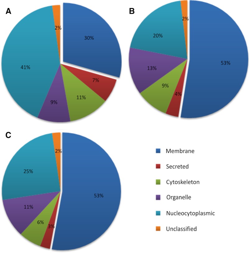
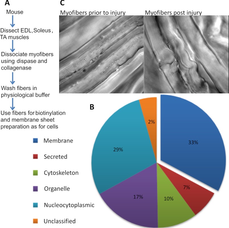
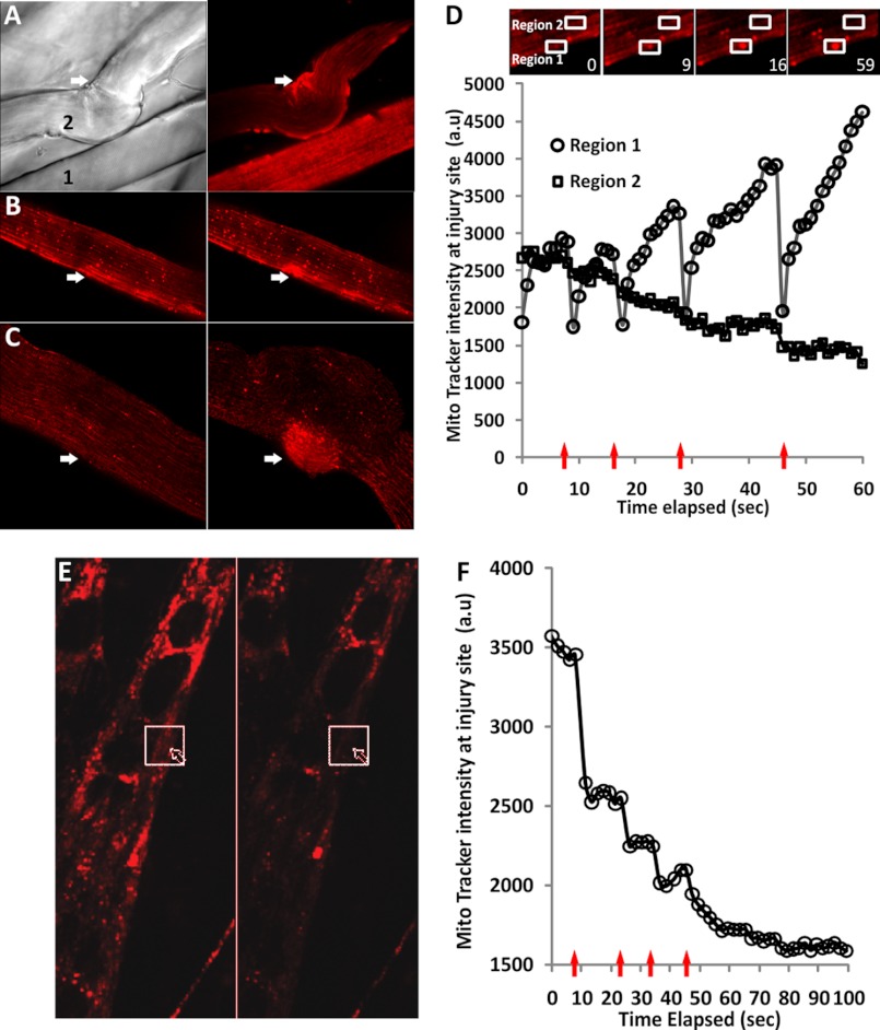
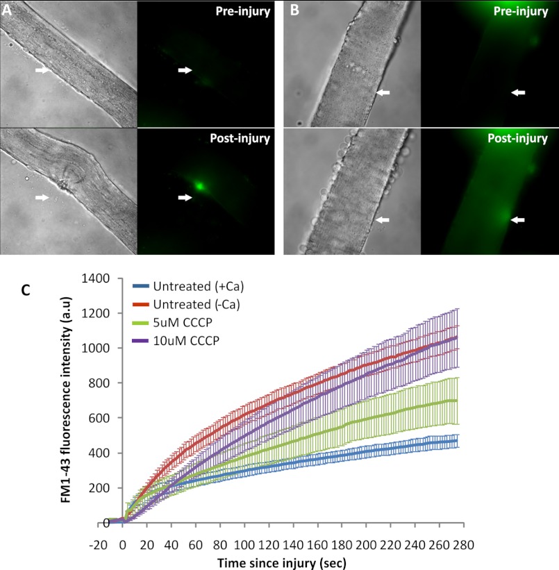
Skeletal muscles are proficient at healing from a variety of injuries. Healing occurs in two phases, early and late phase. Early phase involves healing the injured sarcolemma and restricting the spread of damage to the injured myofiber. Late phase of healing occurs a few days postinjury and involves interaction of injured myofibers with regenerative and inflammatory cells. Of the two phases, cellular and molecular processes involved in the early phase of healing are poorly understood. We have implemented an improved sarcolemmal proteomics approach together with in vivo labeling of proteins with modified amino acids in mice to study acute changes in the sarcolemmal proteome in early phase of myofiber injury. We find that a notable early phase response to muscle injury is an increased association of mitochondria with the injured sarcolemma. Real-time imaging of live myofibers during injury demonstrated that the increased association of mitochondria with the injured sarcolemma involves translocation of mitochondria to the site of injury, a response that is lacking in cultured myoblasts. Inhibiting mitochondrial function at the time of injury inhibited healing of the injured myofibers. This identifies a novel role of mitochondria in the early phase of healing injured myofibers.


Figures

FIGURE 1.
FIGURE 1.
Schematic illustration of detergent and mechanical lysis approaches used to isolate sarcolemmal proteome. Cultured muscle cells were biotinylated, and the cell surface proteins were extracted either by lysing cells with detergent or by mechanical homogenization. Biotinylated cell surface proteins were then purified by binding with magnetic streptavidin beads and sequentially washing with a high salt (50 mm Tris, pH 8.0, and 150 mm NaCl) and high pH buffer (100 mm sodium carbonate, pH 11.5) to remove nonspecifically bound proteins. Detergent lysis allowed isolation of only the biotinylated proteins, whereas mechanical lysis resulted in membrane sheets containing biotinylated and non-biotinylated proteins embedded in or tightly associated with the membrane sheet. The isolated proteins were resolved by SDS-PAGE and analyzed by LC-MS/MS after in-gel digestion with trypsin.
FIGURE 2.
FIGURE 2.
Pie charts showing the subcellular localization of proteins. Shown are proteins identified in the cell surface proteome of C2C12 myoblasts processed using the detergent solubilization approach (A) and from C2C12 (B) and H2K (C) myoblasts using the mechanical lysis approach. Each protein was designated only one subcellular location, and the number of proteins in each compartment has been represented as a percentage of total proteins identified. The subcellular location of the identified proteins was determined by the Gene Ontology database (see the Uniprot Web site) as well as literature search. Data used to construct these pie charts are presented in supplemental Tables S1–S4, which also detail the information on all of the proteins identified in two independent biological replicates, including the percentage of coverage and number of peptide hits.
FIGURE 3.
FIGURE 3.
Sarcolemmal proteomics of healthy and injured myofibers. A, experimental work flow for isolation of the sarcolemmal proteome from isolated muscle fibers. B, pie chart showing the subcellular localization of all proteins identified, which was determined as described in the legend to Fig. 1. Supplemental Table S4 provides the complete list of proteins identified and used in making the pie chart. C, transmitted light images showing a portion of the EDL muscle that has been mildly disaggregated and left uninjured or mechanically injured as described under “Experimental Procedures.”
FIGURE 4.
FIGURE 4.
Effect of myofiber injury on mitochondrial distribution. A, MitoTracker Red (CM-H2XRos)-labeled EDL muscle was loosely dissociated with collagenase and mechanically injured as described under “Experimental Procedures.” Shown are the transmitted light image (left) and MitoTracker staining (right) of uninjured (1) and injured (2) myofibers 10 min following mechanical injury. B, myofibers were labeled with MitoTracker, and the excess dye was washed multiple times. The fibers were then injured using a high intensity pulsed laser, and the images show independent fibers before (left) and 50 s after (right) injury. B, mild injury; C, severe injury; the injury sites are marked by the arrows. D, plot showing MitoTracker intensity at the site of injury (Region 1) and an adjacent region (Region 2) for a fiber that was mildly injured five times (arrows). Due to the use of a high intensity laser for the injury, each injury resulted in quenching of the fluorescence of the mitochondria present at the wound site prior to injury, the recovery of this fluorescence to higher than preinjury value suggesting accumulation of new mitochondria because the MitoTracker used for labeling was removed by multiple washes. The inset shows an enlarged image of the fiber at the start of imaging (0 s) and various time intervals postinjuries. Note the accumulation of the mitochondria at the site of injury (Region 1), whereas a site adjacent to the injury (Region 2) shows decreased mitochondrial fluorescence. E, C2C12 myotubes were labeled with MitoTracker, and following removal of excess dye, the myotube was injured multiple times using a pulsed laser and imaged at 2-s intervals. A myotube before (left) and 100 s after (right) laser injury is shown. The arrow marks the site of injury, and the box marks the region where the MitoTracker intensity was measured. F, plot showing MitoTracker intensity (arbitrary units (a.u.)) at the site of injury. With each of the four injuries (arrows), the myotube healed (indicated by lack of hypercontraction), but the MitoTracker staining intensity decreased irreversibly with each injury. Loss of MitoTracker staining occurred even for mitochondria distal from the site of injury.
FIGURE 5.
FIGURE 5.
Effect of inhibiting mitochondrial function on myofiber healing. Myofibers isolated from EDL muscle were injured using a high intensity pulsed laser in the presence of 2 μm FM1-43 dye. The myofibers were either not treated (A) or treated with 10 μm CCCP for 30 min prior to injury (B). C, plot showing the kinetics of increase in FM1-43 intensity (arbitrary units (a.u.)) following injury averaged for up to 10 myofibers in each condition. The error bars define the S.E. in the signal at each time point.

References

    1. Bansal D., Miyake K., Vogel S. S., Groh S., Chen C. C., Williamson R., McNeil P. L., Campbell K. P. (2003) Defective membrane repair in dysferlin-deficient muscular dystrophy. Nature 423, 168–172 - PubMed
    1. Carpenter S., Karpati G. (1989) Segmental necrosis and its demarcation in experimental micropuncture injury of skeletal muscle fibers. J. Neuropathol. Exp. Neurol. 48, 154–170 - PubMed
    1. Papadimitriou J. M., Robertson T. A., Mitchell C. A., Grounds M. D. (1990) The process of new plasmalemma formation in focally injured skeletal muscle fibers. J. Struct. Biol. 103, 124–134 - PubMed
    1. Faulkner J. A., Brooks S. V., Opiteck J. A. (1993) Injury to skeletal muscle fibers during contractions. Conditions of occurrence and prevention. Phys. Ther. 73, 911–921 - PubMed
    1. Grounds M. D. (1991) Toward understanding skeletal muscle regeneration. Pathol. Res. Pract. 187, 1–22 - PubMed

Publication types

Substances

LinkOut - more resources