. 2012 Aug 31;287(36):30346-57.
doi: 10.1074/jbc.M112.355875. Epub 2012 Jul 9.

Thioredoxin reductase linked to cytoskeleton by focal adhesion kinase reverses actin S-nitrosylation and restores neutrophil β(2) integrin function

Affiliations

Thioredoxin reductase linked to cytoskeleton by focal adhesion kinase reverses actin S-nitrosylation and restores neutrophil β(2) integrin function

Stephen R Thom et al. J Biol Chem. .

Abstract

The investigation goal was to identify mechanisms for reversal of actin S-nitrosylation in neutrophils after exposure to high oxygen partial pressures. Prior work has shown that hyperoxia causes S-nitrosylated actin (SNO-actin) formation, which mediates β(2) integrin dysfunction, and these changes can be reversed by formylmethionylleucylphenylalanine or 8-bromo-cyclic GMP. Herein we show that thioredoxin reductase (TrxR) is responsible for actin denitrosylation. Approximately 80% of cellular TrxR is localized to the cytosol, divided between the G-actin and short filamentous actin (sF-actin) fractions based on Triton solubility of cell lysates. TrxR linkage to sF-actin requires focal adhesion kinase (FAK) based on immunoprecipitation studies. S-Nitrosylation accelerates actin filament turnover (by mechanisms described previously (Thom, S. R., Bhopale, V. M., Yang, M., Bogush, M., Huang, S., and Milovanova, T. (2011) Neutrophil β(2) integrin inhibition by enhanced interactions of vasodilator stimulated phosphoprotein with S-nitrosylated actin. J. Biol. Chem. 286, 32854-32865), which causes FAK to disassociate from sF-actin. TrxR subsequently dissociates from FAK, and the physical separation from actin impedes denitrosylation. If SNO-actin is photochemically reduced with UV light or if actin filament turnover is impeded by incubations with cytochalasin D, latrunculin B, 8-bromo-cGMP, or formylmethionylleucylphenylalanine, FAK and TrxR reassociate with sF-actin and cause SNO-actin removal. FAK-TrxR association can also be demonstrated using isolated enzymes in ex vivo preparations. Uniquely, the FAK kinase domain is the site of TrxR linkage. We conclude that through its scaffold function, FAK influences TrxR activity and actin S-nitrosylation.


Figures

FIGURE 1.
FIGURE 1.
β2 integrin-specific neutrophil adherence. Data show the fraction of neutrophils in a suspension that became adherent after incubation on fibrinogen-coated plates. Adherence was measured using neutrophils exposed to air (control) or 2.0 ATA O2 for 45 min and then incubated for 20 h with control siRNA or siRNA to TrxR1, glutathione reductase, or S-nitrosoglutathione reductase. The next day, suspensions were loaded with calcein AM, divided, and placed in wells containing just PBS or PBS plus agonists to achieve a final concentration of 100 μm 8-Br-cGMP or 100 nm fMLP as described under “Experimental Procedures.” The percentage adherence was calculated and compared against identical samples incubated with blocking antibodies to establish β2 integrin-specific adherence. Data are mean ± S.E. (error bars); n = 10 separate studies using neutrophils from different animals; *, p < 0.05 versus control siRNA air-exposed cells incubated with just PBS.
FIGURE 2.
FIGURE 2.
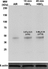
S-Nitrosylated β-actin in neutrophil lysates. Neutrophil suspensions were exposed to air (control) or to 2.0 ATA O2 for 15 or 45 min, centrifuged, resuspended in HEN buffer, and processed for a biotin switch assay as described under “Experimental Procedures.” The blots show a representative experiment; numbers shown on the image represent the -fold increase in SNO-actin band densities relative to control (air only) neutrophil lysates run on the same blots. Values are mean ± S.E. (error bars) for 4–10 independent trials (no significant difference).
FIGURE 3.
FIGURE 3.
S-Nitrosylated β-actin in neutrophil lysates after siRNA treatments. Neutrophil suspensions exposed to air or 45 min at 2.0 ATA O2 were incubated for 20 h with control, scrambled sequence siRNA or siRNA to TrxR1. Samples were centrifuged and resuspended in PBS or PBS containing 100 μm 8-Br-cGMP or 100 nm fMLP. After incubation for 10 min, cells were centrifuged, resuspended in HEN buffer, and processed for the biotin switch assay as described under “Experimental Procedures.” After Western blotting, the densities of biotin-labeled and actin bands were measured, and -fold increase in biotin/actin density for each sample was compared with that measured for air-exposed cells incubated with control siRNA and then PBS that was run on the same Western blot. Data are mean ± S.E. (error bars) -fold increase in ratios of band densities for 4–10 independent samples; *, p < 0.05 (ANOVA). The blots shown at the top are from a representative single experiment.
FIGURE 4.
FIGURE 4.
TrxR content in neutrophil actin fractions. Neutrophil suspensions were exposed to air (control) or 2.0 ATA O2 for 45 min, directly placed into SDS buffer (whole cell lysate), or lysed and separated into Triton-soluble G-actin and sF-actin and Triton-insoluble actin. Western blots were probed for TrxR and β-actin. The figure shows a representative Western blot, and the chart depicts TrxR/actin ratios as mean ± S.E. (error bars) (n = 8 individual samplings). There are no significant differences between air-exposed (control) and HBO2-exposed cell preparations.
FIGURE 5.
FIGURE 5.
TrxR immunoprecipitation Coomassie-stained gel. The figure shows the band pattern for lysates of air-exposed, control, and HBO2-exposed neutrophil lysates immunoprecipitated using anti-TrxR. Proteins listed were identified by MS/MS spectra. For nitric-oxide synthase-2 (iNOS) 36 peptides were identified spanning 32% of the protein; for FAK, 12 peptides were identified spanning 9% of the protein; for heat shock protein-90α (HSP90 α), 20 peptides were identified spanning 24% of the protein; for heat shock protein-90β (HSP90 β), 22 peptides were identified spanning 34% of the protein; for heat shock cognate 71-kDa protein, 19 peptides were identified spanning 29% of the protein; for TrxR, 10 peptides were identified spanning 18% of the protein; for protein-disulfide isomerase A6 precursor, seven peptides were identified spanning 18% of the protein; for actin, 26 peptides were identified spanning 49% of the protein; for tropomyosin α-1 isoform, seven peptides were identified spanning 26% of the protein; for 14-3-3 protein ζ/δ, 12 peptides were identified spanning 44% of the protein; for Ras-related protein Rab-27B, seven peptides were identified spanning 42% of the protein.
FIGURE 6.
FIGURE 6.
FAK-TrxR (A), actin-TrxR (B), and actin-FAK (C) linkage in sF-actin fractions assessed by immunoprecipitation. Neutrophils exposed to air (control) or 2.0 ATA O2 for 45 min were divided and incubated for 10 min with PBS or PBS plus agonists to achieve a final concentration of 100 μm 8-Br-cGMP or 100 nm fMLP. Cells were lysed, and the sF-actin fraction was isolated and subjected to immunoprecipitation using antibodies to TrxR1 or FAK. Western blots at the top show typical results, and data were expressed as the ratio of β-actin (42 kDa) (A) or FAK (110 kDa) (B) band density versus TrxR (55 kDa) band density normalized to the ratio of the air-exposed PBS-incubated cell lysate in each sample. The ratio of actin to FAK when using antibodies to FAK is shown in C. Values are mean ± S.E. (error bars) for six independent trials; *, p < 0.05, ANOVA.
FIGURE 7.
FIGURE 7.
Neutrophil adherence after exposures to hyperoxia up to 45 min. Cells were exposed to 2.0 ATA O2 for 5–45 min, and adherence to fibrinogen-coated plates was quantified. Values are mean ± S.E. (error bars); n = 3–7; *, p < 0.05, ANOVA.
FIGURE 8.
FIGURE 8.
Protein co-localizations with F-actin in neutrophil images. Neutrophils exposed to air or HBO2 ex vivo were placed on fibrinogen-coated slides, permeabilized and stained as described under “Experimental Procedures.” Bar graphs show merged fluorescence intensity, which reflects protein co-localizations. These data were obtained with cells from mice in 3–7 independent experiments by analyzing 30–65 neutrophils in each trial. Values in bar graphs are mean ± S.E. (error bars); *, p < 0.001 versus air-exposed cells.
FIGURE 9.
FIGURE 9.
TrxR protects actin from S-nitrosylation. Neutrophils were isolated and incubated for 20 h with control siRNA or siRNA to TrxR or FAK. Cells were lysed, the sF-actin fraction was isolated and divided into two, and 1 μm auranofin was added to one sample. Both samples were incubated with 200 μm SNAP for 1 h at room temperature, and then S-nitrosylation was assessed by the biotin switch assay. The Western blot at the top demonstrates typical experimental results, and data are expressed as the difference in density of the biotin/actin band ratio of samples incubated in the absence versus presence of auranofin, a TrxR inhibitor. Therefore, values closer to 1.0 demonstrate inability of TrxR in the sample to protect against S-nitrosylation. Values are mean ± S.E. (error bars) for four experiments; *, p < 0.05, ANOVA.
FIGURE 10.
FIGURE 10.
TrxR-FAK ex vivo attachment with isolated enzymes. Purified TrxR was incubated with a His-tagged kinase domain fragment of human FAK as described under “Experimental Procedures.” Where indicated, 5 or 10 μm PF 573228 was added to FAK suspensions 10 min before the addition of TrxR. After 1-h incubations, protein suspensions were passed through cobalt resin, and the bound protein was eluted and then subjected to Western blotting. The pattern shown in the figure was identical in four replicate trials. The first two lanes show blots using only the single protein solutions (TrxR or His-FAK), whereas lanes 3–6 show proteins eluted from cobalt resin. Lane 3 shows His-FAK plus TrxR, and lane 4 shows the result when the TrxR solution is incubated with resin (e.g. no TrxR binds to resin). The last two lanes show results when 5 or 10 μm PF 573228 was added to His-FAK plus TrxR solutions. No TrxR was detected in these suspensions, and at 10 μm PF 573228, the FAK band density was 20 ± 5% (S.E.; n = 3) lower than with no inhibitor or just 5 μm PF 573228.

References

    1. Brown E. J., Lindberg F. P. (1996) Leukocyte adhesion molecules in host defence against infection. Ann. Med. 28, 201–208 - PubMed
    1. Thom S. R. (1993) Functional inhibition of leukocyte B2 integrins by hyperbaric oxygen in carbon monoxide-mediated brain injury in rats. Toxicol. Appl. Pharmacol. 123, 248–256 - PubMed
    1. Thom S. R., Mendiguren I., Hardy K., Bolotin T., Fisher D., Nebolon M., Kilpatrick L. (1997) Inhibition of human neutrophil β2-integrin-dependent adherence by hyperbaric O2. Am. J. Physiol. 272, C770–C777 - PubMed
    1. Kalns J., Lane J., Delgado A., Scruggs J., Ayala E., Gutierrez E., Warren D., Niemeyer D., George Wolf E., Bowden R. A. (2002) Hyperbaric oxygen exposure temporarily reduces Mac-1-mediated functions of human neutrophils. Immunol. Lett. 83, 125–131 - PubMed
    1. Labrouche S., Javorschi S., Leroy D., Gbikpi-Benissan G., Freyburger G. (1999) Influence of hyperbaric oxygen on leukocyte functions and hemostasis in normal volunteer divers. Thromb. Res. 96, 309–315 - PubMed

Publication types

LinkOut - more resources