The cell biology of disease: Acute promyelocytic leukemia, arsenic, and PML bodies
- PMID: 22778276
- PMCID: PMC3392943
- DOI: 10.1083/jcb.201112044
The cell biology of disease: Acute promyelocytic leukemia, arsenic, and PML bodies
Abstract
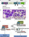
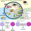
Acute promyelocytic leukemia (APL) is driven by a chromosomal translocation whose product, the PML/retinoic acid (RA) receptor α (RARA) fusion protein, affects both nuclear receptor signaling and PML body assembly. Dissection of APL pathogenesis has led to the rediscovery of PML bodies and revealed their role in cell senescence, disease pathogenesis, and responsiveness to treatment. APL is remarkable because of the fortuitous identification of two clinically effective therapies, RA and arsenic, both of which degrade PML/RARA oncoprotein and, together, cure APL. Analysis of arsenic-induced PML or PML/RARA degradation has implicated oxidative stress in the biogenesis of nuclear bodies and SUMO in their degradation.
Figures


Similar articles
-
Activation of a promyelocytic leukemia-tumor protein 53 axis underlies acute promyelocytic leukemia cure.Nat Med. 2014 Feb;20(2):167-74. doi: 10.1038/nm.3441. Epub 2014 Jan 12. Nat Med. 2014. PMID: 24412926
-
Function of PML-RARA in Acute Promyelocytic Leukemia.Adv Exp Med Biol. 2024;1459:321-339. doi: 10.1007/978-3-031-62731-6_14. Adv Exp Med Biol. 2024. PMID: 39017850 Review.
-
Acute Promyelocytic Leukemia, Retinoic Acid, and Arsenic: A Tale of Dualities.Cold Spring Harb Perspect Med. 2024 Sep 3;14(9):a041582. doi: 10.1101/cshperspect.a041582. Cold Spring Harb Perspect Med. 2024. PMID: 38503502 Review.
-
Autophagy contributes to therapy-induced degradation of the PML/RARA oncoprotein.Blood. 2010 Sep 30;116(13):2324-31. doi: 10.1182/blood-2010-01-261040. Epub 2010 Jun 23. Blood. 2010. PMID: 20574048
-
Novel treatment of acute promyelocytic leukemia: As₂O₃, retinoic acid and retinoid pharmacology.Curr Pharm Biotechnol. 2013;14(9):849-58. doi: 10.2174/1389201015666140113095812. Curr Pharm Biotechnol. 2013. PMID: 24433507 Review.
Cited by
-
Beyond Cisplatin: Combination Therapy with Arsenic Trioxide.Inorganica Chim Acta. 2019 Oct 1;496:119030. doi: 10.1016/j.ica.2019.119030. Epub 2019 Jul 24. Inorganica Chim Acta. 2019. PMID: 32863421 Free PMC article.
-
Control of antioxidative response by the tumor suppressor protein PML through regulating Nrf2 activity.Mol Biol Cell. 2014 Aug 15;25(16):2485-98. doi: 10.1091/mbc.E13-11-0692. Epub 2014 Jun 18. Mol Biol Cell. 2014. PMID: 24943846 Free PMC article.
-
Personalized medicine and the clinical laboratory.Einstein (Sao Paulo). 2014 Sep;12(3):366-73. doi: 10.1590/s1679-45082014rw2859. Einstein (Sao Paulo). 2014. PMID: 25295459 Free PMC article. Review.
-
Protein quality control in the nucleus.Biomolecules. 2014 Jul 9;4(3):646-61. doi: 10.3390/biom4030646. Biomolecules. 2014. PMID: 25010148 Free PMC article. Review.
-
PML protein organizes heterochromatin domains where it regulates histone H3.3 deposition by ATRX/DAXX.Genome Res. 2017 Jun;27(6):913-921. doi: 10.1101/gr.215830.116. Epub 2017 Mar 24. Genome Res. 2017. PMID: 28341773 Free PMC article.
References
-
- Ablain J., Nasr R., Bazarbachi A., de Thé H. 2011. The drug-induced degradation of oncoproteins: an unexpected achilles’ heel of cancer cells? Cancer Discov. 1:117–127 10.1158/2159-8290.CD-11-0087 - DOI - PubMed
Publication types
MeSH terms
Substances
LinkOut - more resources
Full Text Sources
Medical

