. 2012 Aug 14;109(33):E2215-22.
doi: 10.1073/pnas.1120517109. Epub 2012 Jul 9.

Maternally recruited Aurora C kinase is more stable than Aurora B to support mouse oocyte maturation and early development

Affiliations

Maternally recruited Aurora C kinase is more stable than Aurora B to support mouse oocyte maturation and early development

Karen Schindler et al. Proc Natl Acad Sci U S A. .

Abstract

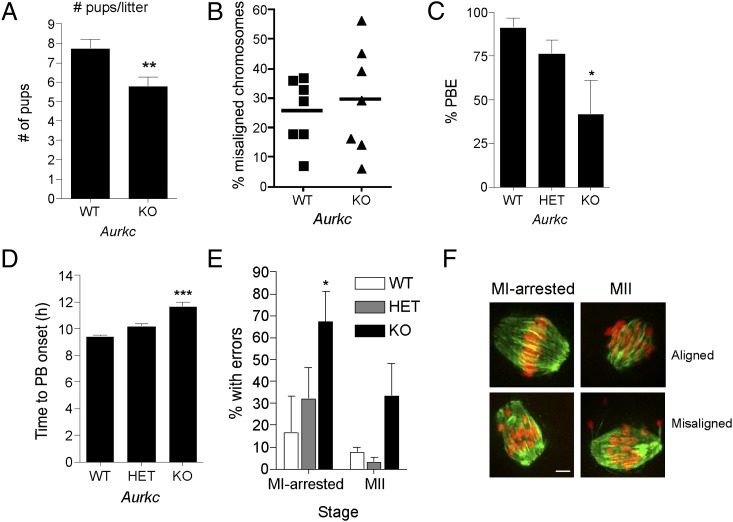
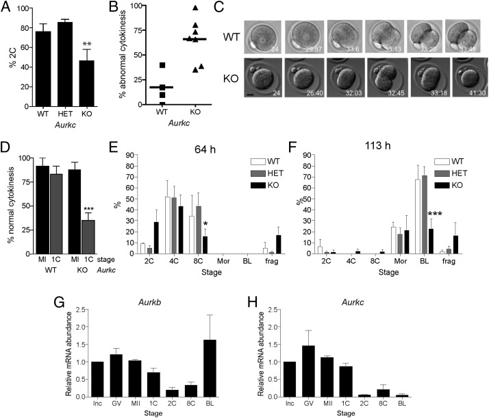
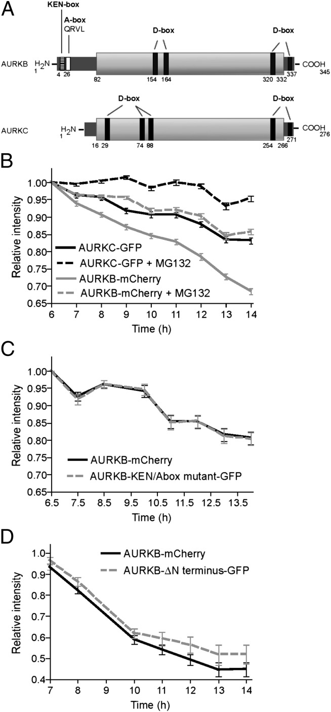
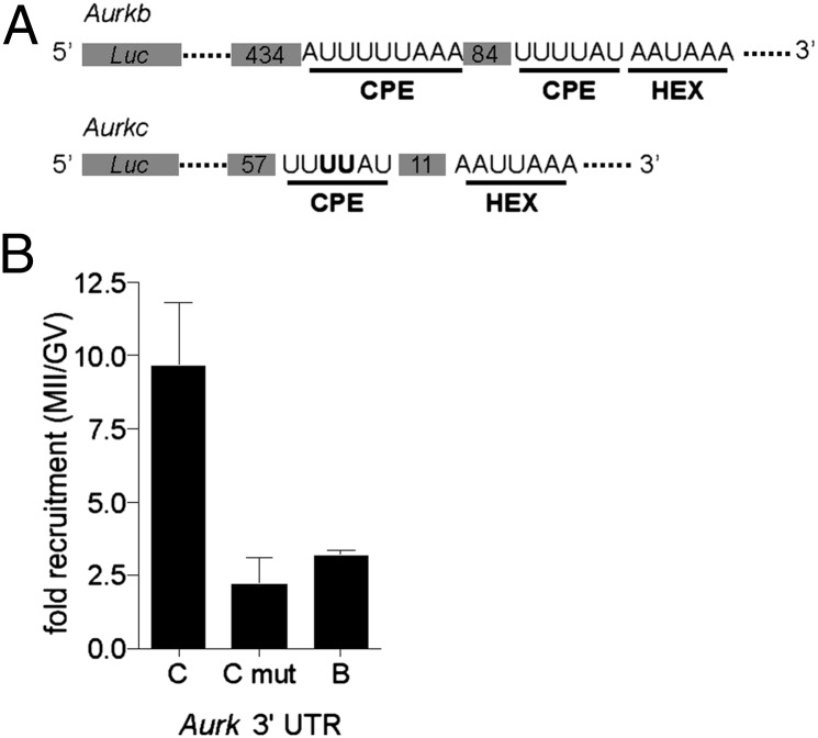
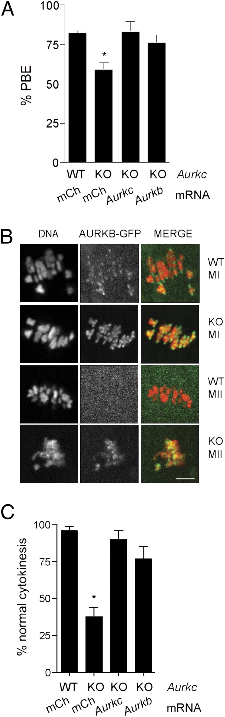
Aurora kinases are highly conserved, essential regulators of cell division. Two Aurora kinase isoforms, A and B (AURKA and AURKB), are expressed ubiquitously in mammals, whereas a third isoform, Aurora C (AURKC), is largely restricted to germ cells. Because AURKC is very similar to AURKB, based on sequence and functional analyses, why germ cells express AURKC is unclear. We report that Aurkc(-/-) females are subfertile, and that AURKB function declines as development progresses based on increasing severity of cytokinesis failure and arrested embryonic development. Furthermore, we find that neither Aurkb nor Aurkc is expressed after the one-cell stage, and that AURKC is more stable during maturation than AURKB using fluorescently tagged reporter proteins. In addition, Aurkc mRNA is recruited during maturation. Because maturation occurs in the absence of transcription, posttranscriptional regulation of Aurkc mRNA, coupled with the greater stability of AURKC protein, provides a means to ensure sufficient Aurora kinase activity, despite loss of AURKB, to support both meiotic and early embryonic cell divisions. These findings suggest a model for the presence of AURKC in oocytes: that AURKC compensates for loss of AURKB through differences in both message recruitment and protein stability.


Conflict of interest statement

The authors declare no conflict of interest.

Figures

Fig. 1.
Fig. 1.
Loss of AURKC leads to meiotic abnormalities. (A) Results of fertility trials. The average number of pups born to females of the indicated genotype over an 8-mo breeding trial is shown. (B) GV-intact oocytes were isolated from mice of the indicated genotype and matured in vitro for 8 h before fixation at MI. Percent of oocytes with misaligned chromosomes was plotted for each mouse analyzed. (C–F) GV-intact oocytes were isolated from mice of the indicated genotype and matured in vitro to determine incidence (B) and timing (C) of polar body extrusion (PBE). Cells were fixed when controls (WT) had reached MII and processed for immunocytochemistry to detect chromosomes and spindles. The percentage of oocytes that contained abnormal chromosome configurations at either MI or MII was determined (E), and representative images are shown (F). Graphs represent mean (± SEM) from at least 30 oocytes from three independent experiments. (Scale bars, 5 μm.) One-way ANOVA was used to analyze the data in B–D. *P < 0.05, **P < 0.01, ***P < 0.001; WT, wild-type; HET, heterozygous; KO, knockout.
Fig. 2.
Fig. 2.
Embryonic development is compromised in Aurkc−/− mice. (A–F) Female mice of the indicated genotype were mated to wild-type males and one-cell embryos were isolated and cultured in vitro. (A) The percentage of one-cell embryos that cleaved to the two-cell embryonic stage. (B–D) Embryos were imaged live by DIC every 5–7 min. In B, the percentage of embryos with abnormal cytokinesis was plotted for each mouse. C contains representative images of a wild-type embryo undergoing normal cytokinesis (Upper) and a KO embryo failing cytokinesis (Lower). The time stamp is h:min after hCG injection. (Scale bar, 5 μm.) (D) The percentage of normal cytokinesis events from Fig. 1C (MI) and panel B (1C) were compared. (E and F) Embryo development was monitored at the indicated times after hCG and mating. These experiments were conducted four times with at least two mice per genotype each time, and the data are expressed as the mean ± SEM. (G and H) mRNA levels were measured by quantitative RT-PCR at the indicated stages. Data were normalized against a probe that detects exogenously added Gfp message. Mean (± SEM) from three independent experiments are shown. One-way ANOVA was used to analyze the data. *P < 0.05; **P < 0.01; ***P < 0.001. BL, blastocyst; frag, fragmented; GV, full grown GV-intact oocyte; Inc, meiotically incompetent oocyte; Mor, morula; 1C, one-cell embryo; 2C, two-cell embryo; 4C, four-cell embryo; 8C, eight-cell embryo.
Fig. 3.
Fig. 3.
AURKC is more stable than AURKB during meiotic maturation. (A–C) GV-intact oocytes were coinjected with the indicated cRNAs and matured to the indicated stage. In each graph, the first time point is 1 h after cycloheximide addition, and fluorescent images were obtained at the indicated times. Below are representative images from each time course. Data represent mean (± SEM) from at least 30 oocytes from two independent experiments. (Scale bars, 5 μm.) (D) Merge of the data from A–C.
Fig. 4.
Fig. 4.
AURKB stability does not depend upon its N terminus. (A) Schematic representation of AURKB and AURKC. KEN, A-, and D-boxes are indicated, and the conserved regions are shaded in light gray. (B–D) GV-intact oocytes were coinjected with the indicated cRNAs and matured to the indicated stage. Cycloheximide was added 1 h before the first time point and fluorescent images were obtained at the indicated times. Data represent mean (± SEM) from at least 30 oocytes from two independent experiments. MG132 was added as indicated to inhibit the proteasome (B).
Fig. 5.
Fig. 5.
Aurkc is a maternally recruited message. (A) Schematic representation of the Firefly luciferase (Luc) fusions to the Aurkb and Aurkc 3′ UTRs. Putative CPE and HEX motifs are underlined and the number of nucleotides between the motifs are indicated in the gray boxes. In Aurkc, the two nucleotides in the CPE that were mutated are in bold. Note that the lengths are not to scale. (B) GV-intact oocytes were coinjected with Luc fused to the indicated 3′ UTR and Renilla luciferase as an injection volume control. Luminescence was measured and quantified as the fold-difference in MII compared with GV. Data represent mean (± SEM) from 10 oocytes from two independent experiments. mut, mutated.
Fig. 6.
Fig. 6.
Ectopic expression of AURKB in oocytes and embryos from Aurkc KO mice rescues their defects. (A and C) GV-intact oocytes or one-cell embryos from a single mouse of the indicated genotype were subdivided and microinjected with the indicated cRNAs and matured to MII to determine incidence of PBE or developed to the two-cell stage to monitor cytokinesis. These experiments were repeated three times with two to four KO mice per experiment. (B) Wild-type or Aurkc KO oocytes were microinjected with Aurkb-Gfp cRNA, matured to MI and MII, and imaged live. Shown are representative images. (Scale bar, 5 μm.) One-way ANOVA was used to analyze the data. *P < 0.05; mCh, mCherry.
Fig. P1.
Fig. P1.
Oocytes contain a third AURK to support oocyte maturation and early development because AURKB is not stable during meiosis and it is not recruited for translation as efficiently. Somatic cells continually cycle from G1 phase through mitosis during their life span. AURKB (orange) localizes to centromeres during metaphase, is destroyed at the end of the cell cycle, and is resynthesized during the next cell cycle. Oocytes contain both AURKB and AURKC (blue) (green where they colocalize). Both proteins (colored circles) are gradually degraded during meiosis, but AURKB does so more rapidly than AURKC does. There is no intervening cell cycle between meiosis I (MI) and meiosis II (MII) and oocytes are transcriptionally quiescent, making this cell type dependent on mRNA recruitment to synthesize adequate amounts of AURK. Aurkc message (colored lines) is recruited for translation more efficiently than Aurkb message is. The boxes around regions of chromosomes refer to the zoomed in Insets that contain cartoon labels representing AURKB and AURKC. A, anaphase; M, mitosis; S, synthesis; T, telophase; *, polar body.

References

    1. Gopalan G, Chan CS, Donovan PJ. A novel mammalian, mitotic spindle-associated kinase is related to yeast and fly chromosome segregation regulators. J Cell Biol. 1997;138:643–656. - PMC - PubMed
    1. Yanai A, Arama E, Kilfin G, Motro B. ayk1, a novel mammalian gene related to Drosophila aurora centrosome separation kinase, is specifically expressed during meiosis. Oncogene. 1997;14:2943–2950. - PubMed
    1. Tseng TC, Chen SH, Hsu YP, Tang TK. Protein kinase profile of sperm and eggs: Cloning and characterization of two novel testis-specific protein kinases (AIE1, AIE2) related to yeast and fly chromosome segregation regulators. DNA Cell Biol. 1998;17:823–833. - PubMed
    1. Baldini E, et al. Aurora kinases are expressed in medullary thyroid carcinoma (MTC) and their inhibition suppresses in vitro growth and tumorigenicity of the MTC derived cell line TT. BMC Cancer. 2011;11:411. - PMC - PubMed
    1. Price DM, Kanyo R, Steinberg N, Chik CL, Ho AK. Nocturnal activation of aurora C in rat pineal gland: Its role in the norepinephrine-induced phosphorylation of histone H3 and gene expression. Endocrinology. 2009;150:2334–2341. - PubMed

Publication types

MeSH terms

LinkOut - more resources