. 2012 Jul 24;109(30):12011-6.
doi: 10.1073/pnas.1204129109. Epub 2012 Jul 9.

A designed ankyrin repeat protein selected to bind to tubulin caps the microtubule plus end

Affiliations

A designed ankyrin repeat protein selected to bind to tubulin caps the microtubule plus end

Ludovic Pecqueur et al. Proc Natl Acad Sci U S A. .

Abstract

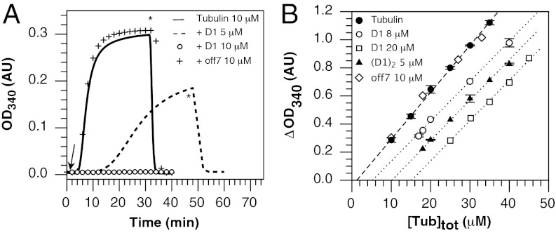
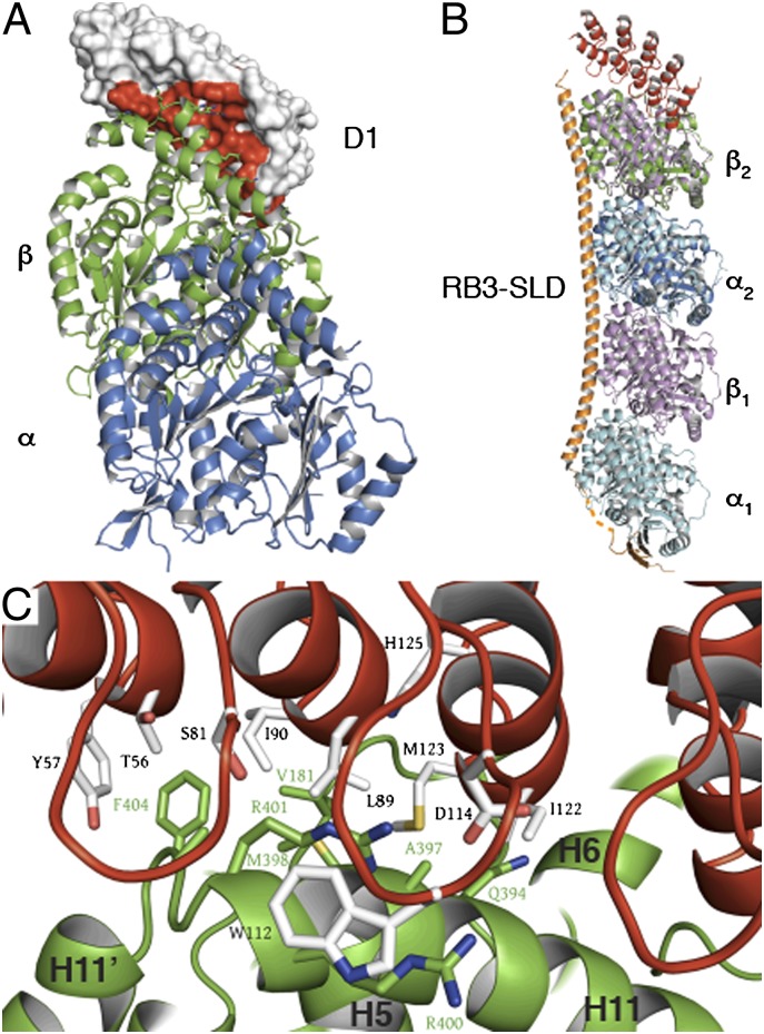
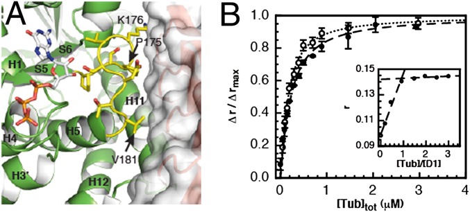
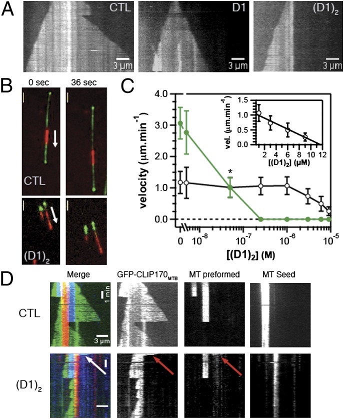
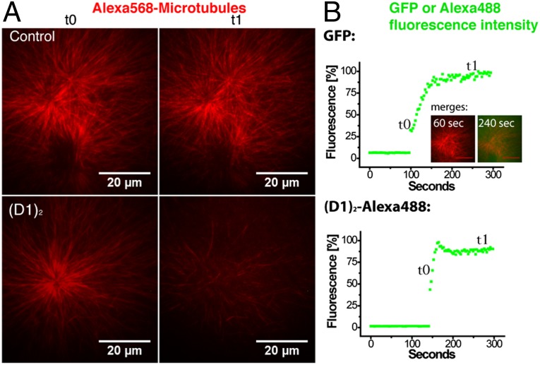
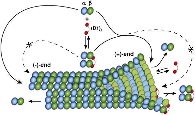
Microtubules are cytoskeleton filaments consisting of αβ-tubulin heterodimers. They switch between phases of growth and shrinkage. The underlying mechanism of this property, called dynamic instability, is not fully understood. Here, we identified a designed ankyrin repeat protein (DARPin) that interferes with microtubule assembly in a unique manner. The X-ray structure of its complex with GTP-tubulin shows that it binds to the β-tubulin surface exposed at microtubule (+) ends. The details of the structure provide insight into the role of GTP in microtubule polymerization and the conformational state of tubulin at the very microtubule end. They show in particular that GTP facilitates the tubulin structural switch that accompanies microtubule assembly but does not trigger it in unpolymerized tubulin. Total internal reflection fluorescence microscopy revealed that the DARPin specifically blocks growth at the microtubule (+) end by a selective end-capping mechanism, ultimately favoring microtubule disassembly from that end. DARPins promise to become designable tools for the dissection of microtubule dynamic properties selective for either of their two different ends.


Conflict of interest statement

The authors declare no conflict of interest.

Figures

Fig. 1.
Fig. 1.
D1 prevents microtubule assembly. (A) In vitro polymerization of 10 μM tubulin alone and in presence of 5 μM or 10 μM D1 and of an unrelated DARPin selected from the same library as D1 (off7, 10 μM), used as a control (20). The arrow indicates the temperature jump from 4 °C to 37 °C and the asterisk indicates the time of the reverse temperature jump. (B) Critical concentration plots of tubulin and of tubulin in presence of 8 μM or 20 μM D1, 10 μM off7, or 5 μM (D1)2. (D1)2 is a D1 tandem repeat; see main text for its complete definition. Error bars represents SDs deduced from at least duplicate experiments.
Fig. 2.
Fig. 2.
The structure of the tubulin–D1 complex. (A) Overall organization. D1 binds to the β-tubulin longitudinal interface. The red surface corresponds to D1 interfacing residues. (B) Superposition of Tub-D1 with T2R (pdb 3RYI). D1 is in red here as well as in C. Tubulin in Tub-D1 (α is in dark blue and β is in green) is superimposed to the α2β2 GDP-bound heterodimer in T2R (α2 is in cyan, as is α1, and β2 is in pink, as is β1); the RB3-SLD is in orange. (C) Residues of the tubulin–D1 interface belong to all five D1 ankyrin repeats and to β-tubulin helices H6, H11, and loop T5 (residues 206–215, 385–397, and 171–181, respectively; for a nomenclature of tubulin secondary structure elements see ref. 45). Residues shown are those that are randomized in the library from which D1 was selected and tubulin residues within 5 Å of them (for a stereoview see Fig. S2).
Fig. 3.
Fig. 3.
D1 binds to GDP- and GTP-tubulin with similar affinities. (A) D1 binds close to the β-tubulin nucleotide-binding site and contacts its T5 loop (yellow). T5 is in its “out conformation,” which is fully populated when tubulin is GTP bound (10). T5 residues in contact with D1 are labeled. (B) Variations of fluorescence anisotropy of labeled D1 (120 nM) upon binding to GDP- (open circles) and GTP-tubulin (solid circles). Errors bars represent SDs from duplicate experiments. The lines represent the fit as described in Methods. Inset shows the titration of 1 μM Oregon-labeled D1 by GDP-tubulin, under conditions where the concentration of D1 was much larger than the dissociation equilibrium constant. Under these conditions, the fluorescence anisotropy reached its maximum for a 1:1 tubulin:D1 ratio, consistent with the 1:1 stoichiometry of the complex.
Fig. 4.
Fig. 4.
(D1)2 efficiently inhibits microtubule plus-end growth. (A) Kymographs of Alexa568-labeled microtubules growing from surface-immobilized microtubule seeds in the absence or presence of 0.25 μM D1 or (D1)2 as indicated, observed by TIRF microscopy; total duration displayed is 5 min. The concentration of Alexa568-labeled tubulin (labeling ratio: 6.5%) was 20 μM. (B) TIRF microscopy images of dynamic microtubules being transported by surface-immobilized (−) end-directed kinesin-14 (XCTK2). Microtubules polymerize from Alexa568-labeled GMPCPP microtubule seeds (red) in the presence of 20 μM tubulin and 70 nM Mal3-GFP (green) and either no (Upper) or 0.5 μM (D1)2 (Lower). (Scale bar: 3 μm.) (C) Growth velocity of microtubule (+) (green symbols) and (−) (black symbols) ends in the presence of 20 μM tubulin as a function of increasing (D1)2 concentrations. The data point for 50 nM (D1)2 (marked with an *) is an average of (+)- and (−) end growth velocity, because the two ends could not be distinguished due to similar growth velocities in this condition. Error bars are SD, each experimental value being the average of at least 20 microtubules from three or more independent experiments. (Inset) (−) end growth velocity as a function of (D1)2 concentrations on a linear scale with a linear fit (black line). Note that the (D1)2 concentration required to depolymerize microtubules in these experiments cannot be directly compared with those in turbidity measurements, because these are different types of experiments that were performed under different conditions (Methods). (D) Kymographs show the effect of 0.5 μM (D1)2 on microtubules prepolymerized in the presence of 15 μM Cy5-labeled tubulin (blue) from immobilized Alexa568-labeled stabilized microtubule seeds (red). Imaging started ca. 30 s after replacing labeled with unlabeled tubulin and addition of 65 nM of a GFP-labeled CLIPMTB (green) (Methods) (Lower). The control without (D1)2 is also presented (Upper). Merged kymographs (Left) and the individual one-channel kymographs are shown as indicated. The arrow shows a (+) end that has not depolymerized at the time imaging started. Total time displayed is 500 s.
Fig. 5.
Fig. 5.
(D1)2 promotes MT depolymerization in Xenopus egg extract. (A) Asters in Xenopus egg extract containing Alexa568-tubulin were adsorbed to glass coverslips and observed by TIRF microscopy. (B) After addition of GFP (Upper) or Alexa488-(D1)2 (Lower) to an estimated final concentration of 0.75 μM, the diffusion into the evanescent field was monitored by measuring the GFP or Alexa488 fluorescence intensity. Alexa568-microtubule asters that are still present when the newly added protein just starts to diffuse into the evanescent field (t0 in A or B), largely disappear 150 s later selectively when Alexa488-(D1)2 was added (t1 in A and B). Inserted images in B show merged dual-color images to visualize how GFP (green) diffuses into the region with microtubule asters (red).
Fig. 6.
Fig. 6.
The mechanism of action of (D1)2. (D1)2 (red) prevents assembly at the (−) end of the one tubulin heterodimer (α, blue; β, green) it is bound to. (D1)2-bound tubulins associate at the (+) end but then block addition of all tubulin heterodimers to a capped protofilament.

References

    1. Desai A, Mitchison TJ. Microtubule polymerization dynamics. Annu Rev Cell Dev Biol. 1997;13:83–117. - PubMed
    1. Goodwin SS, Vale RD. Patronin regulates the microtubule network by protecting microtubule minus ends. Cell. 2010;143:263–274. - PMC - PubMed
    1. Kollman JM, Merdes A, Mourey L, Agard DA. Microtubule nucleation by γ-tubulin complexes. Nat Rev Mol Cell Biol. 2011;12:709–721. - PMC - PubMed
    1. Brouhard GJ, et al. XMAP215 is a processive microtubule polymerase. Cell. 2008;132:79–88. - PMC - PubMed
    1. Bringmann H, et al. A kinesin-like motor inhibits microtubule dynamic instability. Science. 2004;303:1519–1522. - PubMed

Publication types

Associated data