

Magnetic characterization of isolated candidate vertebrate magnetoreceptor cells
- PMID: 22778440
- PMCID: PMC3409731
- DOI: 10.1073/pnas.1205653109
Magnetic characterization of isolated candidate vertebrate magnetoreceptor cells
Abstract
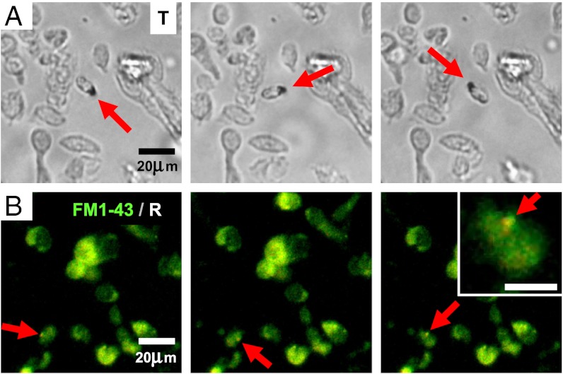
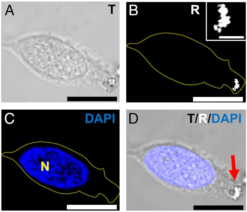
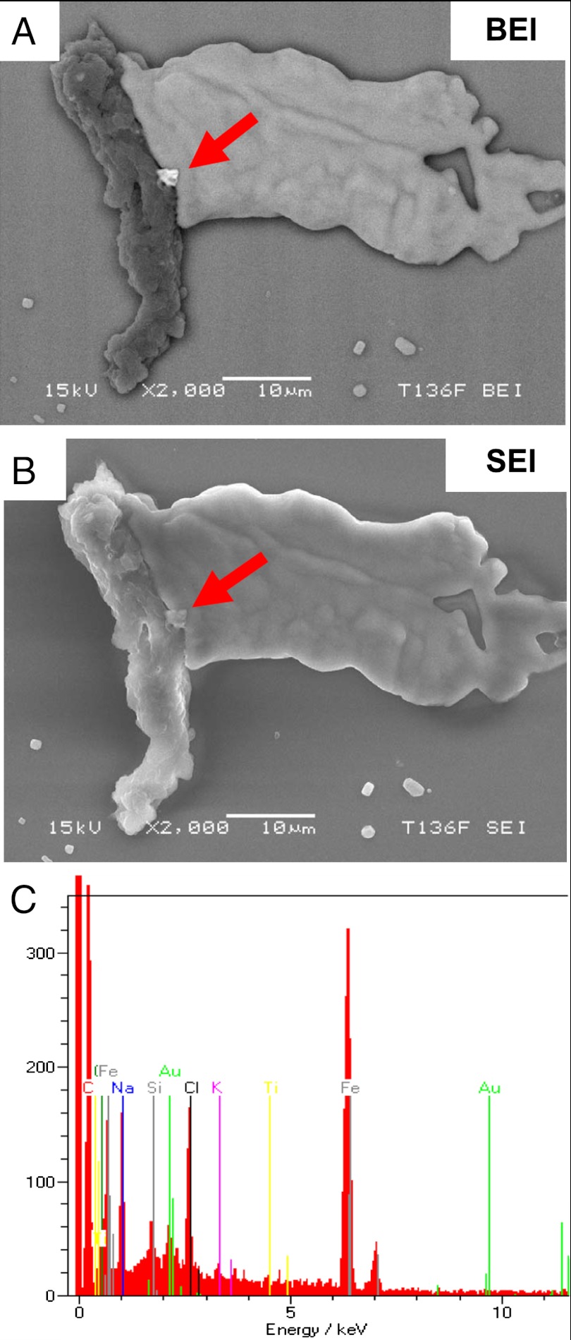
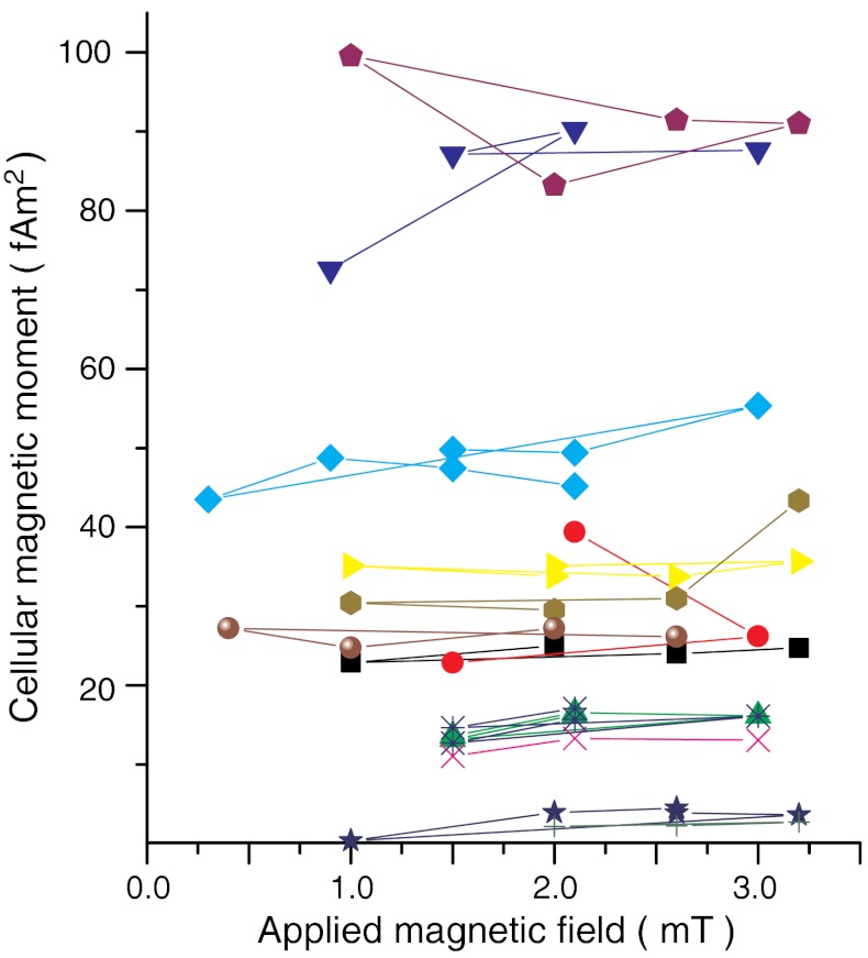
Over the past 50 y, behavioral experiments have produced a large body of evidence for the existence of a magnetic sense in a wide range of animals. However, the underlying sensory physiology remains poorly understood due to the elusiveness of the magnetosensory structures. Here we present an effective method for isolating and characterizing potential magnetite-based magnetoreceptor cells. In essence, a rotating magnetic field is employed to visually identify, within a dissociated tissue preparation, cells that contain magnetic material by their rotational behavior. As a tissue of choice, we selected trout olfactory epithelium that has been previously suggested to host candidate magnetoreceptor cells. We were able to reproducibly detect magnetic cells and to determine their magnetic dipole moment. The obtained values (4 to 100 fAm(2)) greatly exceed previous estimates (0.5 fAm(2)). The magnetism of the cells is due to a μm-sized intracellular structure of iron-rich crystals, most likely single-domain magnetite. In confocal reflectance imaging, these produce bright reflective spots close to the cell membrane. The magnetic inclusions are found to be firmly coupled to the cell membrane, enabling a direct transduction of mechanical stress produced by magnetic torque acting on the cellular dipole in situ. Our results show that the magnetically identified cells clearly meet the physical requirements for a magnetoreceptor capable of rapidly detecting small changes in the external magnetic field. This would also explain interference of ac powerline magnetic fields with magnetoreception, as reported in cattle.
Conflict of interest statement
The authors declare no conflict of interest.
Figures
References
-
- Wiltschko W, Wiltschko R. Magnetic orientation and magnetoreception in birds and other animals. J Comp Physiol A. 2005;191:675–693. - PubMed
-
- Lohmann KJ. Magnetic-field perception. Nature. 2010;464:1140–1142. - PubMed
-
- Kirschvink JL, Gould JL. Biogenic magnetite as a basis for magnetic field detection in animals. BioSystems. 1981;13:181–201. - PubMed
-
- Kirschvink JL. Comment on constraints on biological effects of weak extremely-low-frequency electromagnetic fields. Phys Rev A. 1992;46:2178–2186. - PubMed
Publication types
MeSH terms
Substances
Grants and funding
LinkOut - more resources
Full Text Sources
Other Literature Sources
Medical
Research Materials
Miscellaneous
