

Biliary cystadenomas: a case for complete resection
- PMID: 22778493
- PMCID: PMC3388282
- DOI: 10.1155/2012/501705
Biliary cystadenomas: a case for complete resection
Abstract
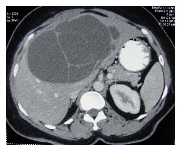
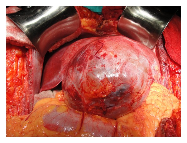
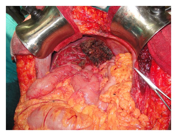
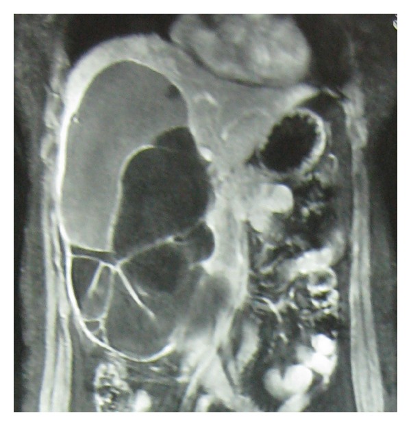
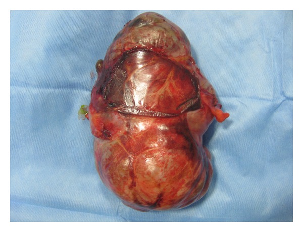
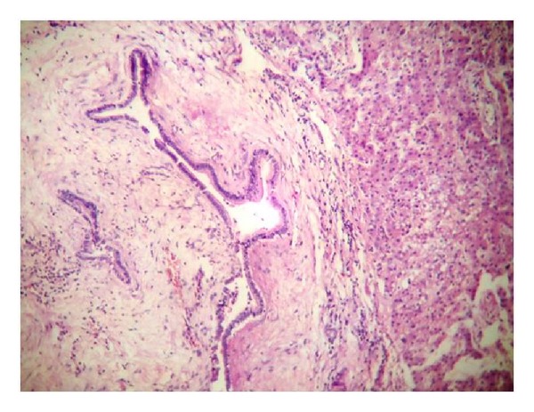
Introduction and Objective. Biliary cystadenoma is a rare benign neoplasm of the liver with less than 200 cases being reported allover the world. We report a series of 13 cases highlighting the radiological findings and problems related to its management. Materials and Methods. Records of thirteen patients who underwent surgery for biliary cystadenomas, between March 2006 and October 2011, were reviewed retrospectively. Results. Majority of the patients were females (11 out of 13), with a median age of 46 (23-65) years. The most frequent symptom was abdominal pain (92%). Seven patients had presented with history of previous surgery for liver lesions. Five patients had presented with recurrence after partial resection for a suspected hydatid cyst and two after surgery for presumed simple liver cyst. Ten of the 13 patients had complete resection of the cyst with enucleation in 3 patients, 2 of whom in addition required T-tube drainage of the bile duct. There has been no recurrence during the follow-up period ranging from 3 months to 5 years. Conclusion. Biliary cystadenoma must be differentiated from other benign cysts. Hepatic resection or cyst enucleation is the recommended treatment option.
Figures
Similar articles
-
Enucleation of Biliary Cystadenomas: a Review.J Gastrointest Surg. 2021 Oct;25(10):2700-2706. doi: 10.1007/s11605-021-05106-x. Epub 2021 Sep 9. J Gastrointest Surg. 2021. PMID: 34505221 Review.
-
Diagnostic and Therapeutic Challenges of Intrahepatic Biliary Cystadenoma and Cystadenocarcinoma: A Report of 10 Cases and Review of the Literature.Int Surg. 2015 Jul;100(7-8):1212-9. doi: 10.9738/INTSURG-D-15-00025.1. Int Surg. 2015. PMID: 26595495 Review.
-
Surgical management of simple liver cysts.Cir Cir. 2012 Jan-Feb;80(1):52-5. Cir Cir. 2012. PMID: 22472153
-
Effective treatment of biliary cystadenoma.Ann Surg. 2005 May;241(5):769-73; discussion 773-5. doi: 10.1097/01.sla.0000161982.57360.1b. Ann Surg. 2005. PMID: 15849512 Free PMC article.
-
Extrahepatic biliary cystadenomas and cystadenocarcinoma. Report of seven cases and review of the literature.Ann Surg. 1995 Nov;222(5):619-25. doi: 10.1097/00000658-199511000-00003. Ann Surg. 1995. PMID: 7487208 Free PMC article. Review.
Cited by
-
Mucinous Cystadenoma: A Rare Hepatic Tumor in a Child.Front Pediatr. 2017 Oct 9;5:215. doi: 10.3389/fped.2017.00215. eCollection 2017. Front Pediatr. 2017. PMID: 29062832 Free PMC article.
-
Extrahepatic biliary cystadenoma: a rare cause of biliary obstruction.Oman Med J. 2015 Jan;30(1):66-8. doi: 10.5001/omj.2015.13. Oman Med J. 2015. PMID: 25830005 Free PMC article.
-
Intrahepatic Biliary Cystadenoma With Colonic Adenomatous Polyps in a Patient With Chronic Hepatitis B: A Case Report and Literature Review.Front Med (Lausanne). 2021 Dec 16;8:760607. doi: 10.3389/fmed.2021.760607. eCollection 2021. Front Med (Lausanne). 2021. PMID: 34977067 Free PMC article.
-
Biliary cystadenoma masquerading as an adnexal cyst in pregnancy.BMJ Case Rep. 2021 Dec 14;14(12):e246392. doi: 10.1136/bcr-2021-246392. BMJ Case Rep. 2021. PMID: 34906958 Free PMC article.
-
Mucinous cystic neoplasm of the liver with extrahepatic growth presenting with ascending cholangitis diagnosed by endoscopic ultrasound features: a case report.J Med Case Rep. 2018 Feb 15;12(1):33. doi: 10.1186/s13256-017-1560-4. J Med Case Rep. 2018. PMID: 29444709 Free PMC article.
References
-
- Wheeler DA, Edmondson HA. Cystadenoma with mesenchymal stroma (CMS) in the liver and bile ducts. A clinicopathologic study of 17 cases, 4 with malignant change. Cancer. 1985;56(6):1434–1445. - PubMed
-
- Tsiftsis D, Christodoulakis M, de Bree E, Sanidas E. Primary intrahepatic biliary cystadenomatous tumors. Journal of Surgical Oncology. 1997;64(4):341–346. - PubMed
-
- Short WF, Nedwich A, Levy HA, Howard JM. Biliary cystadenoma. Report of a case and review of the literature. Archives of Surgery. 1971;102(1):78–80. - PubMed
-
- Ishak KG, Willis GW, Cummins SD, Bullock AA. Biliary cystadenoma and cystadenocarcinoma: report of 14 cases and review of the literature. Cancer. 1977;39(1):322–338. - PubMed
LinkOut - more resources
Full Text Sources
