

Review
doi: 10.1021/cn900006n.
Epub 2009 Oct 9.
Intercellular glutamate signaling in the nervous system and beyond
Affiliations
- PMID: 22778802
- PMCID: PMC3368625
- DOI: 10.1021/cn900006n
Item in Clipboard
Review
Intercellular glutamate signaling in the nervous system and beyond
ACS Chem Neurosci.
.
Abstract
Most intercellular glutamate signaling in the nervous system occurs at synapses. Some intercellular glutamate signaling occurs outside synapses, however, and even outside the nervous system where high ambient extracellular glutamate might be expected to preclude the effectiveness of glutamate as an intercellular signal. Here, I briefly review the types of intercellular glutamate signaling in the nervous system and beyond, with emphasis on the diversity of signaling mechanisms and fundamental unanswered questions.
Keywords: Glutamate; paracrine; receptor; synaptic.
Figures
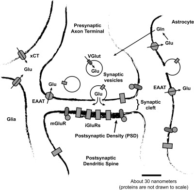
A canonical (but probably atypical) glutamatergic synapse. Cytoplasmic glutamate is put into synaptic vesicles by vesicular glutamate transporter (VGlut) proteins and eventually released into the synaptic cleft when the vesicles fuse with the presynaptic plasma membrane. After being released into the synaptic cleft, glutamate may bind to and activate glutamate receptor proteins. The postsynaptic density (PSD) contains by far the highest concentration of glutamate receptor protein, but receptors are also found in presynaptic terminals, in the neuronal membrane outside the synapse, in astrocytes, and in other cell types. There are two main types of glutamate receptor: ionotropic glutamate receptors (iGluRs) and metabotropic glutamate receptors (mGluRs). Glutamate secreted into the extracellular space is transported back into cells by excitatory amino acid transporters (EAATs). Most EAATs are present in astrocytes, although EAATs also exist in neurons and other cell types. Glutamate taken up by astrocytes is recycled by being converted to glutamine and exported into the extracellular space, where it is then transported into neurons and converted back into glutamate. Glia, neurons, and (to a lesser degree) other cell types also express xCT cystine−glutamate antiport proteins, which comprise the xc-system of amino acid transport. xCT proteins export glutamate from cells and import cystine; they are the largest source of ambient extracellular glutamate in the nervous system. Ambient extracellular glutamate modulates synaptic transmission via constitutive activation of mGluRs or constitutive desensitization of iGluRs (see text).
References
-
- Danbolt N. C. (2001) Glutamate uptake. Prog. Neurobiol. 65, 1–105. - PubMed
-
- Basic Neurochemistry: Molecualar, Cellular, and Medical Aspects (2006) 7th ed., Elsevier Academic Press, Burlington, MA.
-
- Ottersen O. P.; Zhang N.; Walberg F. (1992) Metabolic compartmentation of glutamate and glutamine: morphological evidence obtained by quantitative immunocytochemistry in rat cerebellum. Neuroscience 46, 519–534. - PubMed
-
- Osen K. K.; Storm-Mathisen J.; Ottersen O. P.; Dihle B. (1995) Glutamate is concentrated in and released from parallel fiber terminals in the dorsal cochlear nucleus: a quantitative immunocytochemical analysis in guinea pig. J. Comp. Neurol. 357, 482–500. - PubMed
-
- Bramham C. R.; Torp R.; Zhang N.; Storm-Mathisen J.; Ottersen O. P. (1990) Distribution of glutamate-like immunoreactivity in excitatory hippocampal pathways: a semiquantitative electron microscopic study in rats. Neuroscience 39, 405–417. - PubMed
Publication types
MeSH terms
Substances
LinkOut - more resources
Full Text Sources
