. 2010 Oct 20;1(10):661-78.
doi: 10.1021/cn900019r. Epub 2010 Aug 26.

Effects of peptides derived from terminal modifications of the aβ central hydrophobic core on aβ fibrillization

Effects of peptides derived from terminal modifications of the aβ central hydrophobic core on aβ fibrillization

Cyrus K Bett et al. ACS Chem Neurosci. .

Abstract

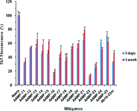
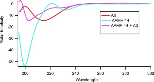
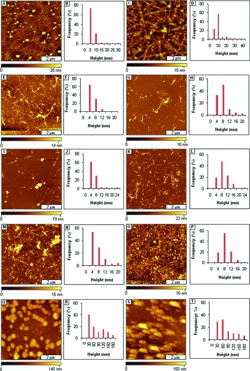
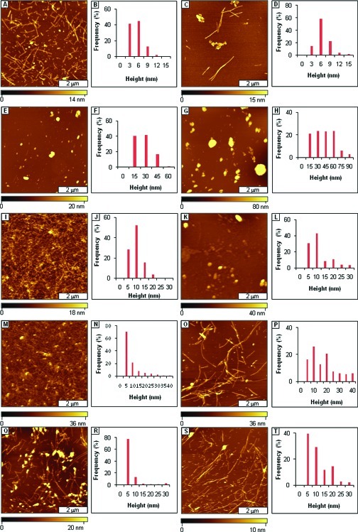
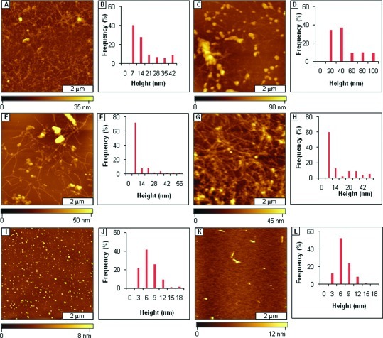
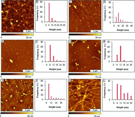
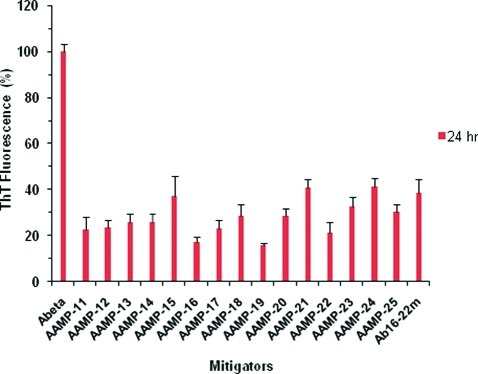
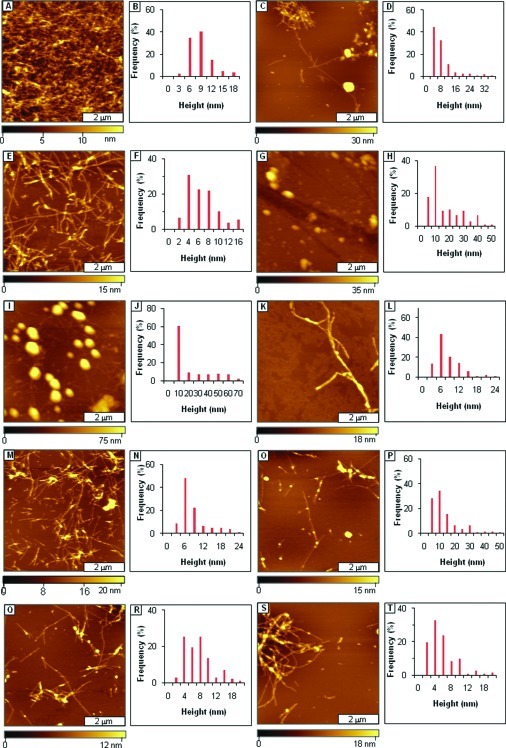
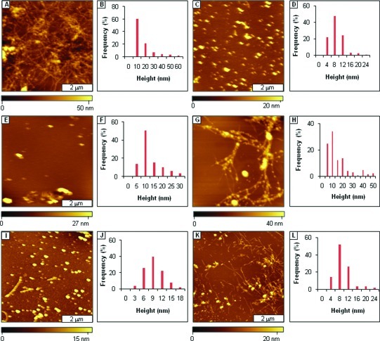
Considerable research effort has focused on the discovery of mitigators that block the toxicity of the β-amyloid peptide (Aβ) by targeting a specific step involved in Aβ fibrillogenesis and subsequent aggregation. Given that aggregation intermediates are hypothesized to be responsible for Aβ toxicity, such compounds could likely prevent or mitigate aggregation, or alternatively cause further association of toxic oligomers into larger nontoxic aggregates. Herein we investigate the effect of modifications of the KLVFF hydrophobic core of Aβ by replacing N- and C-terminal groups with various polar moieties. Several of these terminal modifications were found to disrupt the formation of amyloid fibrils and in some cases induced the disassembly of preformed fibrils. Significantly, mitigators that incorporate MiniPEG polar groups were found to be effective against Aβ(1-40) fibrilligonesis. Previously, we have shown that mitigators incorporating alpha,alpha-disubstituted amino acids (ααAAs) were effective in disrupting fibril formation as well as inducing fibril disassembly. In this work, we further disclose that the number of polar residues (six) and ααAAs (three) in the original mitigator can be reduced without dramatically changing the ability to disrupt Aβ(1-40) fibrillization in vitro.

Keywords: Amyloid peptide (Aβ); assembly; disassembly; fibrils; mitigators; spherical structures.


Figures

Figure 1
Figure 1
Time dependent ThT fluorescence assay of Aβ/AAMPs. (The final concentration was 40 μM for both Aβ1−40 and AAMPs.)
Figure 2
Figure 2
An example of a CD spectra for AAMP-14 alone, Aβ1−40 alone, and an Aβ1−40/AAMP-14 equimolar mixture after 1 week of aging. (Final concentration was 40 μM for both Aβ1−40 and AAMPs.)
Figure 3
Figure 3
Mitigation of Aβ1−40 aggregation by AAMPs with polar groups added to both the C- and N-termini, as viewed by AFM topographs. (A) Fibrils formed after 3 days of incubation by Aβ1−40 alone; (B) height analysis for A; (C) fibrils present after 1 week; (D) height analysis for C; (E) fibrils formed after mitigation for 3 days by AAMP-11; (F) height analysis for E; (G) fibrils formed after 1 week; (H) height distribution histogram for G; (I) mixture of spherical and protofibril structures after 3 days of mitigation by AAMP-12; (J) height analysis for I; (K) spherical and short sized fibrils observed after 1 week; (L) height histogram for K; (M) protofibrils present after 3 days with AAMP-13; (N) height analysis for M; (O) fibrils formed after 1 week; (P) height distribution analysis for O; (Q) spherical structures present after mitigation for 3 days by AAMP-14; (R) height distribution for Q; (S) spherical structures observed after 1 week; (T) height analysis for S. (The final concentration was 40 μM for both Aβ1−40 and AAMPs.)
Figure 4
Figure 4
Disruption of Aβ1−40 fibril formation by AAMPs with polar groups added to the C- or N-terminus, characterized by tapping mode AFM. (A) Views of short fibrils after 3 days aging Aβ1−40 in the presence of AAMP-15; (B) corresponding height distribution for A; (C) after 1 week; (D) height analysis for C; (E) spherical structures detected after 1 week of aging an Aβ1−40/AAMP-16 mixture; (F) corresponding height distribution; (G) after 1 week; (H) height distribution analysis for G; (I) views of spherical and protofibrils formed by an Aβ1−40/AAMP-17 mixture after 3 days of aging; (J) corresponding height analysis; (K) after 1 week; (L) height analysis for K; (M) views of spherical particles and protofibrils formed by Aβ1−40 aggregation mitigation by AAMP-18 after 3 days of aging; (N) corresponding height distribution; (O) fibrils detected after 1 week; (P) height analysis for O. (Q) Spherical aggregates and protofibrils observed after 3 days of Aβ1−40 mitigation by AAMP-19; (R) height distribution histogram for Q; (S) fibrils were present after 1 week of aging; (T) height analysis for S.
Figure 5
Figure 5
Disruption of Aβ1−40 fibril formation by ααAAs-containing AAMPs. (A) Protofibrils and spherical structures observed after 3 days of aging Aβ1−40 in the presence of AAMP-20; (B) height distribution for A; (C) spherical structures and fibrils (background) observed after 1 week; (D) corresponding height analysis for C; (E) views of fibrils with some spherical structures detected after 3 days of aging the AB1−40/AAMP-21 mixture; (F) corresponding height distribution; (G) after 1 week; (H) height distribution analysis for G; (I) spherical structures observed for the Aβ1−40/AAMP-22 mixture after 3 days of aging; (J) corresponding height analysis; (K) spherical and linear aggregates after 1 week; (L) height analysis for K.
Figure 6
Figure 6
Effects of ααAAs-containing AAMPs on Aβ1−40 fibril formation. (A) Protofibrils and spherical structures were observed after 3 days of aging Aβ1−40 in the presence of AAMP-23; (B) height distribution for A; (C) spherical structures and linear structures observed after 1 week; (D) corresponding height analysis; (E) spherical structures observed after 1 week of aging the Aβ1−40/AAMP-24 mixture; (F) height distribution analysis for E; (G) rod-like fibrils and spherical aggregates observed after 1 week; (H) corresponding histogram analysis; (I) spherical and protofibrillar structures observed after aging Aβ1−40/AAMP-25 for 3 days; (J) corresponding height analysis; (K) after 1 week (L); height analysis for K.
Figure 7
Figure 7
Fibril disassembly by various AAMPs as monitored by ThT fluorescence, presented as the percent relative to that of Aβ1−40.
Figure 8
Figure 8
Disassembly of preformed fibrils of Aβ1−40. (A) Control sample of Aβ1−40 fibrils. (B) Corresponding height histogram. (C) Fibrils and spherical structures after disassembly by AAMP-11. (D) Histogram analysis for C. (E) Partial disassembly by AAMP-12. (F) Height histogram analysis for E. (G) Spherical particles formed by AAMP-13 fibril disassembly. (H) Height analysis for G. (I) Spherical aggregates and fibrils (background) formed from disassembly by AAMP-14. (J) Corresponding height analysis. (K) Isolated fibrils present from disassembly by AAMP-15. (L) Height histogram for K. (M) Mixture of short and long fibrils observed from disassembly by AAMP-16. (N) Corresponding height analysis. (O) Fibrils showing beaded sections for Aβ1−40 fibril disassembly by AAMP-17. (P) Corresponding height histogram. (Q) Fibrils showing nucleation units for mature fibrils after disassembly by AAMP-18. (R) Corresponding height analysis. (S) Partial preformed fibril disassembly observed with AAMP-19. (T) Height histogram for O.
Figure 9
Figure 9
Disassembly of Aβ1−40 preformed fibrils imaged using tapping mode AFM. (A) Fibril disassembly by AAMP-20. (B) Corresponding height histogram. (C) Spherical structures were observed with disassembly by AAMP-21. (D) Height analysis for C. (E) Spherical structures with disassembly by AAMP-22. (F) Corresponding height histogram. (G) Fibrils with beaded morphology observed from disassembly by AAMP-23. (H) Height analysis for G. (I) Spherical aggregates and isolated protofibrils formed from disassembly by AAMP-24. (J) Height analysis for I. (K) Fibrils and some spherical species observed from fibril disassembly by AAMP-25. (L) Corresponding height analysis. (The final concentration was 40 μM for both Aβ1−40 and AAMPs.)

Similar articles

Cited by

References

    1. Selkoe D. J. (1991) The molecular pathology of Alzheimers-disease. Neuron 6, 487–498. - PubMed
    1. Selkoe D. J. (2003) Folding proteins in fatal ways. Nature 426, 900–904. - PubMed
    1. Tycko R. (2003) Insights into the amyloid folding problem from solid-state NMR. Biochemistry 42, 3151–3159. - PubMed
    1. Baglioni S.; Casamenti F.; Bucciantini M.; Luheshi L. M.; Taddei N.; Chiti F.; Dobson C. M.; Stefani M. (2006) Prefibrillar amyloid aggregates could be generic toxins in higher organisms. J. Neurosci. 26, 8160–8167. - PMC - PubMed
    1. Chromy B. A.; Nowak R. J.; Lambert M. P.; Viola K. L.; Chang L.; Velasco P. T.; Jones B. W.; Fernandez S. J.; Lacor P. N.; Horowitz P.; Finch C. E.; Krafft G. A.; Klein W. L. (2003) Self-assembly of A beta(1−42) into globular neurotoxins. Biochemistry 42, 12749–12760. - PubMed

MeSH terms